. 1999 Feb;103(4):461-8.
doi: 10.1172/JCI5252.

Explanations for the clinical and microscopic localization of lesions in pemphigus foliaceus and vulgaris

Affiliations

Explanations for the clinical and microscopic localization of lesions in pemphigus foliaceus and vulgaris

M G Mahoney et al. J Clin Invest. 1999 Feb.

Abstract

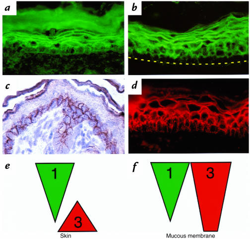
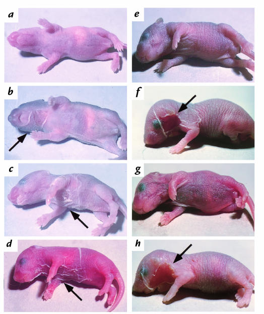
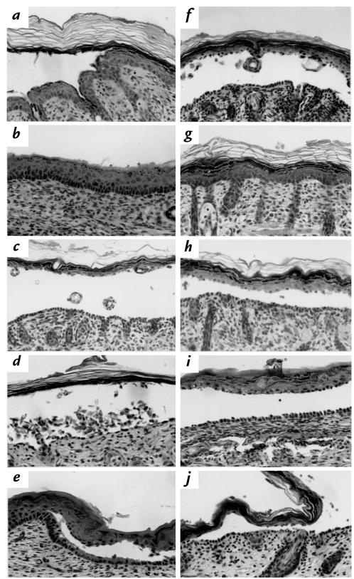
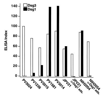
Patients with pemphigus foliaceus (PF) have blisters on skin, but not mucous membranes, whereas patients with pemphigus vulgaris (PV) develop blisters on mucous membranes and/or skin. PF and PV blisters are due to loss of keratinocyte cell-cell adhesion in the superficial and deep epidermis, respectively. PF autoantibodies are directed against desmoglein (Dsg) 1; PV autoantibodies bind Dsg3 or both Dsg3 and Dsg1. In this study, we test the hypothesis that coexpression of Dsg1 and Dsg3 in keratinocytes protects against pathology due to antibody-induced dysfunction of either one alone. Using passive transfer of pemphigus IgG to normal and DSG3(null) neonatal mice, we show that in the areas of epidermis and mucous membrane that coexpress Dsg1 and Dsg3, antibodies against either desmoglein alone do not cause spontaneous blisters, but antibodies against both do. In areas (such as superficial epidermis of normal mice) where Dsg1 without Dsg3 is expressed, anti-Dsg1 antibodies alone can cause blisters. Thus, the anti-desmoglein antibody profiles in pemphigus sera and the normal tissue distributions of Dsg1 and Dsg3 determine the sites of blister formation. These studies suggest that pemphigus autoantibodies inhibit the adhesive function of desmoglein proteins, and demonstrate that either Dsg1 or Dsg3 alone is sufficient to maintain keratinocyte adhesion.


Figures

Figure 1
Figure 1
Distribution of Dsg1 and Dsg3 in neonatal mouse skin and mucous membrane. Immunofluorescence staining of neonatal mouse skin (a) and mucous membrane (b) with human PF1239 serum. Immunoperoxidase (c) and immunofluorescence (d) staining of neonatal mouse skin and mucous membrane with rabbit anti-Dsg3 antibodies. Schematic diagram of the Dsg1 and Dsg3 expression in the neonatal mouse skin (e) and oral mucous membrane (f). Note that Dsg1 is expressed throughout all cell layers, more in the superficial layers and less in the deep layers of both epidermis and mucous membrane. In the epidermis, Dsg3 is expressed only in the deep basal and most immediate suprabasal layers, whereas in the oral mucosa Dsg3 is expressed throughout all cell layers. Dsg, desmoglein.
Figure 2
Figure 2
Neonatal mice injected with PF and PV IgG. (ac) Injection of low-dose (3 mg) PF982 IgG. DSG3+/+ has no detectable blisters (a); DSG3+/– has localized blisters (b); DSG3null shows extensive blisters (c). These results show that the presence of Dsg3 limits blisters due to anti-Dsg1 in PF sera. (d) DSG3null injected with 10 mg of IgG from PV3014 containing both anti-Dsg3 and anti-Dsg1 antibodies shows extensive blisters, demonstrating that anti-Dsg1 antibodies in PV serum are pathogenic. (e) Wild-type mouse injected with high-dose (26 mg) IgG from PV3024 containing only anti-Dsg3 antibodies (without anti-Dsg1 antibodies) developed no skin blisters. (f) Wild-type mouse injected with 9.4 mg from NIH2054, containing anti-Dsg3 and anti-Dsg1 antibodies, shows extensive blisters. (g) Wild-type mouse injected with 12.0 mg of IgG from NIH2054 adsorbed with Dsg1 no longer demonstrates blister formation. (h) Wild-type mouse injected with 10 mg IgG from PV3024 (anti-Dsg3 antibodies) plus 1 mg IgG from PF982 (anti-Dsg1 antibodies) shows extensive blisters. (e–h) These results show the importance of anti-Dsg1 for efficient blister formation by the anti-Dsg3 in PV sera. Arrows mark the sites of blister formation. PF, pemphigus foliaceus; PV, pemphigus vulgaris.
Figure 3
Figure 3
Histology of skin and oral mucosa of neonatal mice injected with PF and PV IgG. (a) Skin from a wild-type mouse injected with (10 mg) PF982 (anti-Dsg1) IgG shows superficial blisters. (b) Mucous membrane from a wild-type mouse injected with (10 mg) PF982 IgG shows no blisters. Skin from a DSG3null mouse injected with (10 mg) PF982 IgG reveals deep suprabasilar blisters (c) and extensive acantholysis (d). (e) Mucous membrane from a DSG3null neonatal mouse injected with (10 mg) PF982 IgG shows deep suprabasilar blisters. (c–e) These results demonstrate that in DSG3null mice, in which there is no Dsg3 to compensate, PF IgG causes deep suprabasilar blisters in both the skin and mucous membranes. (f) Skin from a DSG3null mouse injected with (10 mg) PV3014 (anti-Dsg1 and anti-Dsg3) IgG shows suprabasilar blisters. (g) Skin from a wild-type mouse injected with (26 mg) PV3024 (anti-Dsg3) IgG shows no blisters. (h) Skin from a wild-type mouse injected with (10 mg) PV3014 (anti-Dsg1 and anti-Dsg3) IgG show suprabasilar blisters. (i) Mucous membrane from a wild-type mouse injected with (10 mg) PV3014 IgG shows suprabasilar blisters. (j) Skin from a wild-type mouse injected with (10 mg) PV3024 IgG and (1 mg) PF982 IgG shows suprabasilar blisters. (f–j) These results demonstrate that anti-Dsg1 antibodies in PV are pathogenic and that both anti-Dsg1 and anti-Dsg3 antibodies are necessary for efficient blister formation in PV.
Figure 4
Figure 4
Levels of anti-Dsg1 and anti-Dsg3 antibodies in PV sera measured by ELISA. Dsg1 ads. indicates that the serum was adsorbed with the extracellular domain of Dsg1 produced in baculovirus. The ELISA index is defined in Methods.

References

    1. Stanley, J.R. 1993. Pemphigus. In Dermatology in general medicine. T.B. Fitzpatrick, A.Z. Eisen, K. Wolff, I. M. Freedberg, and K.F. Austen, editors. McGraw-Hill. New York, NY. 606–615.
    1. Lever, W.F. 1965. Pemphigus and pemphigoid. Charles C. Thomas. Springfield, IL. 15–66.
    1. Meurer M, Millns JL, Rogers RS, III, Jordon RE. Oral pemphigus vulgaris. A report of ten cases. Arch Dermatol. 1977;113:1520–1524. - PubMed
    1. Beutner EH, Jordon RE. Demonstration of skin antibodies in sera of pemphigus vulgaris patients by indirect immunofluorescent staining. Proc Soc Exp Biol Med. 1964;117:505–510. - PubMed
    1. Rappersberger K, Roos N, Stanley JR. Immunomorphological and biochemical identification of the pemphigus foliaceus autoantigen within desmosomes. J Invest Dermatol. 1992;99:323–330. - PubMed

Publication types