. 1999 Feb;103(4):543-53.
doi: 10.1172/JCI2517.

Bcl-2 and Bcl-XL serve an anti-inflammatory function in endothelial cells through inhibition of NF-kappaB

Affiliations

Bcl-2 and Bcl-XL serve an anti-inflammatory function in endothelial cells through inhibition of NF-kappaB

A Z Badrichani et al. J Clin Invest. 1999 Feb.

Abstract

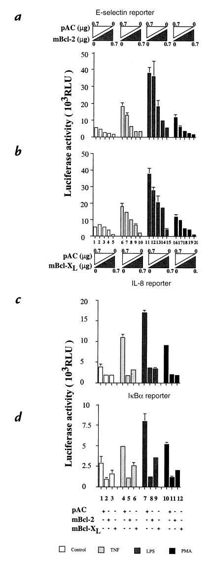
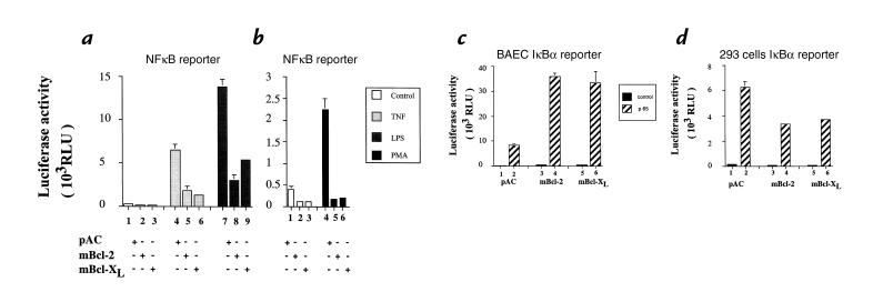
To maintain the integrity of the vascular barrier, endothelial cells (EC) are resistant to cell death. The molecular basis of this resistance may be explained by the function of antiapoptotic genes such as bcl family members. Overexpression of Bcl-2 or Bcl-XL protects EC from tumor necrosis factor (TNF)-mediated apoptosis. In addition, Bcl-2 or Bcl-XL inhibits activation of NF-kappaB and thus upregulation of proinflammatory genes. Bcl-2-mediated inhibition of NF-kappaB in EC occurs upstream of IkappaBalpha degradation without affecting p65-mediated transactivation. Overexpression of bcl genes in EC does not affect other transcription factors. Using deletion mutants of Bcl-2, the NF-kappaB inhibitory function of Bcl-2 was mapped to bcl homology domains BH2 and BH4, whereas all BH domains were required for the antiapoptotic function. These data suggest that Bcl-2 and Bcl-XL belong to a cytoprotective response that counteracts proapoptotic and proinflammatory insults and restores the physiological anti-inflammatory phenotype to the EC. By inhibiting NF-kappaB without sensitizing the cells (as with IkappaBalpha) to TNF-mediated apoptosis, Bcl-2 and Bcl-XL are prime candidates for genetic engineering of EC in pathological conditions where EC loss and unfettered activation are undesirable.


Figures

Figure 1
Figure 1
Expression of Bcl-2 or Bcl-XL in BAEC after Lipofectamine-mediated transfection inhibits TNF-induced apoptosis in CHX-sensitized EC. (a) Immunoblot detection of Bcl-2 (lanes 1 and 2) and Bcl-XL (lanes 3 and 4) in BAEC-transfected cells using polyclonal anti–Bcl-2 and anti–Bcl-XL antibodies. Arrows indicate that transfected BAEC express high levels of Bcl-2 or Bcl-XL as opposed to control nontransfected cells. (b) BAEC were cotransfected with a CMVβ-gal reporter (0.5 μg) and 1 μg of pAC (lanes 1 and 2), mBcl-2 (lanes 3 and 4), or mBcl-XL (lanes 5 and 6). CHX was added to transfected cells (all lanes) that were subsequently stimulated (lanes 2, 4, and 6) or not (lanes 1, 3, and 5) with TNF for 12 h. The percent cell survival was calculated as described in Methods. Expression of Bcl-2 or Bcl-XL rescues CHX-sensitized EC from TNF-mediated apoptosis. Error bars are ± SE. Graph shown is representative of three experiments.BAEC, bovine aortic endothelial cells; β-gal, β-galactosidase; CHX, cycloheximide; EC, endothelial cells; TNF, tumor necrosis factor.
Figure 2
Figure 2
Expression of Bcl-2 or Bcl-XL inhibits the induction by TNF, LPS, and PMA of (a and b) E-selectin, (c) IL-8, and (d) IκBα reporters. E-selectin reporter induction by TNF, LPS, and PMA is inhibited by (a) Bcl-2 and (b) Bcl-XL in a dose-dependent manner. Both mBcl-2 and mBcl-XL were titrated (0, 0.25, 0.5, 0.6, 0.7 μg) with pAC to equal 0.7 μg. The graphs shown are representative of four experiments. Significant inhibition is achieved at doses of 0.5 μg and higher. For (c) IL-8 and (d) IκBα reporters, 0.7 μg of either Bcl-2, Bcl-XL, or pAC expression plasmids was used for BAEC transfection. The graphs shown are representative of four and three experiments, respectively. For all the reporters studies, BAEC were treated with TNF at a concentration of 100 U/ml, LPS at a concentration of 100 ng/ml, or PMA at a concentration of 5 × 10⁻⁸ M. Results are given in RLU. Error bars represent ± SE. RLU, relative light units.
Figure 3
Figure 3
(a and b) Bcl-2 or Bcl-XL expression in BAEC prevents the induction of an NF-κB reporter after TNF, LPS, and PMA stimulation without interfering with (c) p65-mediated transactivation, in contrast with (d) 293 cells. (a and b) BAEC were cotransfected with 0.7 μg of pAC (a; lanes 1, 4, and 7), Bcl-2 (a; lanes 2, 5, and 8), or mBcl-XL (a; lanes 3, 6, and 9) expression plasmids along with 0.6 μg of the NF-κB reporter and 0.3 μg of β-gal reporter. Cells were stimulated with either 100 U/ml TNF (a; lanes 46), 100 ng/ml LPS (a; lanes 79), or 5 × 10⁻⁸ PMA (b; lanes 46). The graphs are representative of at least three experiments performed. Results are given in RLU. Error bars are ± SE. (c) BAEC were cotransfected with 0.3 μg β-gal, 0.6 μg IκBα reporter, 40 ng p65 (RelA), and 0.7 μg of either pAC (lanes 1 and 2), mBcl-2 (lanes 3 and 4), or mBcl-XL (lanes 5 and 6) expression plasmids. Overexpression of Bcl-2 or Bcl-XL does not inhibit the induction of IκBα by p65 (lanes 4 and 6 vs. lane 2). (d) 293 cells were transfected with the same plasmids with the exception of higher concentrations of the expression plasmids (1 μg). Results show that expression of Bcl-2 or Bcl-XL inhibits the ability of p65 (RelA) to induce an IκBα reporter. Data shown are representative of three experiments performed. Results are expressed in RLU. Error bars represent ± SE.
Figure 4
Figure 4
(a) Adenoviral-mediated gene transfer of Bcl-2 to HUVEC achieves high levels of expression of the transgene. (b) Bcl-2 expression inhibits E-selectin, and (c) VCAM-1 inhibits upregulation by inhibiting NF-κB activation at (d) a level upstream of IκBα degradation. (a) 90%-confluent HUVEC monolayers were either noninfected or infected with the control rAd.β-gal or the rAd.hBcl-2 at a MOI of 100. Forty-eight hours after infection, cell extracts were recovered, and 1.5 μg of protein was run on a SDS-PAGE and checked by Western blot analysis for the expression of the transgene using a rabbit polyclonal anti–human Bcl-2 antiserum. Results show high levels of expression of Bcl-2 in HUVEC infected with the rAd.hBcl-2 (arrow). (b and c) Expression of Bcl-2 in HUVEC significantly decreases TNF-mediated upregulation of E-selectin and VCAM-1 as assessed by flow cytometry. In all cases, E-selectin and VCAM-1 expression levels on nonstimulated HUVEC is represented by a light line, expression after TNF treatment is shown by a dark line, and labeling of an isotype-matched control monoclonal antibody is illustrated by a broken line. (d) Western blot analysis of IκBα expression after TNF treatment. Extracts from noninfected (lanes 13), rAd.hBcl-2 (lanes 4–6), and rAd.β-gal (lanes 7–9) were recovered before and 15 min and 2 h after TNF treatment, and assayed for IκBα expression. Results show that Bcl-2 expression in HUVEC inhibits the usual IκBα degradation that occurs 15 min after TNF stimulation (lane 4 vs. lanes 2 and 8). A second, slower migrating form of IκBα is strongly stabilized in the Bcl-2–expressing EC. HUVEC, human umbilical vein cells; MOI, moiety of infection; VCAM-1, vascular cell adhesion molecule-1.
Figure 5
Figure 5
Overexpression of Bcl-2 and Bcl-XL does not affect the transactivation properties of Sp1. BAEC were cotransfected with mBcl-2, mBcl-XL, or pAC together with an HIV-CAT reporter. Induction of this reporter was achieved by cotransfecting the viral protein c-Tat, which requires the Sp1 binding sites of the HIV-CAT reporter. Results show no difference in the c-Tat–mediated induction of the HIV-CAT whether the cells were transfected with pAC (lane 1), Bcl-2 (lane 2), or Bcl-XL (lane 3). Data shown is representative of two experiments performed. Results are expressed in fold induction. CAT, chloramphenicol acetyltransferase.
Figure 6
Figure 6
(a) Structure/function relationships of Bcl-2 deletion mutants. Expression of Bcl-2 deletion mutants in BAEC. (b) The Bcl homology domains BH4 and BH2 are required for the inhibitory effect of Bcl-2 upon NF-κB activation after TNF and LPS stimulation, whereas (c) all BH domains and the NRD are required for the antiapoptotic function of Bcl-2. (a) Immunoblot detection of human wild-type and deletion mutants of Bcl-2 (lanes 2–6) in BAEC-transfected cells, using a polyclonal anti–Bcl-2 antibody. Mutant Δ1 is not recognized by this antibody and was detected using a monoclonal anti–Bcl-2 antibody (lane 9). (b) BAEC were cotransfected with 0.3 μg of β-gal, 0.6 μg of NF-κB reporter along with 0.7 μg of pAC (lanes A1–3), human Bcl-2 (lanes B1–3), Δ1 (lanes C1–3), Δ2 (lanes D1–3), Δ4 (lanes E1–3), Δ8 (lanes F1–3), and Δ12 (lanes G1–3). Cells were stimulated with 100 U/ml TNF (lanes 2) or 100 ng/ml LPS (lanes 3). Overexpression of Δ2, Δ4, or Δ8 inhibits the induction by TNF or LPS of a NF-κB reporter, in contrast to Δ1 or Δ12. Graph shown is representative of four experiments. Results are expressed in RLU. Error bars represent ± SE. (c) BAEC were cotransfected with a CMVβ-gal reporter (0.5 μg) and 1 μg of pAC (lanes 1 and 2), hBcl-2 (lanes 3 and 4), Δ1 (lanes 5 and 6), Δ2 (lanes 7 and 8), Δ4 (lanes 9 and 10), Δ8 (lanes 11 and 12), Δ12 (lanes 13 and 14). CHX was added to transfected cells (all lanes) that were subsequently stimulated (even lanes) or not (odd lanes) with TNF for 12 h. The percent cell survival was calculated as described in Methods. Results demonstrate that all the Bcl-2 deletion mutants lose their ability to protect EC from CHX-TNF mediated apoptosis. Error bars represent ± SE. Graph shown is representative of three experiments. NRD, negative regulatory domain.

References

    1. Pober JS, et al. The potential roles of vascular endothelium in immune reactions [review] Hum Immunol. 1990;28:258–262. - PubMed
    1. Cotran RS, Pober JS. Cytokine-endothelial interactions in inflammation, immunity, and vascular injury. J Am Soc Nephrol. 1990;1:225–235. - PubMed
    1. Bach FH, et al. Endothelial cell activation and thromboregulation during xenograft rejection [review] Immunol Rev. 1994;141:5–30. - PubMed
    1. Gimbrone MAJ. Vascular endothelium: an integrator of pathophysiologic stimuli in atherosclerosis [review] Am J Cardiol. 1995;75:67B–70B. - PubMed
    1. Read MA, Whitley MZ, Williams AJ, Collins T. NF-κB and IκBα — an inducible regulatory system in endothelial activation. J Exp Med. 1994;179:503–512. - PMC - PubMed

MeSH terms