. 1999 Mar;19(3):1661-72.
doi: 10.1128/MCB.19.3.1661.

p50(cdc37) acting in concert with Hsp90 is required for Raf-1 function

Affiliations

p50(cdc37) acting in concert with Hsp90 is required for Raf-1 function

N Grammatikakis et al. Mol Cell Biol. 1999 Mar.

Abstract

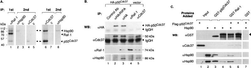
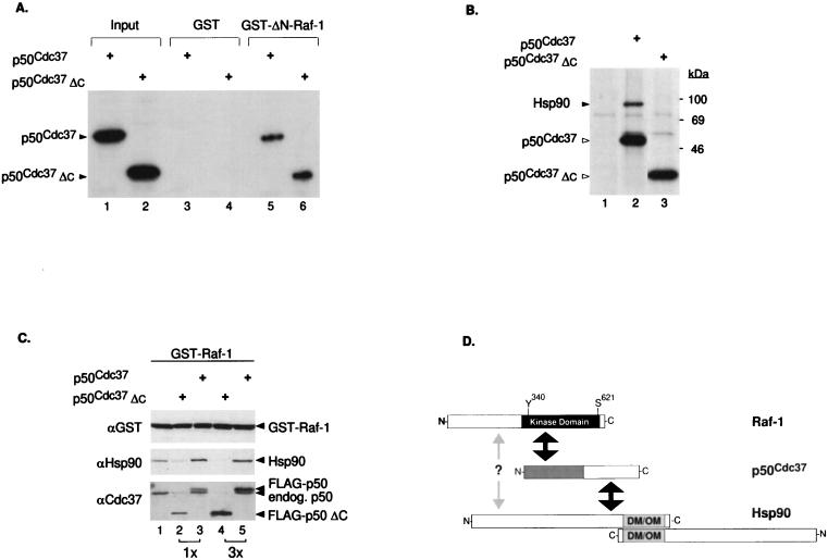
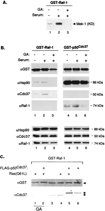
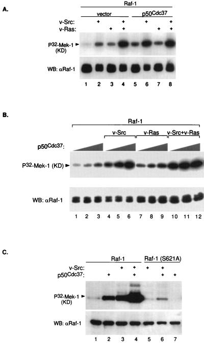
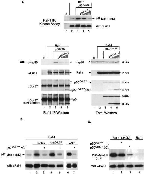
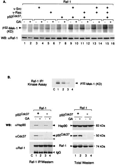
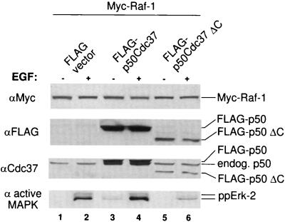
Genetic screens in Drosophila have identified p50(cdc37) to be an essential component of the sevenless receptor/mitogen-activated kinase protein (MAPK) signaling pathway, but neither the function nor the target of p50(cdc37) in this pathway has been defined. In this study, we examined the role of p50(cdc37) and its Hsp90 chaperone partner in Raf/Mek/MAPK signaling biochemically. We found that coexpression of wild-type p50(cdc37) with Raf-1 resulted in robust and dose-dependent activation of Raf-1 in Sf9 cells. In addition, p50(cdc37) greatly potentiated v-Src-mediated Raf-1 activation. Moreover, we found that p50(cdc37) is the primary determinant of Hsp90 recruitment to Raf-1. Overexpression of a p50(cdc37) mutant which is unable to recruit Hsp90 into the Raf-1 complex inhibited Raf-1 and MAPK activation by growth factors. Similarly, pretreatment with geldanamycin (GA), an Hsp90-specific inhibitor, prevented both the association of Raf-1 with the p50(cdc37)-Hsp90 heterodimer and Raf-1 kinase activation by serum. Activation of Raf-1 via baculovirus coexpression with oncogenic Src or Ras in Sf9 cells was also strongly inhibited by dominant negative p50(cdc37) or by GA. Thus, formation of a ternary Raf-1-p50(cdc37)-Hsp90 complex is crucial for Raf-1 activity and MAPK pathway signaling. These results provide the first biochemical evidence for the requirement of the p50(cdc37)-Hsp90 complex in protein kinase regulation and for Raf-1 function in particular.


Figures

FIG. 1
FIG. 1
Association of p50cdc37, Hsp90, and Raf-1 in vivo and in vitro. (A) Lane 1, anti-Raf-1 IP from [35S]methionine-labeled Cos-1 cells. Lanes 2 to 5, after the primary anti-Raf IP was boiled for 2 min in the presence of 0.5% SDS, a second IP was carried out with anti-Hsp90 or control (c) antibody (lanes 2 and 3) or with polyclonal anti-p50cdc37 or nonimmune rabbit (c) antibody (lanes 4 and 5, respectively). Lanes 6 and 7, anti-p50cdc37 primary IPs and nonimmune rabbit serum IPs, respectively, from [35S]methionine-labeled Cos-1 cells. A second IP with anti-Hsp90 antibody (lane 8) was performed with a fraction of the anti-p50cdc37 primary immunoprecipitate identical to that run in lane 6. The relative migration of molecular weight marker proteins is indicated. (B) Plasmids pMT3-HA-p50cdc37 and pMT3-HA were transiently transfected into Cos-1 cells, and extracts were immunoprecipitated with anti-FLAG antibody (Ab) M5 as a control (lane 1) or anti-HA monoclonal antibody 12CA5 under either denaturing or mild conditions (RIPA or NP-40 LB buffer; lanes 2 and 3, respectively) or, to purify endogenous Raf-1 and p50cdc37 proteins, with anti-Raf-1 (lane 4) and anti-p50cdc37 (lane 5) monoclonal antibodies. Immunoprecipitated proteins were examined by Western blotting (WB) and ECL for the presence of transfected HA-p50cdc37 with anti-HA antibody or for the presence of both transfected and endogenous p50cdc37 with anti-p50cdc37 rabbit antisera. Endogenous Raf-1 and Hsp90 proteins were detected with rabbit-anti-Raf-1 antibodies and rat-anti-Hsp90, respectively (top to bottom panels). IgGH, precipitating IgG antibody heavy chains. (C) FLAG-p50cdc37 (immunoaffinity purified from baculovirus-infected Sf9 cells) and Hsp90 (recombinant E. coli; Stressgen) were assayed in vitro for binding to bacterially produced GST-Raf-1, GST-p50cdc37, or GST alone as indicated by GSH-Sepharose pull-down assays and Western blotting (WB) with the indicated antibodies as described in Materials and Methods. Anti-Hsp90 immunoblotting performed with two distinct Hsp90-specific antibodies (SPA-830 and SPA-771) is shown (bottom two panels). The first two lanes indicate the input amounts of purified proteins added. The arrowhead denotes the position of the full-length GST-Raf-1 above the breakdown products.
FIG. 2
FIG. 2
The N-terminal half of p50cdc37 mediates association with the catalytic domain of Raf-1 but is impaired for Hsp90 interaction and accumulation to Raf-1. (A) Plasmids pSG5-p50cdc37 and pSG5-p50cdc37ΔC were transcribed and translated in vitro, using T7 RNA polymerase and a reticulocyte lysate system (Promega); 5 μl of each reaction mixture was either analyzed directly (input lanes) or assayed in vitro for binding to either GST or bacterially purified GST–ΔN-Raf-1(Δ26-309) and visualized by SDS-PAGE and fluorography. Comparable results were obtained with full-length GST–Raf-1 (not shown). (B) Cos-1 cells transfected with pSG5-FLAG vector, pSG5-FLAG-p50cdc37, and pSG5-FLAG-p50cdc37ΔC were [35S]methionine labeled, and anti-FLAG IPs in NP-40 LB of each transfected sample were analyzed by SDS-PAGE and fluorography (lanes 1 to 3, respectively). Proteins at the sizes predicted for overexpressed FLAG-p50cdc37 proteins or associated endogenous Hsp90 are also indicated. (C) Two micrograms of pEBG-GST-Raf-1 was cotransfected with 5 μg of pSG5-FLAG vector (lane 1), pSG5-FLAG-p50cdc37 (lanes 2 and 3), or pSG5-FLAG-p50cdc37ΔC (lanes 4 and 5) at 5 or 15 μg as indicated. After 48 h in DMEM-FBS, all five cultures were harvested and lysed in NP-40 LB, and GST–Raf-1 was GSH-Sepharose purified and tested for associated p50cdc37 or Hsp90 proteins with rabbit anti-p50cdc37 or rat anti-Hsp90 antibody. A control anti-GST immunoblot was also included to detect overexpressed GST–Raf-1 (top panel). (D) Diagram indicating regions of interaction between p50cdc37, Raf-1, and Hsp90. The N-terminal half of p50cdc37 (gray area) which corresponds to p50cdc37ΔC is sufficient for interacting with the C-terminal kinase domain of Raf-1, while its C-terminal half mediates Hsp90 interaction (indicated by black arrows). A distinct weak interaction of Raf-1 directly with Hsp90 through as yet unidentified domains is also proposed and is indicated by the gray arrow. Relative positions of the Y340 and S621 phosphorylation sites present on Raf-1 are also indicated. Since Hsp90 can both homodimerize and form oligomers through its C terminus (DM/OM) (41, 48, 49), higher-order complexes of p50cdc37–Raf-1–Hsp90 can also be envisioned.
FIG. 3
FIG. 3
(A and B) Association of p50cdc37 and Hsp90 with Raf-1 correlates closely with Raf-1 kinase activity. Two micrograms each of pEBG-GST-Raf and pEBG-p50cdc37 were transfected into subconfluent Cos-1 cells, and next day each of the transfected 150-mm-diameter plates was further split into three 100-mm-diameter plates; 16 h later, cultures were fed with serum-free medium for an additional 16 h. GA or only DMSO diluent was then added, followed by serum stimulation as indicated, and the three replicate cultures of each transfection were harvested and solubilized in NP-40 LB. (B, top panels) GST fusion proteins were then purified by GSH affinity chromatography as described in Materials and Methods and analyzed for associated proteins by SDS-PAGE and immunoblotting with the indicated antibodies; (A) 0.2-volume extract portions were similarly processed and tested for GST–Raf-1 kinase activity toward recombinant kinase-defective (KD) Mek-1. (B, bottom panels) Control immunoblots of total cell extracts. Control transfections with empty pEBG vector, followed by GSH pull-down assays and Western blotting, showed that no p50cdc37, Hsp90, or Raf-1 associated with the GST propeptide alone (not shown). (C) pEBG-GST-Raf-1 was transfected into Cos-1 cells alone or with pMT2-Ras(Q61L) and pSG5-FLAG-p50cdc37 as indicated; 48 h later, GST–Raf-1 was isolated from NP-40 LB-solubilized cell extracts and tested by Western blotting and ECL for associated endogenous and overexpressed p50cdc37, using anti-Cdc37 antiserum (bottom). Anti-GST blotting was performed to verify levels of GST–Raf-1 expression and recovery. For lanes 1 and 2, GA (2 μg/ml) was included in the growth medium for 6 h before harvest.
FIG. 4
FIG. 4
Sf9 cell coinfection with p50cdc37 results in Raf-1 activation. (A) Baculoviruses encoding Raf-1, v-Src, v-Ras, or p50cdc37 were infected in Sf9 cells in the combinations indicated; 48 h postinfection, Raf-1 was immunoprecipitated with anti-Raf-1 polyclonal antibody C-12 in RIPA buffer and tested for its ability to phosphorylate recombinant kinase-defective (KD) Mek-1 as described in Materials and Methods (top). As controls, kinase assay reactions were also Western blotted (WB) with the same anti-Raf-1 antibody (bottom). (B) Baculovirus coinfection followed by Raf-1 kinase assay (top) and Western blot (bottom) were performed as for panel A. In each set, increasing amounts of p50cdc37 baculovirus (at 1, 3, and 9×) were added as indicated. (C) Wild-type Raf-1 and Raf-1(S621A) were either expressed alone or coexpressed with indicated v-Src or p50cdc37 baculovirus constructs, immunoprecipitated, and assayed for in vitro kinase activity as for panel A.
FIG. 5
FIG. 5
p50cdc37ΔC disrupts Raf-1–p50cdc37–Hsp90 complex formation and abrogates p50cdc37-mediated Raf-1 activation. (A) A baculovirus encoding p50cdc37ΔC mutant was coinfected at the same MOI or a threefold greater excess MOI with p50cdc37 (lanes 4 and 5) and Raf-1. Control Sf9 cultures included an empty-vector baculovirus infection (C; lane 1) and cultures infected with Raf-1 alone or in combination with p50cdc37 (lanes 2 and 3, respectively); 48 h postinfection, cells were solubilized in NP-40 LB, and a portion of each of the five extracted cultures was harvested, subjected to anti-Raf-1 IPs under nondenaturing conditions using NP-40 LB (see Materials and Methods), and analyzed either for Raf-1 kinase activity toward kinase-defective (KD) recombinant Mek-1 (top) or for p50cdc37- and Hsp90-associated proteins. For assessment of protein expression, control Western blots (WB) of total cellular extracts are shown on the right. (B and C) p50cdc37ΔC inhibits v-Src and v-Ras activation of Raf-1. (B) Raf-1 was immunoprecipitated and analyzed for its activity toward recombinant inactive Mek-1 from Sf9 cells coinfected with the indicated baculoviruses as described for Fig. 4A. The effect of v-Src (lanes 6 and 7) was examined in a separate experiment involving a shorter kinase assay exposure. (C) The effect of p50cdc37ΔC on the constitutively active Raf-1(Y340D) mutant was examined as described above. For comparison, wild-type Raf-1 was subjected to similar analysis and is shown in lane 4.
FIG. 6
FIG. 6
GA inhibits Raf-1 activation in Sf9 cell by disrupting Raf-1–Hsp90–p50cdc37 complex formation. (A) Raf-1 alone or in combination with v-Src, v-Ras, or p50cdc37 was expressed in Sf9 cells, incubated for 48 h, immunoprecipitated with anti-Raf-1 polyclonal antisera in RIPA buffer, and tested for in vitro kinase activity. Even-numbered lanes represent parallel cultures treated with GA (2 μg/ml) for 4 h before being harvested and analyzed similarly. Blotted kinase reactions (top panel) were tested for immunoprecipitated Raf-1 protein levels, using rabbit anti-Raf-1 Western blotting (WB) (bottom). Note that GA-treated Raf-1 migrates slower than nontreated samples (bottom) and is severely deficient in phosphorylating recombinant kinase-defective (KD) Mek-1 (top panel). (B) Sf9 cell cultures coinfected with Raf-1 and p50cdc37 or empty-vector baculovirus were each split into two replicate cultures 24 h postinfection; 24 h later, one replicate culture was treated with GA (2 μg/ml) for 2 h while the other was similarly treated with only DMSO diluent as indicated. Cell extracts in NP-40 LB were subjected to Raf-1 IP followed by Raf-1 kinase assay (top panel) or Western blot analysis (bottom left) or, additionally, directly analyzed for respective Raf-1, p50cdc37, or Hsp90 protein expression (lane C is like lane 3 except that immunoprecipitating Raf-1 antibody was omitted.) Open arrowheads denote positions of immunoprecipitating anti-Raf-1 antibodies.
FIG. 7
FIG. 7
Dominant negative p50cdc37 inhibits MAPK activation. Cos-1 cells transiently transfected by using Targefect with pMT2-Raf-1 and p50cdc37 or p50cdc37ΔC, or with vector alone, were split; one set of duplicates was serum starved, while the other was stimulated with EGF. Solubilized extracts were then analyzed either with anti-active-Erk rabbit antiserum (bottom) or for levels of expression with the indicated antibodies (top three panels).

Similar articles

Cited by

References

    1. Avruch J, Zhang X F, Kyriakis J M. Raf meets Ras: completing the framework of a signal transduction pathway. Trends Biochem Sci. 1994;19:279–283. - PubMed
    1. Borkovich K A, Farrelly F W, Finkelstein D B, Taulien J, Lindquist S. hsp82 is an essential protein that is required in higher concentrations for growth of cells at higher temperatures. Mol Cell Biol. 1989;9:3919–3930. - PMC - PubMed
    1. Bruder J, Heidecker G, Rapp U. Serum-, TPA-, and ras-induced expression from Ap-1/Ets-driven promoters requires Raf-1 kinase. Genes Dev. 1992;6:545–556. - PubMed
    1. Brugge J S. Interaction of the Rous sarcoma virus protein pp60src with the cellular proteins pp50 and pp90. Curr Top Microbiol Immunol. 1986;123:1–22. - PubMed
    1. Chow Y H, Pumiglia K, Jun T H, Dent P, Sturgill T W, Jove R. Functional mapping of the N-terminal regulatory domain in the human Raf-1 protein kinase. J Biol Chem. 1995;270:14100–14106. - PubMed

Publication types

MeSH terms

LinkOut - more resources