. 1999 Mar;19(3):1841-52.
doi: 10.1128/MCB.19.3.1841.

A low-affinity serum response element allows other transcription factors to activate inducible gene expression in cardiac myocytes

Affiliations

A low-affinity serum response element allows other transcription factors to activate inducible gene expression in cardiac myocytes

W A Hines et al. Mol Cell Biol. 1999 Mar.

Abstract

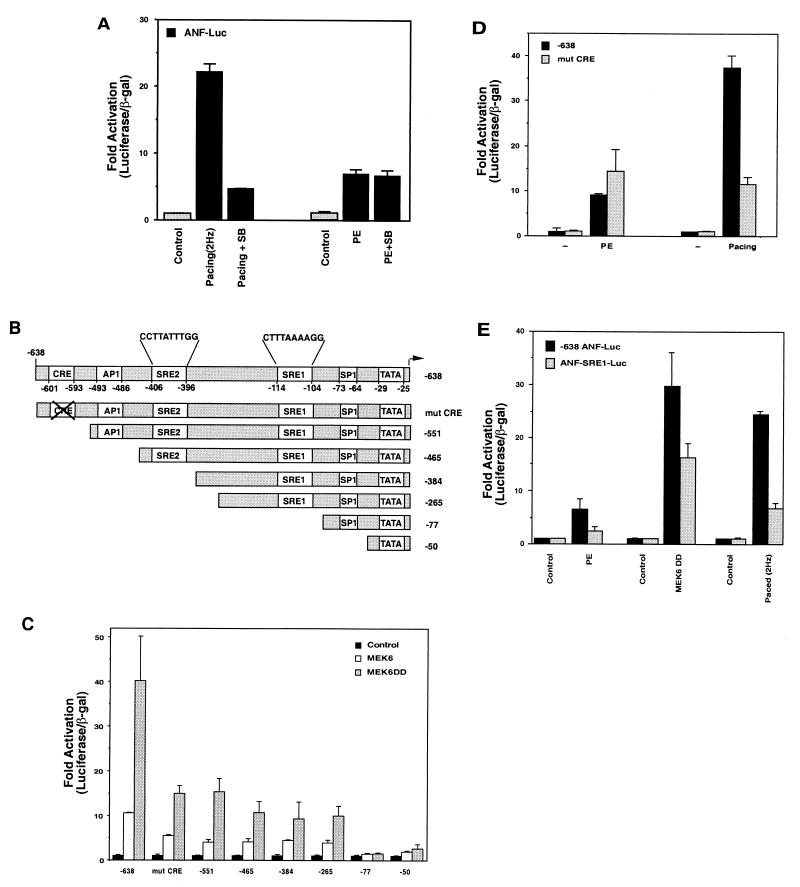
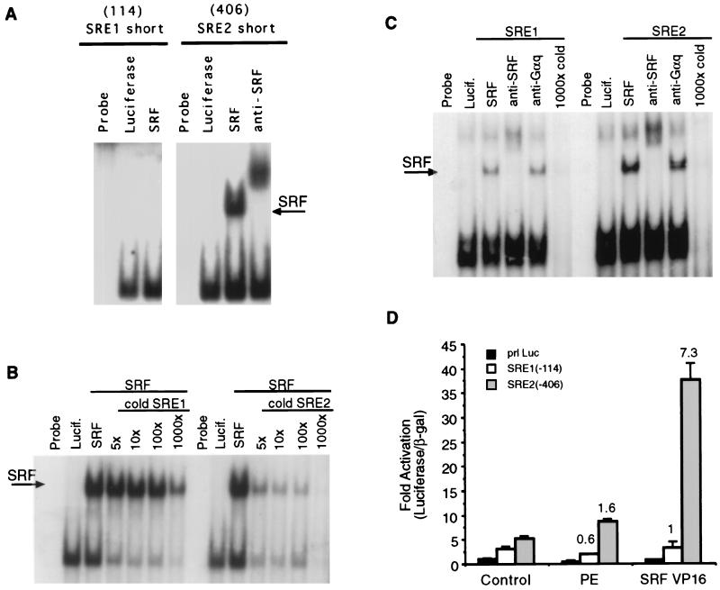
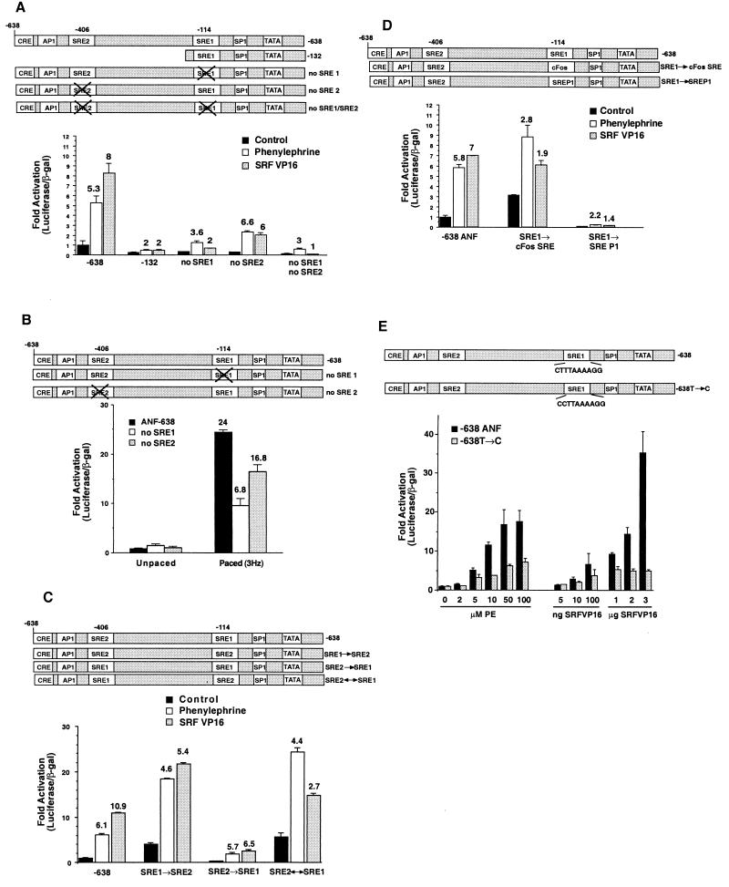
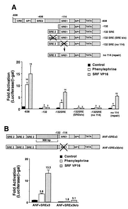
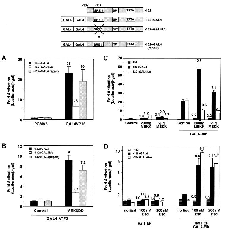
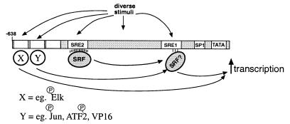
Hypertrophic growth of cardiac muscle cells is induced by a variety of physiological and pathological stimuli and is associated with a number of changes, including activation of genes such as atrial natriuretic factor. We found that two serum response element (SRE)-like DNA elements, one of which does not meet the consensus sequence and binds serum response factor (SRF) with low affinity, regulate the activity of this promoter. Surprisingly, the ability to induce the promoter by two different physiologic stimuli, as well as various activated transcription factors, including SRF-VP16, was primarily dependent upon the nonconsensus rather than the consensus SRE. This SRE controls the induction of gene expression via an unusual mechanism in that it is required to allow some, but not all, active transcription factors at unrelated sites on the promoter to stimulate gene expression. Thus, in addition to regulation of SRF activity by growth stimuli, regulation of a low-affinity SRE element controls inducible gene expression by modulating the ability of other transcription factors to stimulate the transcription machinery.


Figures

FIG. 1
FIG. 1
Pacing- but not phenylephrine-induced ANF expression requires p38. (A) Cells were transfected with ANF-luciferase and then treated with phenylephrine (PE) or electrically paced to induce contraction at 2 Hz in the presence or absence of the p38 inhibitor SB 203580 (SB). Both electrical pacing and phenylephrine-induced ANF-luciferase expression, but inhibition of p38 only reduced pacing-induced expression. (B) Schematic of truncation mutants of the −638 ANF promoter, indicating putative transcriptional elements. (C) Cells were transfected with control vectors, expression vectors for wild-type or activated MEK6, and various truncations of the ANF promoter driving luciferase or a mutation in a CRE-like element. Note that MEK6 activity induces the ANF promoter and that this is partially inhibited by mutation or truncations that remove the CRE-like element. Complete inhibition of activation occurs when an SRE-like element is deleted (−77). (D) Cells were transfected with the wild-type −638 promoter, or one which contains the CRE mutation, and then stimulated by phenylephrine or pacing. Both stimuli induced the wild-type promoter and while the CRE mutation significantly inhibited pacing-induced expression, it had no effect on phenylephrine-stimulated expression. (E) Cells were transfected with the −638 promoter or a mutant promoter in the SRE1 sequence (ANF-SRE1-Luc) and treated with phenylephrine, cotransfected with active MEK6, or electrically paced. Activation of the SRE1 mutant promoter is compromised for all stimuli.
FIG. 2
FIG. 2
SRF binding to SRE1 and SRE2. (A) In vitro binding reactions with SRF or luciferase from reticulocyte lysate in the presence of anti-SRF antibody as indicated, along with the core SRE1 (SRE1 short) and SRE2 (SRE2 short) probes. SRF binds more efficiently to SRE2 than to SRE1. (B) Competition assay with SRE2 as a probe along with excess cold SRE1 or SRE2. (C) Binding reactions similar to those in panel A but with longer oligonucleotides that contained 15 bp of the native ANF flanking sequence on either side of the two core SRE elements and the indicated antibodies. Although both SREs can bind to SRF, SRE2 binds much more efficiently than did SRE1. (D) Minimal promoters driven by single copies of SRE1 or SRE2 were transfected into cardiac muscle cells along with SRF-VP16 or empty vector and treated 100 μM phenylephrine (PE). The SRE2-luciferase plasmid is stimulated by SRF-VP16 but not by phenylephrine. SRE1-luciferase is not significantly stimulated by either phenylephrine or SRF-VP16.
FIG. 3
FIG. 3
SRE1 and SRE2 regulate basal activity of the ANF promoter but only SRE1 regulates induction. (A) Activation by phenylephrine or SRF-VP16 was determined for the −638 promoter, a truncated promoter (−132), or the −638 promoter with mutations in SRE1, SRE2, or both SRE1 and SRE2. All promoters were normalized to unstimulated wild-type or mutant promoter. The numbers above the bars represent the fold activation by phenylephrine or SRF-VP16 for each individual promoter compared to untreated cells. Mutations in either SRE reduced the basal activity of the promoter; however, only mutation in the SRE1 element significantly reduced induction by phenylephrine or SRF-VP16. (B) Wild-type −638 promoter or the SRE1 or SRE2 mutation were assayed in control cells or in cells that were stimulated by electrical pacing. Contraction-induced gene expression was significantly inhibited by the SRE1 mutation and only moderately inhibited by the SRE2 mutation. (C) Activation by phenylephrine or SRF-VP16 was determined for the wild-type promoter or for mutant promoters where the SRE1 element was mutated so that it was identical to SRE2 (SRE1 → SRE2), the SRE2 element was mutated to be identical to SRE1 (SRE2 → SRE1), or where the SREs were reversed (SRE2 ↔ SRE1). The basal activity was increased for the SRE1 → SRE2 mutant; however, induction by phenylephrine or SRF-VP16 was reduced. The mutant containing two SRE1 elements had slightly reduced induction by SRF-VP16 but virtually no reduction in phenylephrine induction. The SRE2 ↔ SRE1 had an expression level similar to that of SRE1 → SRE2. (D) Activation by phenylephrine or SRF-VP16 of wild-type −638 promoter or mutant promoters, where SRE1 was mutated to the c-Fos SRE sequence or the low-affinity SREP1 sequence (23). Mutation of SRE1 to the c-Fos SRE increased the basal promoter activity but reduced the induction by either phenylephrine or SRF-VP16. Mutation to the poorly binding SREP1 sequence reduced both basal activity and induction. (E) Point mutation of the SRE1 element to more closely match a consensus SRE sequence (638T → C) increased the basal activity of the promoter 6.6-fold but reduced induction by phenylephrine or SRF-VP16 at all doses of the two agonists. Note that the data for this set of transfections was normalized to each unstimulated promoter.
FIG. 4
FIG. 4
Cooperation between SRE2 and SRE1. (A) The wild-type −638 promoter or the −132 truncation (−132), the −132 promoter with a functional SRE2 element fused to the end (−132 SRE), −132 mutants containing mutated SRE2 (SREk/o) or SRE1 (no114), or repairs of the mutated sequence (no 114 repair) were induced by phenylephrine or SRF-VP16. All transfections were normalized to the untreated controls for each promoter. The fold activation for each promoter (from its unstimulated control) is given by the numbers above the bars. Note that the −132 promoter is not significantly activated by either phenylephrine or SRF-VP16, while the promoter with a single copy of the SRE2 element fused to the end of −132 regains wild-type activation by SRF-VP16 and partially rescues activation by phenylephrine. Mutation of either SRE element reduces activation by both stimuli, while remutation to repair the SRE1 mutant rescues activation by both stimuli. (B) Three SRE2 elements were fused to the end of a 300-bp DNA fragment from the Kan gene on the end of the −132 promoter. The SRE1 element was mutated in this construct, and stimulation was assessed after treatment with phenylephrine or SRF-VP16. Note that mutation of the SRE1 element reduces basal promoter activity and induction by both phenylephrine and SRF-VP16.
FIG. 5
FIG. 5
Activation of transcription by a heterologous transcription factor requires SRE1 activity. (A) Two consensus GAL4-binding sequences were fused to the end of the −132 promoter (−132+GAL4), to a version with the SRE1 element mutated (−132+GAL4k/o), or to a repaired version of this molecule with a reconstructed wild-type SRE1 (repair). Cells were transfected with control vector or a GAL4-VP16 expression plasmid. Note that GAL4-VP16 activates the parental plasmid but that mutation of the SRE1 element in this molecule reduces activation, while a repaired version of this molecule has rescued induction. (B) The GAL4-containing promoters were cotransfected with an expression plasmid encoding GAL4-ATF2, along with a control plasmid or an activated MEK6 expression plasmid, which phosphorylates ATF2. MEK6 induces GAL4-ATF2-dependent gene expression in an SRE1-dependent manner. (C) −132-Luc, GAL4-132-Luc, or the GAL4-132-Luc molecule with mutated SRE1 (−132+GAL4k/o) were transfected with increasing amounts of a MEKK1 expression plasmid with or without a GAL4-Jun expression plasmid. Note that GAL4-Jun increased the expression of the GAL4-containing promoters even in the absence of MEKK1 activity and that this occurs irrespective of the presence or absence of an intact SRE1 element. Expression of active MEKK1 further stimulated GAL4-Jun-dependent expression; however, this stimulation was compromised when the SRE1 element was mutated. (D) Luciferase reporters as in panel C were transfected with a Raf1:ER expression plasmid and with or without a GAL4-Elk expression plasmid. Raf activity was stimulated by adding increasing doses of estradiol to activate GAL4-Elk. In this case, mutation of the SRE1 element had no effect on the level of gene expression induced by the active GAL4-Elk protein.
FIG. 6
FIG. 6
Model for transcriptional induction of the ANF promoter by diverse stimuli. The SRE1 sequence in the ANF promoter could function as an “activator bridge” to promote productive interactions between activated transcription factors at other sites and the basal transcription machinery.

Similar articles

Cited by

References

    1. Akhter S A, Luttrell L M, Rockman H A, Iaccarino G, Lefkowitz R J, Koch W J. Targeting the receptor-Gq interface to inhibit in vivo pressure overload myocardial hypertrophy. Science. 1998;280:574–577. - PubMed
    1. Alberts A S, Geneste O, Treisman R. Activation of SRF-regulated chromosomal templates by Rho-family GTPases requires a signal that also induces H4 hyperacetylation. Cell. 1998;92:475–487. - PubMed
    1. Allo S N, McDermott P J, Carl L L, Morgan H E. Phorbol ester stimulation of protein kinase C activity and ribosomal DNA transcription: role in hypertrophic growth of cultured cardiomyocytes. J Biol Chem. 1991;266:22003–22009. - PubMed
    1. Ardati A, Nemer M. A nuclear pathway for α1-adrenergic receptor signaling in cardiac cells. EMBO J. 1993;12:5131–5139. - PMC - PubMed
    1. Bogoyevitch M A, Gillespie-Brown J, Ketterman A J, Fuller S J, Ben-Levy R, Ashworth A, Marshall C J, Sugden P H. Stimulation of the stress-activated mitogen-activated protein kinase subfamilies in perfused heart. Circ Res. 1996;79:162–173. - PubMed

Publication types

MeSH terms

LinkOut - more resources