. 1999 Mar;19(3):1853-63.
doi: 10.1128/MCB.19.3.1853.

Substrate specificities of SR proteins in constitutive splicing are determined by their RNA recognition motifs and composite pre-mRNA exonic elements

Affiliations

Substrate specificities of SR proteins in constitutive splicing are determined by their RNA recognition motifs and composite pre-mRNA exonic elements

A Mayeda et al. Mol Cell Biol. 1999 Mar.

Abstract

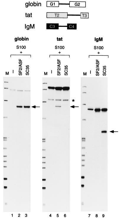
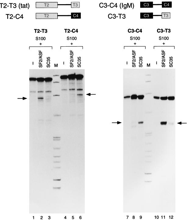
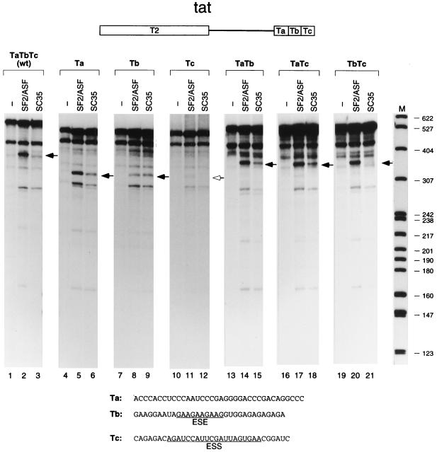
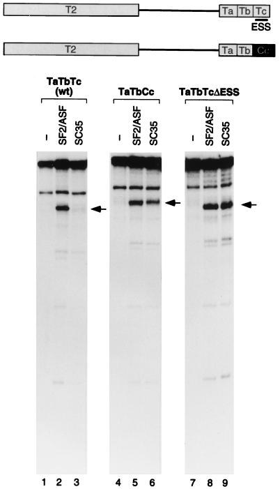
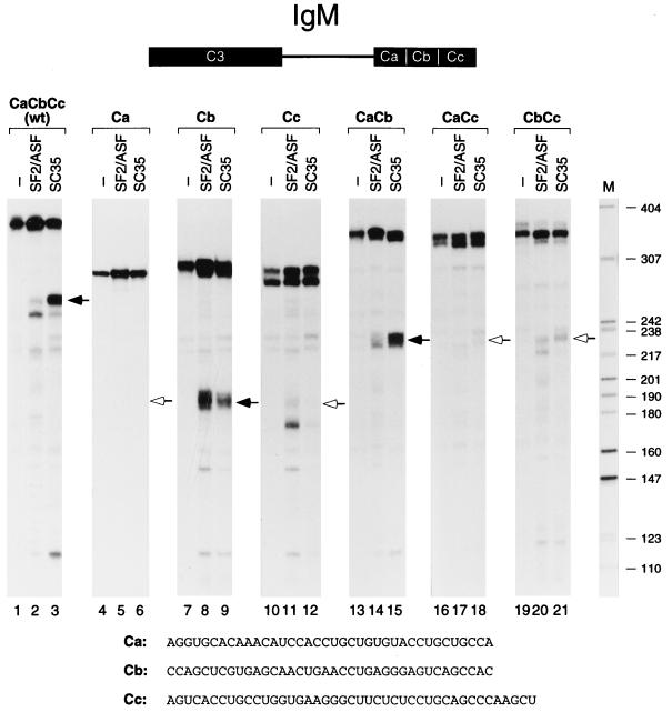
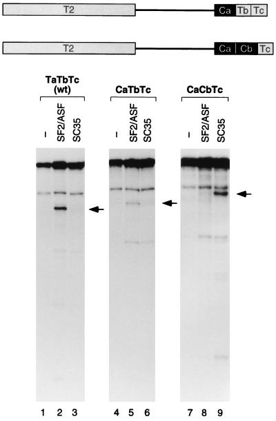
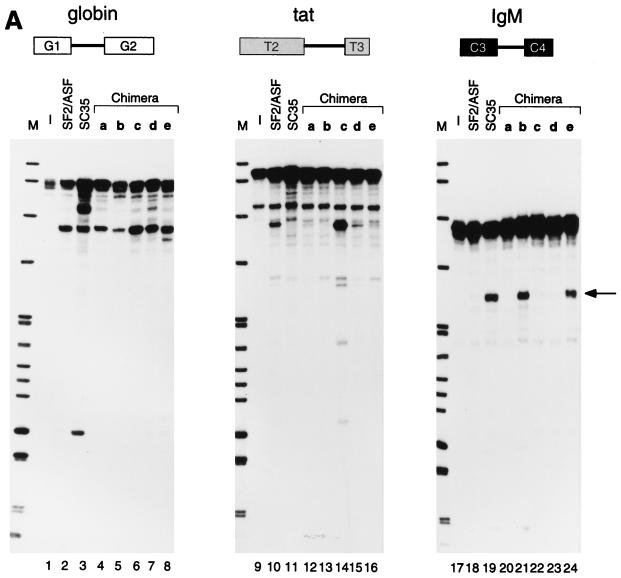
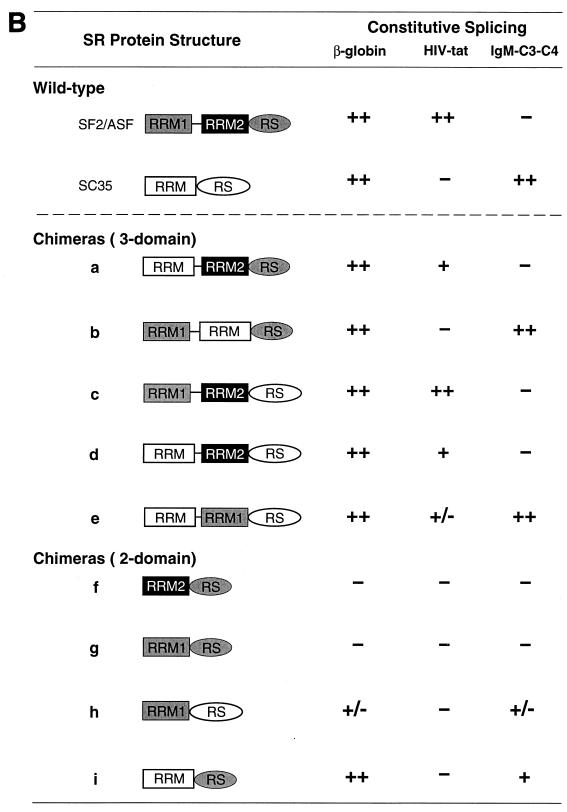
We report striking differences in the substrate specificities of two human SR proteins, SF2/ASF and SC35, in constitutive splicing. beta-Globin pre-mRNA (exons 1 and 2) is spliced indiscriminately with either SR protein. Human immunodeficiency virus tat pre-mRNA (exons 2 and 3) and immunoglobulin mu-chain (IgM) pre-mRNA (exons C3 and C4) are preferentially spliced with SF2/ASF and SC35, respectively. Using in vitro splicing with mutated or chimeric derivatives of the tat and IgM pre-mRNAs, we defined specific combinations of segments in the downstream exons, which mediate either positive or negative effects to confer SR protein specificity. A series of recombinant chimeric proteins consisting of domains of SF2/ASF and SC35 in various combinations was used to localize trans-acting domains responsible for substrate specificity. The RS domains of SF2/ASF and SC35 can be exchanged without effect on substrate specificity. The RNA recognition motifs (RRMs) of SF2/ASF are active only in the context of a two-RRM structure, and RRM2 has a dominant role in substrate specificity. In contrast, the single RRM of SC35 can function alone, but its substrate specificity can be influenced by the presence of an additional RRM. The RRMs behave as modules that, when present in different combinations, can have positive, neutral, or negative effects on splicing, depending upon the specific substrate. We conclude that SR protein-specific recognition of specific positive and negative pre-mRNA exonic elements via one or more RRMs is a crucial determinant of the substrate specificity of SR proteins in constitutive splicing.


Figures

FIG. 1
FIG. 1
In vitro splicing of three representative pre-mRNAs in S100 extract complemented with SF2/ASF or SC35. The structures of the β-globin, tat, and IgM minigene pre-mRNAs (see Materials and Methods) are shown schematically at the top. The positions of the spliced mRNAs are indicated by arrows. The asterisk indicates a cleavage product unrelated to splicing (18). The splicing products were previously characterized in detail (18, 30, 43). pBR322/HpaII DNA size markers are shown (lanes M).
FIG. 2
FIG. 2
In vitro splicing of hybrid tat and IgM pre-mRNAs with swapped downstream exons. The structures of the control wild-type minigene pre-mRNAs (tat and IgM) and swap pre-mRNAs (T2-C4 and C3-T3) are shown schematically at the top. The positions of the spliced mRNAs are indicated by arrows. DNA size markers (lanes M) are as described for Fig. 1.
FIG. 3
FIG. 3
In vitro splicing of tat pre-mRNA derivatives with downstream exon deletions. The structure of the wild-type (wt) tat minigene pre-mRNA, with the T3 exon divided into three segments, Ta, Tb, and Tc, is shown schematically at the top. The RNA sequence of each segment is shown at the bottom. Previously identified ESE and ESS elements are underlined (1, 33, 34). The positions of the spliced mRNAs are indicated by arrows; the open arrowhead indicates the expected position in the case of an mRNA that is at the limit of detection. DNA size markers (lane M) are as described for Fig. 1 (sizes in nucleotides).
FIG. 4
FIG. 4
In vitro splicing of tat pre-mRNA derivatives with replacement or internal deletion of exon segment Tc. The structures of the pre-mRNA derivatives are shown schematically at the top. The deleted ESS element (Fig. 3) is indicated by a horizontal bar, and the black box shows the Cc segment of the IgM C4 exon (Fig. 5) replacing the Tc segment. The positions of the spliced mRNAs are indicated by arrows. wt, wild type.
FIG. 5
FIG. 5
In vitro splicing of IgM pre-mRNA derivatives with downstream exon deletions. The structure of the wild-type (wt) IgM minigene pre-mRNA, with the C4 exon divided into three segments, Ca, Cb, and Cc, is shown schematically at the top. The RNA sequence of each segment is shown at the bottom. The positions of the spliced mRNAs are indicated by arrows; the open arrowheads indicate the expected positions in the cases of mRNAs that are at or below the limit of detection. The identity of the aberrant processing product in lane 11 has not been determined. DNA size markers (lane M) are as described for Fig. 1 (sizes in nucleotides).
FIG. 6
FIG. 6
In vitro splicing of tat pre-mRNA derivatives with replacements of downstream exon segments. The structures of the pre-mRNAs are shown schematically at the top. The positions of the spliced mRNAs are indicated by arrows. wt, wild type.
FIG. 7
FIG. 7
(A) In vitro splicing of β-globin, tat, and IgM pre-mRNAs with wild-type or chimeric glutathione S-transferase-tagged SR proteins assembled from different combinations of SF2/ASF and SC35 domains. See panel B for the designation of each protein. The structures of the pre-mRNAs are shown schematically at the top. The positions of the spliced mRNAs are indicated by arrows. DNA size markers (lanes M) are as described for Fig. 1. (B) Summary of the splicing activities of the chimeric SR proteins with each pre-mRNA. The domain structure of each protein is shown schematically, with the designation used in panel A. The relative splicing complementation activities are indicated (−, no detectable activity; +/−, trace activity; +, weak activity; ++, strong activity) and are based on two independent experiments, one of which is shown in panel A.
FIG. 7
FIG. 7
(A) In vitro splicing of β-globin, tat, and IgM pre-mRNAs with wild-type or chimeric glutathione S-transferase-tagged SR proteins assembled from different combinations of SF2/ASF and SC35 domains. See panel B for the designation of each protein. The structures of the pre-mRNAs are shown schematically at the top. The positions of the spliced mRNAs are indicated by arrows. DNA size markers (lanes M) are as described for Fig. 1. (B) Summary of the splicing activities of the chimeric SR proteins with each pre-mRNA. The domain structure of each protein is shown schematically, with the designation used in panel A. The relative splicing complementation activities are indicated (−, no detectable activity; +/−, trace activity; +, weak activity; ++, strong activity) and are based on two independent experiments, one of which is shown in panel A.

References

    1. Amendt B A, Si Z H, Stoltzfus C M. Presence of exon splicing silencers within human immunodeficiency virus type 1 tat exon 2 and tat-rev exon 3: evidence for inhibition mediated by cellular factors. Mol Cell Biol. 1995;15:4606–4615. . (Erratum, 15:5480.) - PMC - PubMed
    1. Birney E, Kumar S, Krainer A R. Analysis of the RNA-recognition motif and RS and RGG domains: conservation in metazoan pre-mRNA splicing factors. Nucleic Acids Res. 1993;21:5803–5816. - PMC - PubMed
    1. Cáceres J F, Krainer A R. Functional analysis of pre-mRNA splicing factor SF2/ASF structural domains. EMBO J. 1993;12:4715–4726. - PMC - PubMed
    1. Cáceres J F, Krainer A R. Mammalian pre-mRNA splicing factors. In: Krainer A R, editor. Eukaryotic mRNA processing. Oxford, United Kingdom: Oxford University Press; 1997. pp. 174–212.
    1. Cáceres J F, Misteli T, Screaton G R, Spector D L, Krainer A R. Role of the modular domains of SR proteins in subnuclear localization and alternative splicing specificity. J Cell Biol. 1997;138:225–238. - PMC - PubMed

Publication types