. 1999 Mar;19(3):1871-80.
doi: 10.1128/MCB.19.3.1871.

Phosphorylation of the cap-binding protein eukaryotic translation initiation factor 4E by protein kinase Mnk1 in vivo

Affiliations

Phosphorylation of the cap-binding protein eukaryotic translation initiation factor 4E by protein kinase Mnk1 in vivo

A J Waskiewicz et al. Mol Cell Biol. 1999 Mar.

Abstract

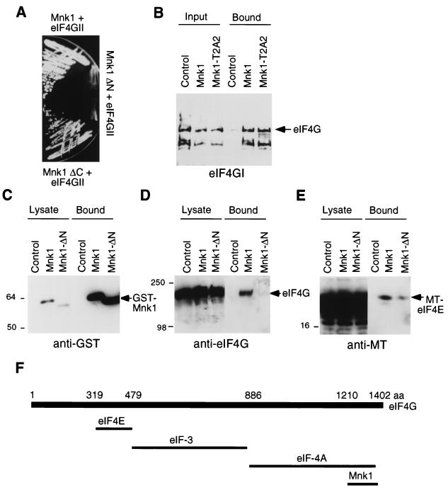
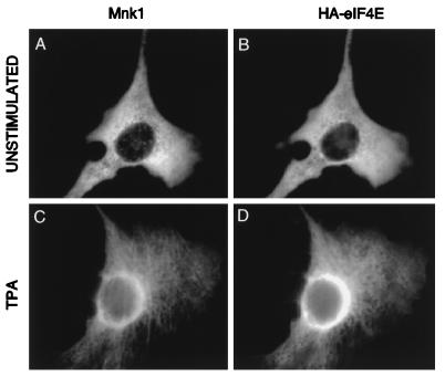
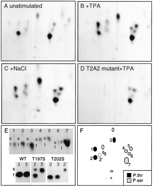
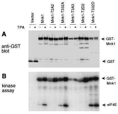
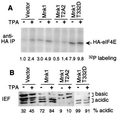
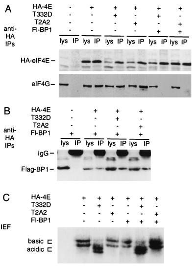
Eukaryotic translation initiation factor 4E (eIF4E) binds to the mRNA 5' cap and brings the mRNA into a complex with other protein synthesis initiation factors and ribosomes. The activity of mammalian eIF4E is important for the translation of capped mRNAs and is thought to be regulated by two mechanisms. First, eIF4E is sequestered by binding proteins, such as 4EBP1, in quiescent cells. Mitogens induce the release of eIF4E by stimulating the phosphorylation of 4EBP1. Second, mitogens and stresses induce the phosphorylation of eIF4E at Ser 209, increasing the affinity of eIF4E for capped mRNA and for an associated scaffolding protein, eIF4G. We previously showed that a mitogen- and stress-activated kinase, Mnk1, phosphorylates eIF4E in vitro at the physiological site. Here we show that Mnk1 regulates eIF4E phosphorylation in vivo. Mnk1 binds directly to eIF4G and copurifies with eIF4G and eIF4E. We identified activating phosphorylation sites in Mnk1 and developed dominant-negative and activated mutants. Expression of dominant-negative Mnk1 reduces mitogen-induced eIF4E phosphorylation, while expression of activated Mnk1 increases basal eIF4E phosphorylation. Activated mutant Mnk1 also induces extensive phosphorylation of eIF4E in cells overexpressing 4EBP1. This suggests that phosphorylation of eIF4E is catalyzed by Mnk1 or a very similar kinase in cells and is independent of other mitogenic signals that release eIF4E from 4EBP1.


Figures

FIG. 1
FIG. 1
(A) Growth of yeast strain L40 expressing LexA-Mnk1 and VP16-eIF4GII (top), LexA-Mnk1-ΔN and VP16-eIF4GII (middle), and LexA-Mnk1-ΔC and VP16-eIF4GII (bottom) on medium lacking histidine. Growth indicates transactivation of the HIS3 reporter gene. (B) GST-Mnk1 and GST-Mnk1-T2A2 (T197A/T202A) were purified from E. coli and incubated with purified mammalian eIF4G. GST fusion proteins were isolated, and bound eIF4G was eluted and analyzed by SDS-PAGE and Western blotting. Input lanes represent one-fifth of the sample used for binding. (C to E) 293 cells were transiently transfected with pEBG, pEBG-Mnk1, or pEBG-Mnk1ΔN, together with pCS3+eIF4E. GST-Mnk1 was purified with glutathione-Sepharose, and bound proteins were visualized with GST antibody (C), eIF4G antibody (D), or 9E10 anti-MT antibody (E). Lysate lanes represent 1/10 of the sample used for purification. (F) Diagram of characterized eIF4G binding sites. See the text for details. aa, amino acids.
FIG. 2
FIG. 2
Subcellular localization of Mnk1 and eIF4E. NIH 3T3 cells were transfected with pCS2-Mnk1 and pSRα-eIF4E. Untagged Mnk1 and HA-eIF4E were visualized with an affinity-purified chicken-anti-Mnk1 antibody and 12CA5 (anti-HA) ascites fluid, followed by fluorescein-conjugated anti-chicken antibody and Texas red-conjugated anti-mouse antibody. (A and C) Mnk1 immunofluorescence; (B and D) HA-eIF4E. Cells were either serum starved (A and B) or treated with phorbol ester for 60 min (C and D).
FIG. 3
FIG. 3
Phosphopeptide analysis of Mnk1. 293 cells were transfected with wild-type Mnk1 (A to C and E), T197A/T202A (T2A2) mutant Mnk1 (D), or various mutants (E) of EBG-Mnk1 and metabolically labeled with [32P]orthophosphate. The cells were serum starved (A and E) or stimulated with 100 nM TPA for 15 min (B, D, and E) or 0.4 M NaCl (C) for 30 min. GST-Mnk1 was purified by glutathione-Sepharose and SDS-PAGE and digested with trypsin and thermolysin. Phosphopeptides were resolved by electrophoresis and chromatography and detected by autoradiography (A to D). (E) The Phosphoamino acid content of individual phosphopeptides was determined. The top panel shows phosphoamino acid analysis of phosphopeptides 1 to 7; the bottom panel shows phosphoamino acid analysis of phosphopeptides 2 and 3 from wild-type (WT), T197S, and T202S mutants of Mnk1. Radioactive phosphoamino acids were detected by autoradiography, and the positions of nonradioactive internal standards, phosphoserine (s), and phosphothreonine (t) were detected with ninhydrin. (F) Schematic showing phosphoserine-containing (grey symbols), phosphothreonine-containing (black symbols), and unanalyzed (open symbols) phosphopeptides. Phosphopeptide 2′ was detected in maps of T202S mutant only.
FIG. 4
FIG. 4
Expression and kinase activity of Mnk1 mutants. GST-Mnk1 and mutants T2A2 (T197A/T202A), T332A, T3A3 (T197A/T202A/T332A), T2D2 (T197D/T202D), and T332D were synthesized in 293 cells. Transfected cells were serum starved and treated with TPA or left untreated. Mnk1 was purified with glutathione-Sepharose. (A) Samples were immunoblotted with polyclonal anti-GST antibody. (B) The same samples were incubated with radiolabeled ATP and eIF4E. Products were detected by SDS-PAGE and autoradiography.
FIG. 5
FIG. 5
Phosphorylation of eIF4E in cells overexpressing Mnk1. 293 cells were transfected with pSRα-eIF4E and either vector pEBG, wild-type Mnk1, Mnk1-T2A2 (T197A/T202A), or Mnk1-T332D. (A) Cells were serum starved, metabolically labeled with 32P, and treated with TPA or left untreated. HA-eIF4E was isolated by immunoprecipitation (IP) and detected by SDS-PAGE and autoradiography. Incorporation was quantified with a PhosphorImager and normalized to incorporation in unstimulated cells expressing vector DNA. (B) Cells were serum starved and treated with TPA or left untreated. HA-eIF4E was purified with m7GTP-Sepharose and subjected to one-dimensional isoelectric focusing (IEF) and Western blotting with anti-HA antibodies. The amounts of the two basic and two acidic species were quantified, yielding a percentage value for acidic species for each sample (indicated below the gel). The inhibition of HA-eIF4E phosphorylation by T2A2 mutant Mnk1 was confirmed by using the pCS3+MT vector, which expresses MT-Mnk1-T2A2 (data not shown).
FIG. 6
FIG. 6
Effect of Mnk1 overexpression on complex formation and phosphorylation of eIF4E. 293 cells were transfected with vectors encoding HA-eIF4E, Flag-4EBP1, and GST-Mnk1-T2A2 (T197A/T202A) or GST-Mnk1-T332D. Proteins were purified by immunoprecipitation (IP) with antibody to HA (12CA5 monoclonal antibody) or with m7GTP-Sepharose and subjected to SDS-PAGE (A and B) or isoelectric focusing (IEF) (C) and Western blotting with antibodies to HA (A and C), eIF4G (A), or Flag (B). All samples are from the same experiment, which was repeated with similar results. Note that basal eIF4E phosphorylation was low, so that inhibitory effects of GST-Mnk1-T2A2 were not detectable. lys, lysate.

References

    1. Alvarez E, Northwood I C, Gonzalez E A, Latour D A, Seth A, Abate C, Curran T. Pro-Leu-Ser/Thr-Pro is a consensus primary sequence for substrate phosphorylation. Characterization of the phosphorylation of c-myc and c-jun proteins by an epidermal growth factor receptor threonine 669 kinase. J Biol Chem. 1991;266:15277–15285. - PubMed
    1. Ben-Levy R, Leighton I A, Doza Y N, Attwood P, Morrice N, Marshall C J, Cohen P. Identification of novel phosphorylation sites required for activation of MAPKAP kinase-2. EMBO J. 1995;14:5920–5930. - PMC - PubMed
    1. Boyle W J, van der Geer P, Hunter T. Phosphopeptide mapping and phosphoamino acid analysis by two-dimensional separation on thin-layer cellulose plates. Methods Enzymol. 1991;201:110–148. - PubMed
    1. Brunn G J, Hudson C C, Sekulic A, Williams J M, Hosoi H, Houghton P J, Lawrence J C, Jr, Abraham R T. Phosphorylation of the translational repressor PHAS-I by the mammalian target of rapamycin. Science. 1997;277:99–101. - PubMed
    1. Bu X, Haas D W, Hagedorn C H. Novel phosphorylation sites of eukaryotic initiation factor-4F and evidence that phosphorylation stabilizes interactions of the p25 and p220 subunits. J Biol Chem. 1993;268:4975–4978. - PubMed

Publication types

MeSH terms

Substances