. 1999 Mar;19(3):1938-49.
doi: 10.1128/MCB.19.3.1938.

The Jun kinase 2 isoform is preferentially required for epidermal growth factor-induced transformation of human A549 lung carcinoma cells

Affiliations

The Jun kinase 2 isoform is preferentially required for epidermal growth factor-induced transformation of human A549 lung carcinoma cells

F Bost et al. Mol Cell Biol. 1999 Mar.

Abstract

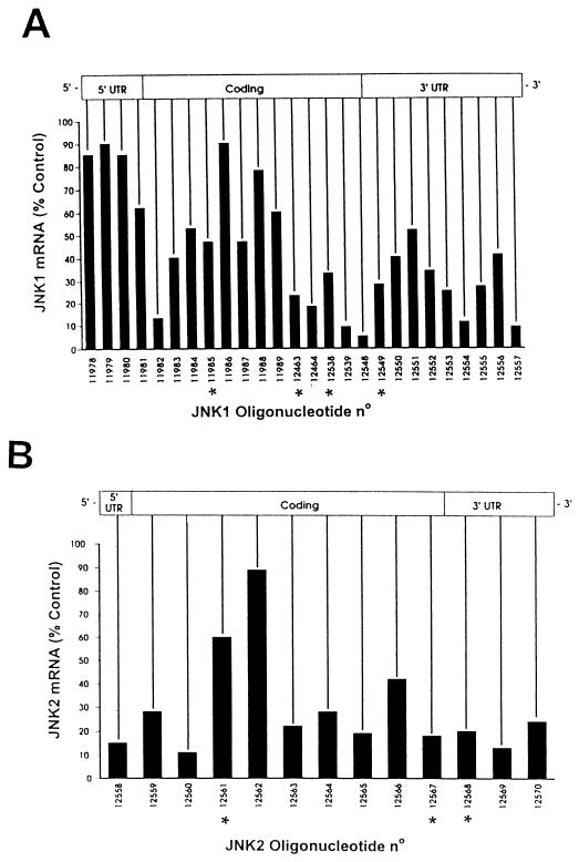
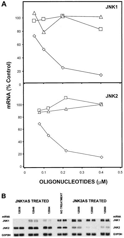
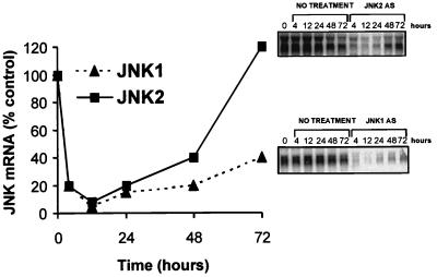
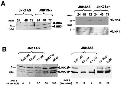
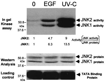
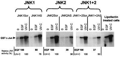
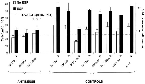
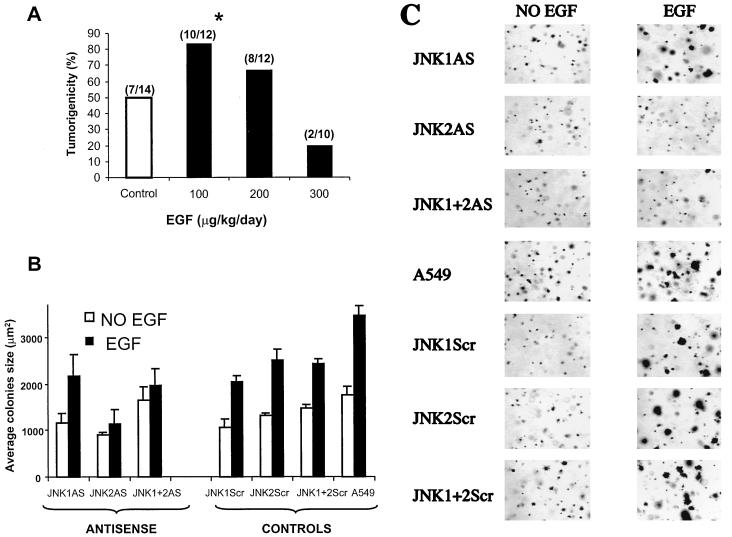
We have previously found that epidermal growth factor (EGF) mediates growth through the Jun N-terminal kinase/stress-activated kinase (JNK/SAPK) pathway in A549 human lung carcinoma cells. As observed here, EGF treatment also greatly enhances the tumorigenicity of A549 cells, suggesting an important role for JNK in cancer cell growth (F. Bost, R. McKay, N. Dean, and D. Mercola, J. Biol. Chem. 272:33422-33429, 1997). Several isoforms families of JNK, JNK1, JNK2, and JNK3, have been isolated; they arise from alternative splicing of three different genes and have distinct substrate binding properties. Here we have used specific phosphorothioate oligonucleotides targeted against the two major isoforms, JNK1 and JNK2, to discriminate their roles in EGF-induced transformation. Multiple antisense sequences have been screened, and two high-affinity and specific candidates have been identified. Antisense JNK1 eliminated steady-state mRNA and JNK1 protein expression with a 50% effective concentration (EC50) of <0.1 microM but did not alter JNK2 mRNA or protein levels. Conversely, antisense JNK2 specifically eliminated JNK2 steady-state mRNA and protein expression with an EC50 of 0.1 microM. Antisense JNK1 and antisense JNK2 inhibited by 40 and 70%, respectively, EGF-induced total JNK activity, whereas sense and scrambled-sequence control oligonucleotides had no effect. The elimination of mRNA, protein, and JNK activities lasted 48 and 72 h following a single Lipofectin treatment with antisense JNK1 and JNK2, respectively, indicating sufficient duration for examining the impact of specific elimination on the phenotype. Direct proliferation assays demonstrated that antisense JNK2 inhibited EGF-induced doubling of growth as well as the combination of active antisense oligonucleotides did. EGF treatment also induced colony formation in soft agar. This effect was completely inhibited by antisense JNK2 and combined-antisense treatment but not altered by antisense JNK1 alone. These results show that EGF doubles the proliferation (growth in soft agar as well as tumorigenicity in athymic mice) of A549 lung carcinoma cells and that the JNK2 isoform but not JNK1 is utilized for mediating the effects of EGF. This study represents the first demonstration of a cellular phenotype regulated by a JNK isoform family, JNK2.


Figures

FIG. 1
FIG. 1
Messenger walk survey for the elimination of JNK1 and JNK2 mRNA levels following treatment with phosphorothioate antisense oligonucleotides targeted to JNK1 or JNK2 mRNA. (A) JNK1 mRNA steady-state levels in A549 cells following treatment with 26 different phosphorothioate antisense oligonucleotides targeted to JNK1 mRNA. JNK1ASISIS12539 gave the most consistent results for the elimination of JNK1 mRNA steady-state levels. (B) A similar experiment was performed with 13 phosphorothioate antisense oligonucleotides complementary to the indicated regions of the JNK2 mRNA. JNK2ASISIS12560 yielded the most consistent results for the elimination of JNK2 mRNA steady-state levels. All oligonucleotides are arrayed relative to their complementary sequence along the JNK transcript. The asterisks indicate the oligonucleotides having a nucleotide composition (2 to 4 bases) very similar to that of the leading compound. The screenings were repeated two times with similar results.
FIG. 2
FIG. 2
Dose-dependent reduction of JNK1 and JNK2 mRNAs and specific elimination of JNK1AS and JNK2AS steady-state mRNA levels. (A) Dose-response curve for JNK1 mRNA level after treatment of A549 cells with different concentrations of JNK1ASISIS12539 (◊), JNK1ScrISIS14321 (□), and JNK1SeISIS14320 (▵). The mRNAs were prepared 24 h after a 4-h transfection, examined by Northern analysis, quantified, and normalized to JNK1 mRNA levels in untreated A549 cells. A similar experiment was carried out for JNK2 mRNA with JNK2ASISIS12560 (◊), JNK2ScrISIS14319 (□) and JNK2SeISIS14318 (▵). (B) A549 cells were treated with three different antisense oligonucleotides complementary to JNK1 mRNA (including the active antisense oligonucleotide JNK1ASISIS12539) and three different antisense oligonucleotides complementary to JNK2 mRNA (including the active antisense oligonucleotide JNK2ASISIS12560). Twenty-four hours after a 4-h transfection with 0.4 μM antisense oligonucleotide, mRNA was prepared and examined by Northern analysis; the same membrane was hybridized successively with JNK1 probe, JNK2 probe, and the G3PDH probe.
FIG. 3
FIG. 3
Duration of elimination of JNK1 and JNK2 mRNA in A549 cells. A549 cells were transfected with 0.4 μM JNK1ASISIS12539 or JNK2ASISIS12560. The mRNAs were prepared at the indicated times after 4, 12, 24, 48, and 72 h and examined by Northern analysis. Quantification was performed as described in Material and Methods.
FIG. 4
FIG. 4
Determination of the duration of antisense effect and dose-dependent inhibition of expression of JNK1 and JNK2 protein after treatment with antisense oligonucleotides. (A) Duration of antisense effect. A549 cells were transfected with 0.4 μM JNK1ASISIS12539 or JNK1ScrISIS14321 for 4.5 h, and cell extracts were prepared at the indicated times. Western analysis was performed with anti-JNK1 antibodies (SC-571). A similar experiment was carried out with JNK2ASISIS12560 and JNK2ScrISIS14319 as a control. Western analysis was performed with anti-JNK2 antibodies (SC-827), and the same membrane was reprobed with JNK1 antibodies (SC-571) (bottom). (B) A549 cells were treated with the indicated concentrations of phosphorothioate 2′-O-methoxyethyl-modified antisense oligonucleotides JNK1ASISIS15346 or JNK2ASISIS15353 and 0.4 μM their respective control oligonucleotides, JNK1ScrISIS18076 and JNK2ScrISIS18078. Cell extracts were prepared 36 h after the transfection and examined by Western analysis with anti-JNK1 antibodies (SC-571). The protein levels (shown below the gels) were determined by comparison to the respective protein levels in untreated cells by using the Electrophoresis Documentation and analysis System 120 (Kodak Digital Science).
FIG. 5
FIG. 5
Activation of JNK1 and JNK2 by rhEGF and UV-C. A549 cells were stimulated with 0.1 μM rhEGF or UV-C at 100 J/m2. Twenty minutes after stimulation, cell extracts were prepared and used for in-gel kinase assay with GST–c-Jun as a substrate, as described in Materials and Methods. The activation of each isoform (Activity) was calculated by normalization to the corresponding amount of JNK protein shown in the lower panel (Western Analysis).
FIG. 6
FIG. 6
Differential inhibition of JNK activity by JNK1AS and JNK2AS. A549 cells were transfected for 4.5 h with the same active antisense or control phosphorothioate 2′-O-methoxyethyl-modified oligonucleotides (0.2 μM) used in the experiments shown in Fig. 4B. Thirty-six hours after transfection, the cells were stimulated with 0.1 μM rhEGF or UV-C at 100 J/m2 or were not stimulated. Twenty minutes after stimulation, cell extracts were prepared and used for a Jun kinase assay, as described in Materials and Methods. The activation is given relative to the activation obtained with rhEGF or UV-C in the scrambled-oligonucleotide-treated cells.
FIG. 7
FIG. 7
JNK2AS preferentially inhibits EGF-induced proliferation in A549. Proliferation assays were performed as described in Materials and Methods with the unmodified oligonucleotides. The cells were maintained in 0.5% FBS during the experiment. Five days after the treatment with 0.1 μM rhEGF, the cells were counted with a Coulter counter. The dashed line indicates the growth attained by A549 cells that stably express the dominant-negative inhibitor c-Jun(S63A,S73A) which is known to be inhibited from responding to rhEGF (7). The proliferation data shown here are the average of two identical and independent experiments, each carried out in triplicate. Fold increase in cell number is given considering 1 as the average number of cells grown in the absence of rhEGF. The standard errors (error bars) are given as √(ς12 + ς22), where ς1 and ς2 are the standard errors of the replicate experiments. Statistical analyses were carried out with the combined data of both replicates by analysis of variance implemented with Systat software. The statistical significance for EGF-induced growth was P < 0.002 (⋆) or P < 0.05 (★).
FIG. 8
FIG. 8
EGF-induced tumorigenicity in nude mice and inhibition of growth on soft agar by antisense oligonucleotides. (A) A549 cells (5 × 10⁶) were injected into nude mice and analyzed for their ability to develop tumors in animals treated daily with saline solution (control) (open bar) or 100, 200, or 300 μg of rhEGF/kg (solid bars). After 8 days, visible tumors were scored as the ratio of the number of tumors over the number of sites injected (indicated above the bars). The asterisk indicates that the tumorigenicity increase at the 100-μg/kg/day dose is significant, with a P value of <0.021 (χ2 test). (B) A549 cells were transfected with the indicated unmodified oligonucleotides (0.4 μM or 0.2 plus 0.2 μM [for the combination]). Eighteen hours after transfection, the cells were transferred to 0.3% agarose containing 0.8 or 0.4 μM (for the combination) the indicated oligonucleotides. The cells were stained with p-iodotetrazolium violet and analyzed with IPLabSpectrum software. The error bars indicate the standard deviation. (C) Morphologies of A549 colonies treated with 0.1 μM rhEGF (right) or untreated (left) and transfected with the indicated oligonucleotides.

References

    1. Adamson E D, Wiley L M. The EGFR gene family in embryonic cell activities. Curr Top Dev Biol. 1997;35:71–120. - PubMed
    1. Adler V, Polotskaya A, Kim J, Dolan L, Davis R, Pincus M, Ronai Z. Dose rate and mode of exposure are key factors in JNK activation by UV irradiation. Carcinogenesis. 1996;9:2073–2076. - PubMed
    1. Angel P, Karin M. The role of Jun, Fos and the AP-1 complex in cell-proliferation and transformation. Biochem Biophys Acta. 1991;1072:129–157. - PubMed
    1. Antonyak M A, Moscatello D K, Wong A J. Constitutive activation of c-Jun N-terminal kinase by a mutant epidermal growth factor receptor. J Biol Chem. 1998;273:2817–2822. - PubMed
    1. Baldassarre G, Bianco C, Tortora G, Ruggiero A, Moasser M, Dmitrovsky E, Bianco A R, Ciardiello F. Transfection with a cripto antisense plasmid suppresses endogenous cripto expression and inhibits transformation in a human embryonal carcinoma cell line. Int J Cancer. 1996;66:538–543. - PubMed

Publication types

MeSH terms