. 1999 Mar;19(3):1950-60.
doi: 10.1128/MCB.19.3.1950.

Reactive oxygen intermediate-dependent NF-kappaB activation by interleukin-1beta requires 5-lipoxygenase or NADPH oxidase activity

Affiliations

Reactive oxygen intermediate-dependent NF-kappaB activation by interleukin-1beta requires 5-lipoxygenase or NADPH oxidase activity

G Bonizzi et al. Mol Cell Biol. 1999 Mar.

Abstract

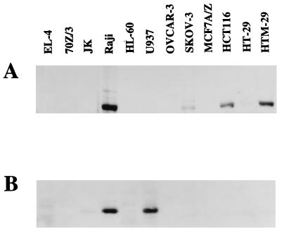
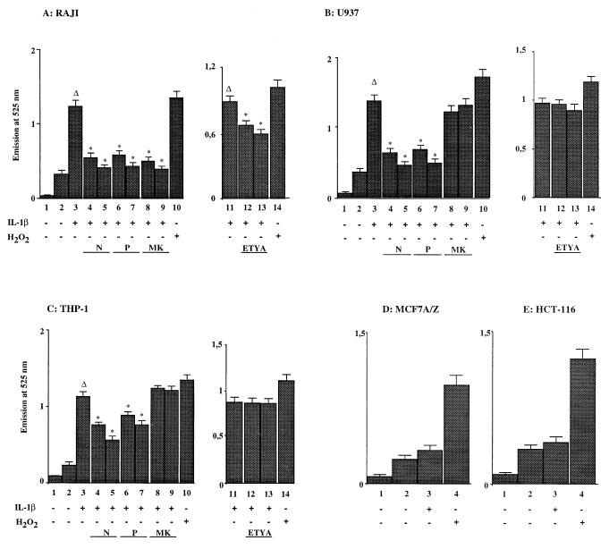
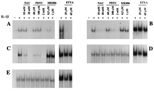
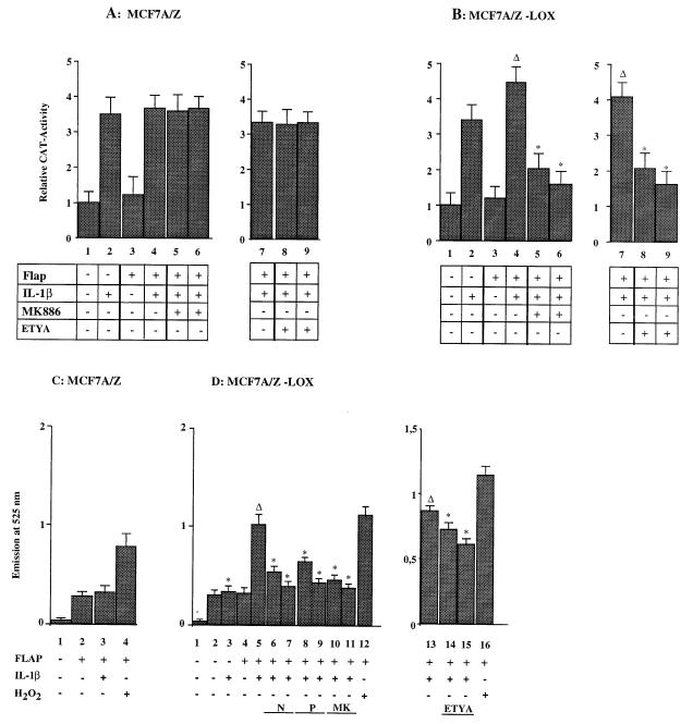
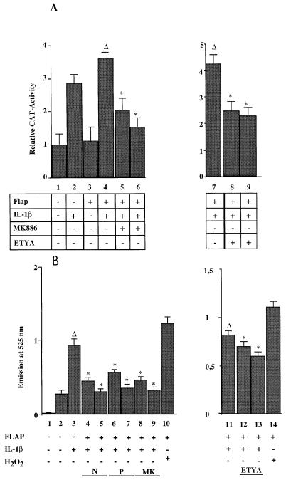
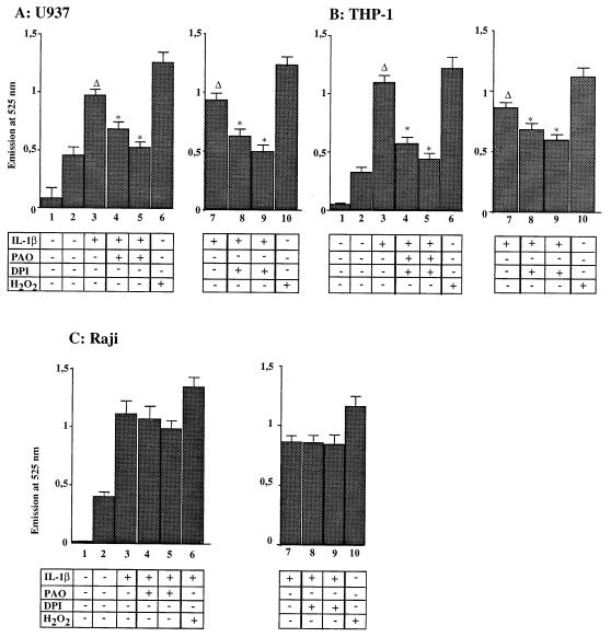
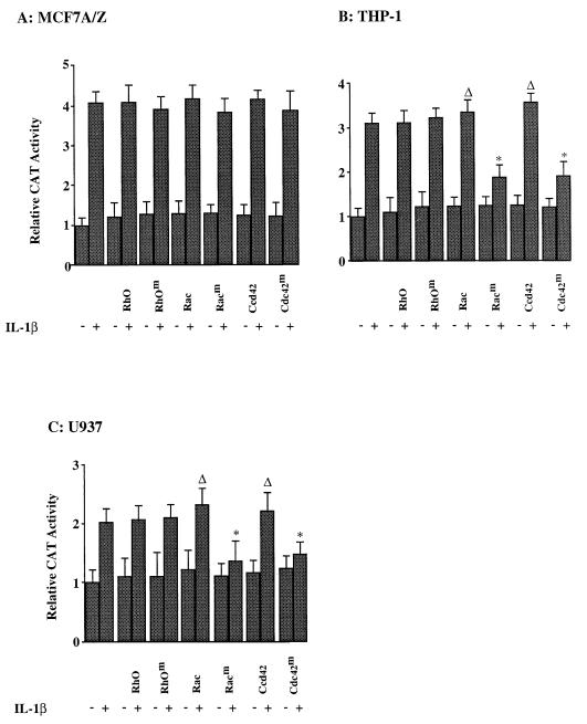
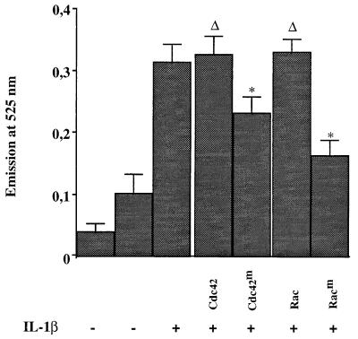
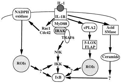
We previously reported that the role of reactive oxygen intermediates (ROIs) in NF-kappaB activation by proinflammatory cytokines was cell specific. However, the sources for ROIs in various cell types are yet to be determined and might include 5-lipoxygenase (5-LOX) and NADPH oxidase. 5-LOX and 5-LOX activating protein (FLAP) are coexpressed in lymphoid cells but not in monocytic or epithelial cells. Stimulation of lymphoid cells with interleukin-1beta (IL-1beta) led to ROI production and NF-kappaB activation, which could both be blocked by antioxidants or FLAP inhibitors, confirming that 5-LOX was the source of ROIs and was required for NF-kappaB activation in these cells. IL-1beta stimulation of epithelial cells did not generate any ROIs and NF-kappaB induction was not influenced by 5-LOX inhibitors. However, reintroduction of a functional 5-LOX system in these cells allowed ROI production and 5-LOX-dependent NF-kappaB activation. In monocytic cells, IL-1beta treatment led to a production of ROIs which is independent of the 5-LOX enzyme but requires the NADPH oxidase activity. This pathway involves the Rac1 and Cdc42 GTPases, two enzymes which are not required for NF-kappaB activation by IL-1beta in epithelial cells. In conclusion, three different cell-specific pathways lead to NF-kappaB activation by IL-1beta: a pathway dependent on ROI production by 5-LOX in lymphoid cells, an ROI- and 5-LOX-independent pathway in epithelial cells, and a pathway requiring ROI production by NADPH oxidase in monocytic cells.


Figures

FIG. 1
FIG. 1
5-LOX and FLAP expression in cell lines. Protein extracts from various cell lines were prepared and analyzed for 5-LOX (A) and FLAP (B) expression by immunoblots revealed with specific antibodies. The 5-LOX antibody was directed against the human protein and could not detect the murine enzyme in EL-4 and 70Z/3 cells. The FLAP antibody could recognize both the human and the murine proteins, and the murine protein gave a faint and more slowly migrating band in EL-4 and 70Z/3 cells, which is visible after longer exposures of the gel.
FIG. 2
FIG. 2
Production of ROIs following IL-1β stimulation. Formation of ROIs was measured using a DFCH probe in unstimulated and IL-1β-stimulated cells. Panels A, B, and C show the relative fluorescence emission at 525 nm of the DFCH probe in Raji, U937, and THP-1 cells. Columns 1, background (no DFCH probe); columns 2, unstimulated cells; columns 3, stimulation with IL-1β (50 U/ml); columns 4, as in columns 3 plus NAC (10 mM); columns 5, as in columns 3 plus NAC (20 mM); columns 6, as in columns 3 plus PDTC (60 μM); columns 7, as in columns 3 plus PDTC (100 μM); columns 8, as in columns 3 plus MK886 (0.5 μM); columns 9, as in columns 3 plus MK886 (1 μM); columns 11, stimulation with IL-1β (50 U/ml); columns 12, as in columns 11 plus ETYA (35 μM); columns 13, as in columns 11 plus ETYA (65 μM); columns 10 and 14, stimulation with H2O2 (250 μM). Panels D and E show the relative fluorescence emission at 525 nm of the DFCH probe in MCF7 A/Z and HCT-116 cells. Columns 1, background (no DFCH probe); columns 2, unstimulated cells; columns 3, stimulation with IL-1β (50 U/ml); columns 4, stimulation with H2O2 (250 μM). Asterisks indicate that the values were statistically different from the reference values (Δ).
FIG. 3
FIG. 3
ROI or 5-LOX inhibitors blocked NF-κB activation by IL-1β in a cell-specific manner. Nuclear extracts were prepared from Raji (A), U937 (B), THP-1 (C), HCT116 (D), or MCF7 A/Z (E) cells, either untreated or stimulated with IL-1β (50 U/ml). The same cells were preincubated prior to IL-1β stimulation with increasing concentrations of NAC, PDTC, MK886, or ETYA, as indicated in the figure. These extracts were analyzed by EMSA for binding to a specific κB probe.
FIG. 4
FIG. 4
Transfection of 5-LOX and FLAP expression vectors restored ROI production and ROI-dependent NF-κB activity in MCF7 A/Z cells. (A) MCF A/Z cells were transfected with the HIV-κB-CAT reporter plasmid alone or together with a FLAP expression vector, and CAT activities were measured in unstimulated cells or in cells stimulated with IL-1β for 6 h (50 U/ml), as indicated in the figure. The IL-1β treatment was performed in the absence or in the presence of either the FLAP inhibitor MK886 at 0.5 μM (column 5) and 1 μM (column 6) or the 5-LOX inhibitor ETYA at 35 μM (column 8) and 65 μM (column 9). Each column represents the mean of three independent experiments (± SD). We did not observe any statistically significant differences between cells stimulated by IL-1β in the presence or absence of the inhibitors. The total amount of transfected DNA was kept constant throughout the experiment by addition of appropriate amounts of the expression vector without insert. (B) MCF A/Z cells stably transfected with the 5-LOX expression vector (MCF A/Z-LOX) were transfected with the HIV-κB-CAT reporter plasmid alone or together with a FLAP expression vector, and CAT activities were measured in unstimulated cells or in cells stimulated with IL-1β as described for panel A. Asterisks indicate that values were statistically different from the reference values (Δ). (C) Formation of ROIs was measured by using DFCH in MCF7 A/Z cells transiently transfected with the FLAP expression vector. Column 1, background (no DFCH probe); column 2, unstimulated cells; column 3, stimulation with IL-1β (50 U/ml); column 4, stimulation with H2O2 (250 μM). (D) ROI production in MCF7 A/Z-LOX cells either unmodified or transiently transfected with the FLAP expression vector. Column 1, background (no DFCH probe); column 2, unstimulated cells; column 3, stimulation with IL-1β (50 U/ml); column 4, FLAP-transfected unstimulated cells; column 5, FLAP-transfected cells stimulated with IL-1β (50 U/ml); column 6, as in column 5 plus NAC (10 mM); column 7, as in column 5 plus NAC (20 mM); column 8, as in column 5 plus PDTC (60 μM); column 9, as in column 5 plus PDTC (100 μM); column 10, as in column 5 plus MK886 (0.5 μM); column 11, as in column 5 plus MK886 (1 μM); column 13, stimulation with IL-1β (50 U/ml); column 14, as in column 13 plus ETYA (35 μM); column 15, as in column 13 plus ETYA (65 μM); columns 12 and 16, stimulation with H2O2 (250 μM). Asterisks indicate that values were statistically different from the reference values (Δ).
FIG. 5
FIG. 5
Transfection of the FLAP expression vector restored ROI production and ROI-dependent NF-κB activity in HCT-116 cells. (A) HCT-116 cells were transfected with the HIV-κB-CAT reporter plasmid alone or together with a FLAP expression vector, and CAT activities were measured in unstimulated cells or in cells stimulated with IL-1β for 6 h (50 U/ml), as indicated in the figure. The IL-1β treatment was performed in the absence or in the presence of either the FLAP inhibitor MK886 at 0.5 μM (column 5) and 1 μM (column 6) or the 5-LOX inhibitor ETYA at 35 μM (column 8) and 65 μM (column 9). Each column represents the mean of three independent experiments (± SD). Asterisks indicate that values were statistically different from the reference values (Δ). (B) Formation of ROIs in HCT-116 cells transiently transfected with the FLAP expression vector. Column 1, background (no DFCH probe); column 2, unstimulated, FLAP transfected cells; column 3, stimulation with IL-1β (50 U/ml); column 4, as in column 3 plus NAC (10 mM); column 5, as in column 3 plus NAC (20 mM); column 6, as in column 3 plus PDTC (60 μM); column 7, as in column 3 plus PDTC (100 μM); column 8, as in column 3 plus MK886 (0.5 μM); column 9, as in column 3 plus MK886 (1 μM); column 11, stimulation with IL-1β (50 U/ml); column 12, as in column 11 plus ETYA (35 μM); column 13, as in column 11 plus ETYA (65 μM); columns 10 and 14, stimulation with H2O2 (250 μM). Asterisks indicate that values are statistically different from the reference values (Δ).
FIG. 6
FIG. 6
NADPH oxidase inhibitors blocked ROI production induced by IL-1β stimulation in monocytic cells. Formation of ROIs was measured by using a DFCH probe in U937 (A), THP-1 (B), and Raji (C) cells after IL-1β stimulation, with and without preincubation in the presence of the NADPH oxidase inhibitors DPI and PAO. Columns 1, background (no DFCH probe); columns 2, unstimulated cells; columns 3, stimulation with IL-1β (50 U/ml); columns 4, as in columns 3 plus PAO (30 μg/ml); columns 5, as in columns 3 plus PAO (60 μg/ml); columns 7, stimulation with IL-1β (50 U/ml); columns 8, as in columns 7 plus DPI (10 μM); columns 9, as in columns 7 plus DPI (30 μM); columns 6 and 10, stimulation with H2O2 (250 μM). Asterisks indicate that values are statistically different from the reference values (Δ).
FIG. 7
FIG. 7
NADPH oxidase inhibitors blocked NF-κB activation by IL-1β in monocytic cells. Nuclear extracts were prepared from Raji (A), U937 (B), THP-1 (C), HCT116 (D), or MCF7 A/Z (E) cells, either untreated or stimulated with IL-1β (50 U/ml). The same cells were preincubated for 1 h prior to IL-1β stimulation with increasing concentrations of DPI or PAO, as indicated in the figure. These extracts were analyzed by EMSA for binding to a specific κB probe.
FIG. 8
FIG. 8
GTPases are required for NF-κB transcriptional activity in monocytic cells. MCF7 A/Z (A), THP-1 (B), or U937 (C) cells were transfected with the HIV-κB-CAT reporter plasmid alone or together with expression vectors for wild-type or dominant-negative RhoA, Rac1, and Cdc42. Cells were either untreated (−) or stimulated with IL-1β (+) for 6 h, and CAT activities were measured. Each column represents the mean of three independent experiments (± SD). Asterisks indicate that values are statistically different from the reference values (Δ).
FIG. 9
FIG. 9
GTPases are required for ROI production in response to IL-1β in monocytic cells. U937 cells were transfected with the CD20 expression vector and with plasmids coding for wild-type (Cdc42 and Rac) or dominant-negative (Cdc42m and Racm) Rac1 and Cdc42. CD20-expressing transfected cells were positively selected with an anti-CD20 monoclonal antibody through MACS and flow cytometry cell sorting. Selected cells were either untreated (−) or stimulated with IL-1β (+) for 15 min, and formation of ROIs was measured by using a DFCH probe. Asterisks indicate that values are statistically different from the reference values (Δ).
FIG. 10
FIG. 10
Signaling pathways for NF-κB activation by IL-1β. Following the interaction of IL-1 with the type 1 IL-1 receptor, the NIK-IKK pathway can be activated through the MyD88, IRAK, and TRAF6 signaling proteins. Alternatively, Rac1 and Cdc42 can activate the NADPH oxidase complex and induce NF-κB activity through the production of ROIs. Other pathways involve, in lymphoid cells, the cytoplasmic phospholipase A2, the 5-LOX, and FLAP complex and the production of ROIs or, in epithelial cells, the acid sphingomyelinase (SMase) and the production of ceramide. CPLA2, cytosolic phospholipase A2.

References

    1. Abo A, Boyhan A, West I, Thrasher A J, Segal A W. Reconstitution of neutrophil NADPH oxidase activity in the cell-free system by four components: p67-phox, p47-phox, p21rac1 and cytochrome b-245. J Biol Chem. 1992;267:16767–16770. - PubMed
    1. Baldwin A S. The NF-κB and IκB proteins: new discoveries and insights. Annu Rev Immunol. 1996;14:649–681. - PubMed
    1. Barnes P J, Karin M. Nuclear factor-κB—a pivotal transcription factor in chronic inflammatory diseases. N Engl J Med. 1997;336:1066–1071. - PubMed
    1. Beg A A, Finco T S, Nantermet P V, Baldwin A S., Jr Tumor necrosis factor and interleukin-1 lead to phosphorylation and loss of IκBα: a mechanism for NF-κB activation. Mol Cell Biol. 1993;13:3301–3310. - PMC - PubMed
    1. Bokoch G M. Regulation of the phagocyte respiratory burst by small GTP-binding proteins. Trends Cell Biol. 1995;5:109–113. - PubMed

Publication types

MeSH terms