. 1999 Mar;19(3):1961-72.
doi: 10.1128/MCB.19.3.1961.

Neu differentiation factor stimulates phosphorylation and activation of the Sp1 transcription factor

Affiliations

Neu differentiation factor stimulates phosphorylation and activation of the Sp1 transcription factor

I Alroy et al. Mol Cell Biol. 1999 Mar.

Abstract

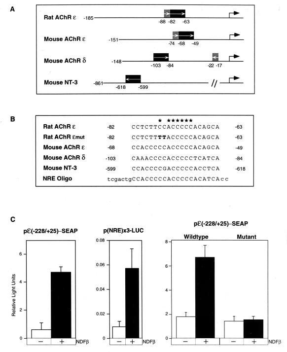
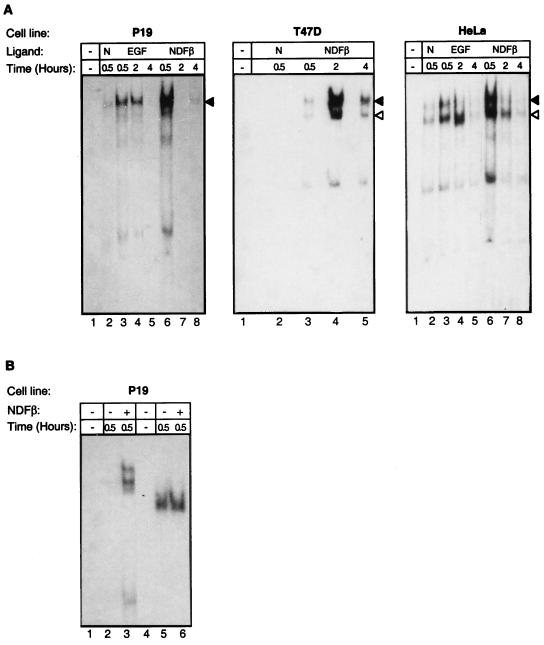
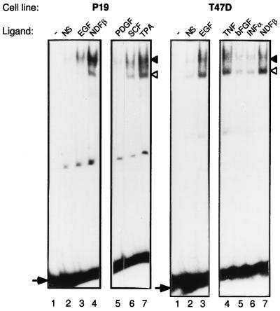
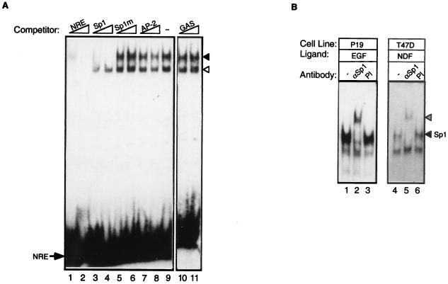
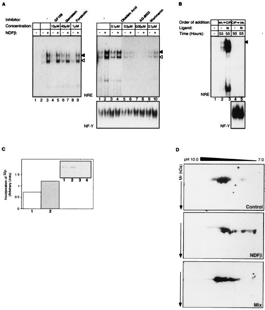
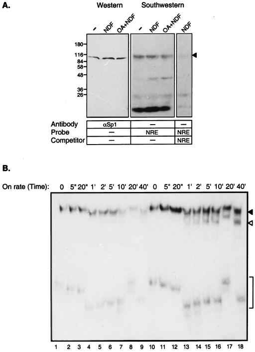
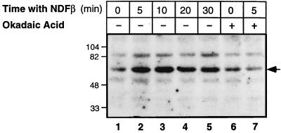
Neu differentiation factors (NDFs), or neuregulins, are epidermal growth factor-like growth factors which bind to two tyrosine kinase receptors, ErbB-3 and ErbB-4. The transcription of several genes is regulated by neuregulins, including genes encoding specific subunits of the acetylcholine receptor at the neuromuscular junction. Here, we have examined the promoter of the acetylcholine receptor epsilon subunit and delineated a minimal CA-rich sequence which mediates transcriptional activation by NDF (NDF-response element [NRE]). Using gel mobility shift analysis with an NRE oligonucleotide, we detected two complexes that are induced by treatment with neuregulin and other growth factors and identified Sp1, a constitutively expressed zinc finger phosphoprotein, as a component of one of these complexes. Phosphatase treatment, two-dimensional gel electrophoresis, and an in-gel kinase assay indicated that Sp1 is phosphorylated by a 60-kDa kinase in response to NDF-induced signals. Moreover, Sp1 seems to act downstream of all members of the ErbB family and thus may funnel the signaling of the ErbB network into the nucleus.


Figures

FIG. 1
FIG. 1
Mapping of the NRE in the promoter of the AChR ɛ gene. (A) Minimal promoter regions needed for muscle-specific expression of the rat AChR ɛ gene (nucleotides −185 to +25), the mouse AChR ɛ gene (−151 to +84), and the mouse AChR δ gene (−148 to +24), as well as the region in the promoter of the NT-3 gene encompassing the putative NRE. The E-box element, which confers muscle-specific expression, is indicated by a shaded box, and the region that confers responsiveness to neuregulin is indicated by a solid box; arrows within the boxes denote the orientation of this element. The mRNA start sites are indicated by large arrows. (B) Sequence comparison of the CA-rich elements found in the promoters of the NDF-inducible genes (asterisks indicate bases that are strictly conserved). Also shown is the sequence of an oligonucleotide (NRE Oligo) that we used to construct a reporter plasmid and perform the EMSA. A mutation we introduced in the rat AChR ɛ reporter is also listed (Rat AChR ɛmut). (C) NDF-induced transcriptional activation through NRE. The following constructs were used to assay transcriptional activation of reporter genes (to cotransfect P-19 cells together with a β-galactosidase reporter as an internal control): a minimal promoter of the rat AChR ɛ gene fused to the SEAP gene [pɛ(−228/+25)-SEAP], three copies of the NRE oligonucleotide cloned upstream of a luciferase reporter gene [p(NRE)×3-LUC], and two versions, a wild-type and a mutant form (Rat AChR ɛmut [B]), of pɛ(−228/+25)-SEAP. At 24 h posttransfection, cells were untreated (−) or treated (+) with NDFβ (100 ng/ml). At 16 h later, the cells were harvested for luciferase and β-galactosidase assays, or their medium was assayed for SEAP activity. Signals obtained with the reporter genes were normalized to β-galactosidase activity.
FIG. 2
FIG. 2
NDFβ and EGF induce the formation of two NRE binding complexes. (A) An oligonucleotide duplex (top strand sequence, 5′-TCGACTGCCACCCCCACCCCCACATCACC-3′) was used as a probe and incubated without (−) or with 5 μg of whole-cell extract prepared from P-19, T47D, or HeLa cells. The cells were untreated (N) or treated for the indicated time intervals with NDFβ or EGF, as indicated, and NRE binding was assayed by EMSA. Arrowheads indicate the positions of the two protein-DNA complexes. Note that the complex whose mobility is greater (open arrowhead) is absent in P-19 cells. (B) Whole-cell extracts prepared from NDF-treated or untreated P-19 cells were used for EMSA with the NRE oligonucleotide (lanes 1 to 3) or with the NF-Y oligonucleotide probe as a control (lanes 4 to 6). Note the absence of an NDF effect on NF-Y.
FIG. 3
FIG. 3
Growth factor induction of protein-DNA complexes on the NRE probe. P-19 (left) or T47D (right) cells were treated for 30 min with the indicated growth factors or for 2 h with TPA, and whole-cell extracts were prepared. Extracts were incubated with a labeled NRE probe, and protein-DNA complexes were separated on a nondenaturing polyacrylamide gel. The arrows denote the positions of the free oligonucleotide probe, and the arrowheads denote the positions of protein-DNA complexes (a slow-migrating complex [closed arrowhead] and a fast-migrating complex [open arrowhead]). Abbreviations: NS, nonstimulated; PDGF, platelet-derived growth factor; SCF, stem cell factor; TNF, tumor necrosis factor; bFGF, basic fibroblast growth factor; INFα, alpha interferon.
FIG. 4
FIG. 4
Identification of factors interacting with the NRE. (A) Competition between NRE and consensus oligonucleotides. Whole-cell extracts were prepared from NDFβ-treated T47D cells and incubated with the NRE probe in the absence (−) or presence of the indicated unlabeled competitor oligonucleotides at either 100-fold excess (lanes 1, 3, 5, 7, and 10) or 500-fold excess (lanes 2, 4, 6, 8, and 11). An arrow denotes the position of the free NRE probe, and arrowheads denote the positions of the two protein-DNA complexes (the solid arrowhead indicates the slower-migrating complex). (B) The slower-migrating protein-DNA complex is supershifted with an Sp1-specific antibody. Whole-cell extracts prepared from EGF-treated P-19 cells or NDF-treated T47D cells were preincubated with an Sp1-specific antibody (αSp1). As a control, extracts were treated with a preimmune serum (PI) or with no antibody (−). A labeled NRE probe was then added, and protein-DNA complexes were separated on nondenaturing polyacrylamide gels. The positions of the Sp1-specific complex (solid arrowhead) and the supershifted complex (hatched arrowhead) are marked. Note that the lower NRE complex underwent no supershift.
FIG. 5
FIG. 5
Phosphorylation events are important for the regulation of Sp1 binding to the NRE site. (A) Effects of kinase and phosphatase inhibitors on NRE binding activity in NDFβ-treated T47D cells. Prior to EMSA with an NRE probe, the cells were preincubated for 20 min with the inhibitors GF109203X (GF109, a protein kinase C-specific inhibitor), genistein (a general inhibitor of tyrosine kinases), forskolin (an inhibitor of cyclic AMP phosphodiesterases), okadaic acid (OA, an inhibitor of protein phosphatase 2A), AG2002 (an inhibitor of the ErbB tyrosine kinase activity), and wortmannin (a phosphatidylinositol 3′-kinase inhibitor) and then treated with NDF. Control cultures were not treated with an inhibitor. The positions of the two protein-DNA complexes are denoted by arrowheads (closed arrowhead, Sp1-specific complex; open arrowhead, faster-migrating complex). As a control, EMSA was performed with the NF-Y probe (lower panel). (B) Inhibition of Sp1 DNA binding by in vitro treatment with CIP. The NRE (upper panel) and NF-Y (lower panel) probes were incubated with 5 μg of extracts prepared from P-19 cells that were incubated for the indicated time intervals with NDFβ (N). Control cultures were not treated with a growth factor (−). Prior to EMSA, extracts were incubated with CIP and then the phosphatase was inhibited by addition of specific inhibitors (a mixture of NaF, sodium vanadate, and potassium pyrophosphate [lanes 4 and 5]). Alternatively, the inhibitors were added prior to treatment with CIP (lanes 2 and 3). The position of the Sp1-specific complex is indicated by an arrowhead. (C) Sp1 is a phosphoprotein whose phosphorylation is increased by NDF. P-19 cells were preincubated with a medium containing [32P]orthophosphate and then treated (lanes 1 and 3) or not treated (lanes 2 and 4) with NDF. Whole-cell extracts were prepared and incubated with an anti-Sp1 antiserum (lanes 1 and 2) or an anti-Egr1 antibody (lanes 3 and 4). The immunoprecipitates were resolved by SDS-PAGE and electrophoretically transferred to a nitrocellulose filter. The filter was subjected to autoradiography (inset) and then immunoblotted with an antibody to Sp1 (data not shown). Both signals were quantified, and their ratio is presented in a histogram. Numbers below the bars correspond to gel lanes. (D) Analysis of NDF-induced phosphorylation of Sp1 by two-dimensional gel electrophoresis. Nuclear extracts (5 μg of protein) were prepared from untreated (Control), or P-19 cells treated with 100 ng of NDFβ per ml. As a control, a mixture of the two nuclear extracts (2.5 μg each) was analyzed (MIX). Extracts were separated by isoelectric focusing in the first dimension (pH values are indicated) and by SDS-PAGE in the second dimension (the locations of marker proteins are indicated in kilodaltons). The proteins were then transferred to nitrocellulose filters, and the Sp1 protein was detected by immunoblotting with an anti-Sp1 antibody.
FIG. 6
FIG. 6
Sp1 binding and kinetics of complex formation on the NRE. (A) Southwestern analysis. Whole-cell extracts were prepared from P-19 cells that were untreated (−) or treated for 30 min with NDFβ. A third culture was incubated for 20 min with okadaic acid (OA) prior to treatment with NDF. Duplicate extracts (60 μg) were separated by SDS-PAGE and then subjected to either Western blot analysis with an antibody to Sp1 (left) or to Southwestern analysis with a labeled NRE probe (center and right). For control of NRE binding specificity, the Southwestern analysis was also performed in the presence of an excess of the unlabeled NRE probe (right). The Sp1 band is labeled by an arrowhead. (B) On-rate kinetics of complex formation on the NRE. Whole P-19 cell extracts prepared from untreated (lanes 1 to 9) or NDF-treated (lanes 10 to 18) cells were incubated in EMSA binding buffer. Equal volumes were removed and incubated for the indicated time intervals with a labeled NRE probe. At the end of the incubation, samples were loaded on a running mobility shift gel. The positions of the NRE-specific complexes are marked by a solid arrowhead (the slowest-migrating complex), an open arrowhead, and brackets (the fastest-migrating complex). Time is marked as seconds (“) or minutes (’). Note that due to continuous electrophoresis while loading samples on the gel, all of the bracket-labeled bands represent the same complex.
FIG. 7
FIG. 7
The kinase that enhances Sp1 binding to NRE is NDFβ inducible and okadaic acid sensitive. P-19 cells were treated with NDFβ for the indicated time intervals in the absence (−) or presence (+) of 0.5 μM okadaic acid (added 20 min prior to NDFβ). Cell extracts were then analyzed for kinase activity by using a recombinant form of the human Sp1 protein and an in-gel kinase assay as detailed in Materials and Methods. The position of the kinase which is induced by NDF and inhibited by okadaic acid is marked by an arrow. The locations of molecular weight marker proteins are indicated in thousands.

References

    1. Alroy I, Towers T L, Freedman L P. Transcriptional repression of the interleukin-2 gene by vitamin D3: direct inhibition of NFATp/AP-1 complex formation by a nuclear hormone receptor. Mol Cell Biol. 1995;15:5789–5799. - PMC - PubMed
    1. Alroy I, Yarden Y. The ErbB signaling network in embryogenesis and oncogenesis: signal diversification through combinatorial ligand-receptor interactions. FEBS Lett. 1997;410:83–86. - PubMed
    1. Altiok N, Altiok X, Changeux J P. Heregulin-stimulated acetylcholine receptor gene expression in muscle: requirement for MAP kinase and evidence for a parallel inhibitory pathway independent of electrical activity. EMBO J. 1997;16:717–725. - PMC - PubMed
    1. Armstrong S A, Barry D A, Leggett R W, Mueller C R. Casein kinase II-mediated phosphorylation of the C terminus of Sp1 decreases its DNA binding activity. J Biol Chem. 1997;272:13489–13495. - PubMed
    1. Baldwin T J, Burden S J. Isolation and characterization of the mouse acetylcholine receptor delta subunit gene: identification of a 148-bp cis-acting region that confers myotube-specific expression. J Cell Biol. 1988;107:2271–2279. - PMC - PubMed

Publication types