. 1999 Mar;19(3):2098-108.
doi: 10.1128/MCB.19.3.2098.

Activation-dependent transcriptional regulation of the human Fas promoter requires NF-kappaB p50-p65 recruitment

Affiliations

Activation-dependent transcriptional regulation of the human Fas promoter requires NF-kappaB p50-p65 recruitment

H Chan et al. Mol Cell Biol. 1999 Mar.

Abstract

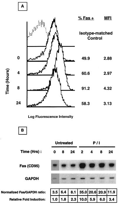
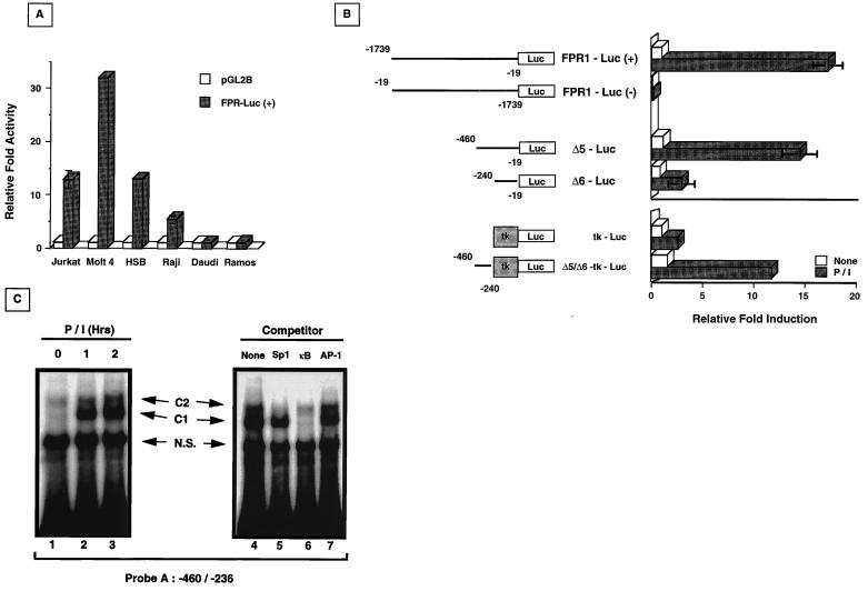
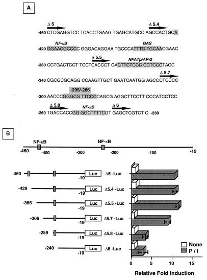
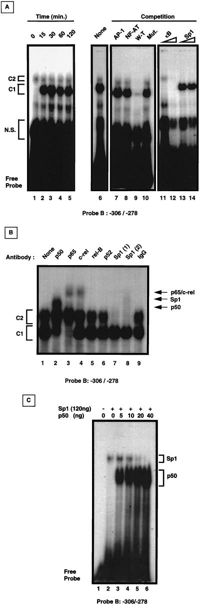
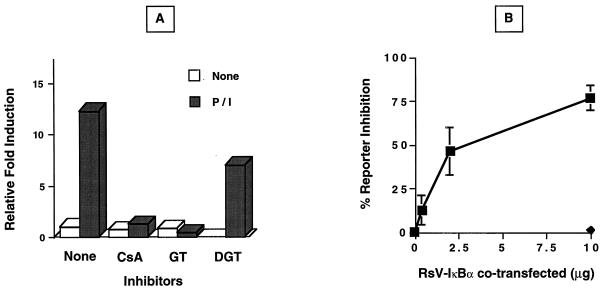
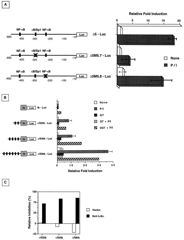
Fas (CD95) and Fas ligand (CD95L) are an interacting receptor-ligand pair required for immune homeostasis. Lymphocyte activation results in the upregulation of Fas expression and the acquisition of sensitivity to FasL-mediated apoptosis. Although Fas upregulation is central to the preservation of immunologic tolerance, little is known about the molecular machinery underlying this process. To investigate the events involved in activation-induced Fas upregulation, we have examined mRNA accumulation, fas promoter activity, and protein expression in the Jurkat T-cell line treated with phorbol myristate acetate and ionomycin (P/I), pharmacological mimics of T-cell receptor activation. Although resting Jurkat cells express Fas, Fas mRNA was induced approximately 10-fold in 2 h upon P/I stimulation. Using sequential deletion mutants of the human fas promoter in transient transfection assays, we identified a 47-bp sequence (positions -306 to -260 relative to the ATG) required for activation-driven fas upregulation. Sequence analysis revealed the presence of a previously unrecognized composite binding site for both the Sp1 and NF-kappaB transcription factors at positions -295 to -286. Electrophoretic mobility shift assay (EMSA) and supershift analyses of this region documented constitutive binding of Sp1 in unactivated nuclear extracts and inducible binding of p50-p65 NF-kappaB heterodimers after P/I activation. Sp1 and NF-kappaB transcription factor binding was shown to be mutually exclusive by EMSA displacement studies with purified recombinant Sp1 and recombinant p50. The functional contribution of the kappaB-Sp1 composite site in P/I-inducible fas promoter activation was verified by using kappaB-Sp1 concatamers (-295 to -286) in a thymidine kinase promoter-driven reporter construct and native promoter constructs in Jurkat cells overexpressing IkappaB-alpha. Site-directed mutagenesis of the critical guanine nucleotides in the kappaB-Sp1 element documented the essential role of this site in activation-dependent fas promoter induction.


Figures

FIG. 1
FIG. 1
Time course induction of Fas upregulation in P/I-treated Jurkat cells. (A) Cells were harvested at the indicated time, and Fas expression was analyzed by flow cytometry. Isotype-matched control antibody staining is represented as a dashed line, and Fas staining is represented as solid lines. The percentage of specific Fas staining and the mean fluorescence intensity (MFI) of Jurkat cells at each time point are shown to the right. (B) Cells were harvested at the indicated time, total RNA was isolated, and Fas mRNA accumulation was determined by RNAse protection analysis. Scanning densitometry was used to normalize Fas mRNA levels and to calculate the relative fold induction compared to that of the untreated, time zero sample.
FIG. 2
FIG. 2
Basal and inducible fas promoter activity and in vitro EMSA analysis. (A) FPR1-Luc(+) containing the sequence from −1739 to −19 is active in T-cell lines and the B-lymphocyte line Raji, but inactive in Daudi or Ramos cells. Cells were transfected with 10 μg of either the empty reporter (pGL2B [□]) or the fas promoter reporter vector (FPR1-Luc [■]); 10 ng of pRL-tk vector was cotransfected for normalization. Extracts were taken after 40 h, and luciferase activity was determined. Relative fold activity was calculated as the normalized FPR1-Luc(+) activity divided by the normalized pGL2B value. (B) fas promoter sequences −460 to −240 are required for inducible fas reporter activity. FPR1-Luc in both orientations (forward and reverse), Δ5-Luc, Δ6-Luc, tk-Luc, and Δ5/Δ6-tk-Luc constructs were transiently transfected into Jurkat cells with pRL-tk for normalization, and the cells were then left untreated (□) or were activated with P/I (■) for 8 h before harvesting of the extracts for measurement of luciferase activity. The values shown are averages of three independent experiments (± standard deviation) and are expressed as relative fold induction over normalized luciferase values from untreated Jurkat cells containing FPR1-Luc(+). For tk-Luc and Δ5/Δ6-tk-Luc, the relative fold induction was calculated separately as the normalized luciferase value divided by the untreated, normalized tk-Luc luciferase value. (C) Specific protein complexes bind the enhancer region between −460 and −236. 32P-labeled probe A (50,000 cpm) was mixed with nuclear extracts from untreated or P/I-activated Jurkat cells (left panel, lanes 1 to 3). Specific complexes (C1 and C2) are indicated. N.S., nonspecific complexes. Cold competition mixtures with a 25-fold excess of the indicated consensus elements (right panel, lanes 4 to 7) were mixed with extracts from Jurkat cells activated with P/I for 1 h prior to addition of labeled probe A.
FIG. 3
FIG. 3
Localization of activation-dependent fas promoter activity. (A) The fas nucleotide sequence between −460 and −230 is shown with known consensus transcription factor elements indicated (shaded areas). The location and designation of the various deletion constructs are indicated by the solid arrow. (B) P/I inducibility of the fas promoter localizes between nucleotides −306 and −260. Jurkat cells transiently transfected with 10 μg of the various deletion constructs and 10 ng of pRL-tk as the normalizing vector were left untreated (□) or activated with P/I (■) for 8 h before extracts were prepared for luciferase activity measurements. The values shown are averages of three independent experiments ± standard deviation and are expressed as the relative fold induction of the various deletion constructs over the untreated, normalized Δ5-Luc luciferase value.
FIG. 4
FIG. 4
In vitro analysis of a potential κB-Sp1 site at −295 to −286. (A) Time course analysis of complex binding to probe B (−306 to −278). Jurkat cells were activated with P/I for the indicated time, nuclear extracts were harvested, and EMSA analysis was performed with 32P-labeled probe B (left panel, lanes 1 to 5). Cold competition was performed with 1-h-activated Jurkat cell nuclear extracts with a 25-fold excess of consensus oligonucleotides for the transcription factors AP-1, NF-AT, wild-type probe B (W-T), or mutated probe B (Mut.) added prior to incubation with 32P-labeled probe B (lanes 7 to 10). For NF-κB and Sp1 competition, a 25-fold excess (lanes 11 and 13) and 100-fold excess (lanes 12 and 14) of consensus oligonucleotides were added to equivalent amounts of P/I-activated nuclear extracts prior to addition of 32P-labeled probe B. N.S., nonspecific binding. (B) Supershift analyses of P/I-activated Jurkat cell nuclear extracts. Nuclear extracts were subjected to EMSA analysis in the absence (lane 1) or presence of the indicated specific antisera (lanes 2 to 8), or preimmune IgG (lane 9). With two different anti-Sp1 antisera, probe B binding to the upper C2 complex was shown to be either shifted or inhibited (lanes 7 and 8). Arrows indicate the identity of the shifted complexes. (C) Mutual exclusive binding of Sp1 and NF-κB p50 to probe B. Recombinant Sp1 (4 fpu [120 ng]) was preincubated with 12,500 cpm of radiolabeled probe B in the absence (lane 2) or presence of increasing amounts (0 to 40 ng) of recombinant NF-κB p50 (lanes 3 to 6) prior to EMSA analysis. Brackets indicate the Sp1-probe B and p50-probe B complexes.
FIG. 5
FIG. 5
Inhibition of activation-dependent fas promoter upregulation. (A) Pharmacological inhibition of Δ5-Luc induction by P/I. Jurkat cells transiently transfected with 10 μg of Δ5-Luc and 10 ng of pRL-tk were preincubated in the absence (None) or presence of cyclosporin A (CsA [1 μg/ml]), gliotoxin (GT [1 μg/ml]), or bis-dethio-bis(methylthio)gliotoxin (DGT [3 μg/ml]) for 30 min prior to P/I stimulation for 8 h. Following incubation, cells were harvested for extract preparation, and luciferase activity was determined. The values shown are representative of two independent experiments ± standard deviation and are expressed as the relative fold induction over normalized untreated Δ5-Luc activity. (B) Dose-dependent inhibition of P/I-inducible Δ5-Luc reporter activity by IκBα. Increasing amounts of wild-type RsV-IκBα (■) or vector control (⧫) expression plasmid were cotransfected into Jurkat cells with 10 μg of Δ5-Luc and 10 ng of pRL-tk 40 h prior to P/I activation. Cells were harvested for extract preparation after 8 h of P/I exposure, and luciferase activity was measured. The values shown are averages from two independent experiments ± standard deviation and are expressed as percent inhibition of P/I-induced, normalized Δ5-Luc reporter activity.
FIG. 6
FIG. 6
The κB-Sp1 enhancer element is required for P/I-inducible Δ5-Luc reporter activity. (A) Nucleotide mutagenesis of Δ5-Luc at either the κB-Sp1 element (GGG→CCC; −295 to −293 [Δ5M5.7-Luc]) or the downstream NF-κB element (GGG→CCY; −252 to −249 [Δ5M5.8-Luc]) was introduced by PCR. Wild-type or mutagenized constructs were cotransfected into Jurkat cells with 10 ng of pRL-tk, and the cells were activated with P/I for 8 h prior to being harvested for extract preparation and luciferase activity measurements. The values shown are averages of three independent experiments ± standard deviations and are expressed as the relative fold increase over untreated, normalized Δ5-Luc luciferase activity. (B) Concatemer constructs containing either none (tk-Luc), two (κ/S2tk-Luc), four (κ/S4tk-Luc), or six (κ/S6tk-Luc) copies of the κB-Sp1 element (−295 to −286) were juxtaposed upstream of a thymidine kinase-driven luciferase reporter and cotransfected into Jurkat cells as mentioned above before activation with P/I and luciferase measurements. Inhibition of NF-κB activity were analyzed by preincubation of transfected cells with either gliotoxin (GT [1 μg/ml]) or bis-dethio-bis(methylthio)gliotoxin (DGT [1 μg/ml]) as described in the legend to Fig. 5 prior to activation with P/I. Relative fold induction is calculated against the untreated, normalized tk-Luc reporter luciferase activity. (C) Specific inhibition of NF-κB-dependent upregulation of the κB-Sp1 motif concatemers by coexpression of RsV-IκBα. Multimerized constructs were cotransfected with 5 μg of either RsV-IκBα or vector-only expression plasmids before activation with P/I as described above. Extracts were taken for luciferase measurements, and relative inhibition is calculated as the percentage of P/I-induced luciferase activity of the corresponding reporter constructs.

Similar articles

Cited by

References

    1. Alderson M R, Tough T W, Davis-Smith T, Braddy S, Falk B, Schooley K A, Goodwin R G, Smith C A, Ramsdell F, Lynch D H. Fas ligand mediates activation-induced cell death in human T lymphocytes. J Exp Med. 1995;181:71–77. - PMC - PubMed
    1. Aries S, Scgaaf B, Muller C, Dennin R, Dalhoff K. Fas (CD95) expression of CD4+ T cells from HIV-infected patients increases with disease progression. J Mol Med. 1995;73:591–593. - PubMed
    1. Azizkhan J C, Jensen D E, Pierce A J, Wade M. Transcription from TATA-less promoters: dihydrofolate reductase as a model. Crit Rev Eukaryot Gene Expr. 1993;3:229–254. - PubMed
    1. Banda N K, Bernier J, Kurahara D K, Kurrle R, Haligwood N, Sekaly R P, Finkel T H. Crosslinking CD4 by human immunodeficiency virus gp120 primes T cells for activation-induced apoptosis. J Exp Med. 1992;176:1099–1106. - PMC - PubMed
    1. Behrmann I, Walczak H, Krammer P H. Structure of the human APO-1 gene. Eur J Immunol. 1994;24:3057–3062. - PubMed

Publication types