. 1999 Mar;19(3):2155-68.
doi: 10.1128/MCB.19.3.2155.

Different regulation of the p53 core domain activities 3'-to-5' exonuclease and sequence-specific DNA binding

Affiliations

Different regulation of the p53 core domain activities 3'-to-5' exonuclease and sequence-specific DNA binding

F Janus et al. Mol Cell Biol. 1999 Mar.

Abstract

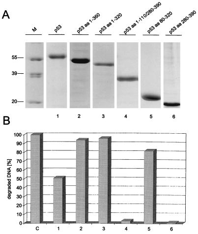
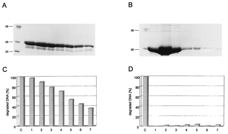
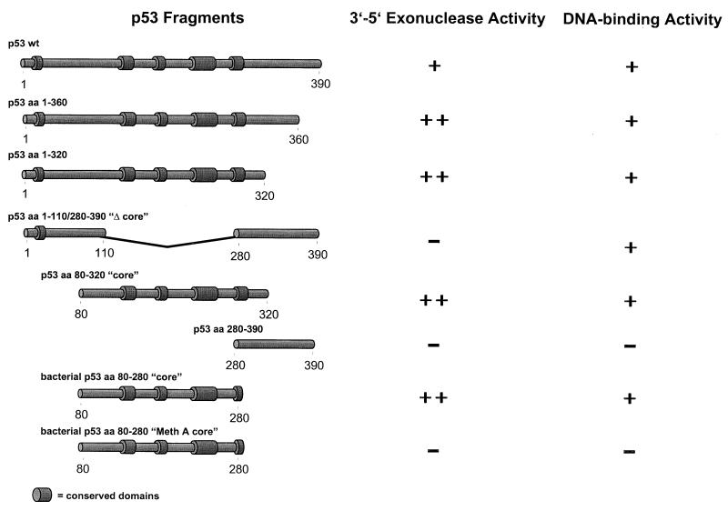
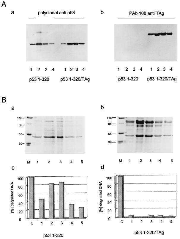
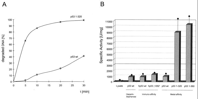
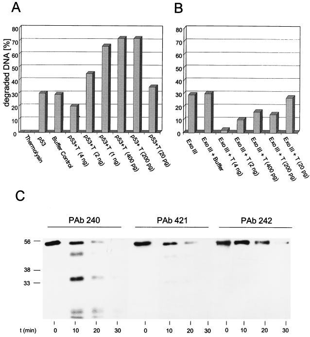
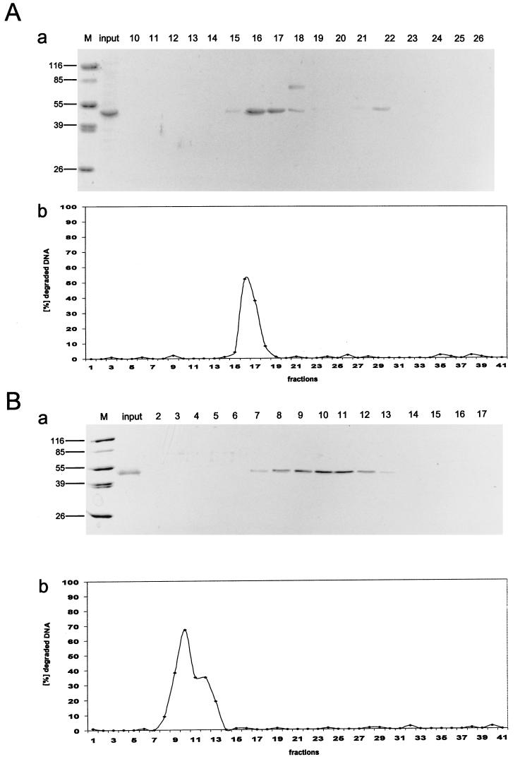
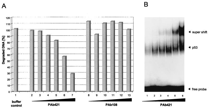
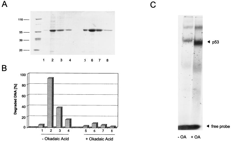
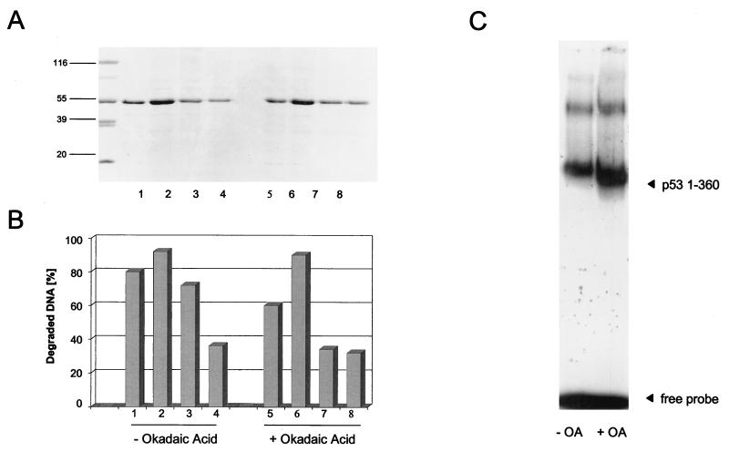
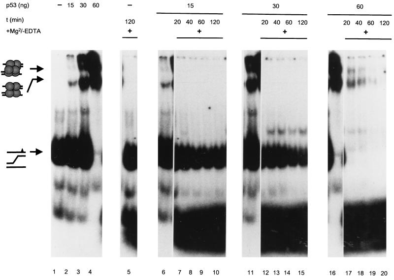
In this study we further characterized the 3'-5' exonuclease activity intrinsic to wild-type p53. We showed that this activity, like sequence-specific DNA binding, is mediated by the p53 core domain. Truncation of the C-terminal 30 amino acids of the p53 molecule enhanced the p53 exonuclease activity by at least 10-fold, indicating that this activity, like sequence-specific DNA binding, is negatively regulated by the C-terminal basic regulatory domain of p53. However, treatments which activated sequence-specific DNA binding of p53, like binding of the monoclonal antibody PAb421, which recognizes a C-terminal epitope on p53, or a higher phosphorylation status, strongly inhibited the p53 exonuclease activity. This suggests that at least on full-length p53, sequence-specific DNA binding and exonuclease activities are subject to different and seemingly opposing regulatory mechanisms. Following up the recent discovery in our laboratory that p53 recognizes and binds with high affinity to three-stranded DNA substrates mimicking early recombination intermediates (C. Dudenhoeffer, G. Rohaly, K. Will, W. Deppert, and L. Wiesmueller, Mol. Cell. Biol. 18:5332-5342), we asked whether such substrates might be degraded by the p53 exonuclease. Addition of Mg2+ ions to the binding assay indeed started the p53 exonuclease and promoted rapid degradation of the bound, but not of the unbound, substrate, indicating that specifically recognized targets can be subjected to exonucleolytic degradation by p53 under defined conditions.


Figures

FIG. 1
FIG. 1
Exonuclease activities of p53 and p53 deletion mutants. wt p53 and p53 deletion mutants were expressed in High Five insect cells infected with recombinant baculovirus and purified by metal affinity chromatography as described in Materials and Methods. Peak fractions of all proteins were analyzed by SDS-PAGE (A), and 150 ng of each protein was tested for exonuclease activity by the 3H filter retention assay (B). Exonuclease III was used as a control. aa, amino acids. Lane M, markers. Numbers on the left in panel A are molecular masses in kilodaltons.
FIG. 2
FIG. 2
wt but not mutant p53 core domain is necessary and sufficient for p53 exonuclease activity. wt (A and C) and MethA (B and D) p53 core fragments (amino acids 80 to 280) were expressed in bacteria and purified by metal affinity chromatography. Column fractions of both proteins were analyzed by SDS-PAGE (A and B) and tested for exonuclease activity by the 3H filter retention assay (C and D). Exonuclease III (lane C) was used as a control. Numbers on the left in panels A and B are molecular masses in kilodaltons.
FIG. 3
FIG. 3
Summary of p53 exonuclease activity mapping data. p53 fragments and their corresponding exonuclease activities are shown schematically. aa, amino acids.
FIG. 4
FIG. 4
SV40 T-Ag inhibits p53 exonuclease activity. High Five insect cells were infected with recombinant baculovirus coding for the p531-320 fragment or SV40 T-Ag. Cells were lysed at 44 h pi, and the p531-320-containing lysate was split. One half was purified by metal affinity chromatography, and the other half was mixed with SV40 T-Ag-containing lysate and also purified for His-tagged p531-320 by metal affinity chromatography. Column fractions of both preparations were analyzed by SDS-PAGE (B, panels a and b) and Western blotting (A) and tested for exonuclease activity (B, panels c and d). Numbers on the left in panel B (panels a and b) are molecular masses in kilodaltons. Lanes M, markers.
FIG. 5
FIG. 5
p53 exonuclease activity is negatively regulated by the C-terminal basic regulatory domain. Full-length wt p53 and p531-320 were tested for exonuclease activity, and their relative activities were compared in a time course (A). Specific exonuclease activities were determined for wt p53 purified by heparin-Sepharose, immunoaffinity, or metal affinity chromatography and were compared to those of the oligomerization-defective mutant p53 1262 and deletion mutants p531-320 and p531-360 (B). Standard deviations are indicated by error bars. One unit corresponds to the degradation of 60 pmol of DNA per 10 min at 37°C. hp53, human p53.
FIG. 6
FIG. 6
Protease treatment of p53 activates its exonucleolytic activity. (A and B) Wild-type p53 (A) or exonuclease III (Ex. III) (B) (150 ng) was digested with various amounts of thermolysin (40 U/mg), ranging from 4 ng to 20 pg. Exonuclease activity was measured after 15 min of digestion. The amount of exonuclease III was chosen to match the exonuclease activity of 150 ng of wt p53. Incubation of p53 with buffer alone for 15 min (p53 buffer control) did not affect the p53 exonuclease activity. (C) Western blot analysis of p53 fragments resulting from thermolysin digestion after 0, 10, 20, and 30 min of incubation. Digestions were performed with 1 ng of thermolysin, corresponding to bar 6 in panel A. After 10 min of thermolysin digestion, lower-migrating forms of p53 were detected with PAb240, directed against an epitope in the p53 core domain. Those forms could not be detected with monoclonal antibodies directed against the p53 N terminus (PAb242) or the p53 C terminus (PAb 421). Numbers on the left in panel C are molecular masses in kilodaltons.
FIG. 7
FIG. 7
Copurification of the p53 exonuclease activity with the p531-320 fragment. The p531-320 fragment was first purified by metal affinity chromatography and then further purified either by anion-exchange chromatography (UNO Q) (A) or by heparin-Sepharose affinity chromatography (B). p531-320 was eluted from the columns with a KCl gradient (see Materials and Methods for details). All fractions were analyzed for exonuclease activity (panels b), and peak fractions were analyzed by SDS-PAGE and Coomassie blue staining (panels a). The purity of the input metal affinity-purified p531-320 protein is shown in panels a, lanes input. Note that the exonuclease activity copurifies with the p531-320 protein in all purification schemes. For quantitative evaluation of these data, see Table 1. Lanes M, markers. Numbers on the left in panels a are molecular masses in kilodaltons.
FIG. 8
FIG. 8
PAb421 influences p53 exonuclease and sequence-specific DNA binding activities in opposite manners. Metal affinity-purified full-length p53 was incubated with increasing amounts of PAb421 and tested for exonuclease activity (A, lanes 2 to 7) or sequence-specific DNA binding (B). p53 was also incubated with a non-p53-specific antibody (PAb108, specific for SV40 T-Ag) and tested for exonuclease activity (A, lanes 8 to 13).
FIG. 9
FIG. 9
Okadaic acid treatment influences p53 exonuclease and sequence-specific DNA binding activities in opposite manners. High Five insect cells were infected with recombinant baculovirus coding for p53. The cells were split, and one half was treated with okadaic acid (OA). p53 from both samples was purified by metal affinity chromatography. Column fractions were analyzed by SDS-PAGE (A) and tested for exonuclease activity (B) and sequence-specific DNA binding (C). Numbers on the left in panel A are molecular masses in thousands.
FIG. 10
FIG. 10
The exonuclease activity of C-terminally truncated p53 is not influenced by okadaic acid treatment. High Five insect cells were infected with recombinant baculovirus coding for the C-terminally truncated p531-360 fragment. The cells were split, and one half was treated with okadaic acid (OA). The p531-360 fragment from both samples was purified by metal affinity chromatography, analyzed by SDS-PAGE (A), and tested for exonuclease activity (B) and sequence-specific DNA binding (C). Numbers on the left in panel A are molecular masses in kilodaltons.
FIG. 11
FIG. 11
DNA recombination intermediates are degraded effectively only in complex with p53. Purified wt p53 (0, 15, 30, and 60 ng) was incubated with a three-stranded DNA substrate mimicking an early recombination intermediate and containing an A-G mismatch (schematically outlined on the left) in the presence (lanes 5, 7 to 10, 12 to 15, and 17 to 20) or absence (lanes 1 to 4, 6, 11, and 16) of 5 mM Mg2+, which is required for switching on the p53 exonuclease (lanes 6, 11, and 16 are identical to lanes 2, 3, and 4, respectively). Incubation reactions were performed for 20, 40, 60, and 120 min at room temperature. Lanes 1 to 4 show a protein concentration-dependent shift of the p53-substrate complexes. Addition of Mg2+ to the binding reaction mixture (lanes 7 to 10, 12 to 15, and 17 to 20) led to complete degradation of the bound (lanes 17 to 20) but not the unbound (lanes 7 to 10 and 12 to 15) substrate in a time- and p53-dependent fashion. A slight decrease in the amount of unbound substrate (lanes 12 to 15) reflects the recruitment of new substrate by p53 after complete digestion of the bound substrate.

Similar articles

Cited by

References

    1. Bakalkin G, Selivanova G, Yakovleva T, Kiseleva E, Kashuba E, Magnusson K P, Szekely L, Klein G, Terenius L, Wiman K G. p53 binds single-stranded DNA ends through the C-terminal domain and internal DNA segments via the middle domain. Nucleic Acids Res. 1995;23:362–369. - PMC - PubMed
    1. Bakalkin G, Yakovleva T, Selivanova G, Magnusson K P, Szekely L, Kiseleva E, Klein G, Terenius L, Wiman K G. p53 binds single-stranded DNA ends and catalyzes DNA renaturation and strand transfer. Proc Natl Acad Sci USA. 1994;91:413–417. - PMC - PubMed
    1. Barak Y, Juven T, Haffner R, Oren M. mdm2 expression is induced by wild-type p53 activity. EMBO J. 1993;12:461–468. - PMC - PubMed
    1. Bargonetti J, Manfredi J J, Chen X, Marshak D R, Prives C. A proteolytic fragment from the central region of p53 has marked sequence-specific DNA-binding activity when generated from wild-type but not from oncogenic mutant p53 protein. Genes Dev. 1993;7:2565–2574. - PubMed
    1. Bargonetti J, Reynisdottir I, Friedman P N, Prives C. Site-specific binding of wild-type p53 to cellular DNA is inhibited by SV40 T antigen and mutant p53. Genes Dev. 1992;6:1886–1898. - PubMed

Publication types