. 1999 Mar;19(3):2242-50.
doi: 10.1128/MCB.19.3.2242.

Mutations of oncoprotein 18/stathmin identify tubulin-directed regulatory activities distinct from tubulin association

Affiliations

Mutations of oncoprotein 18/stathmin identify tubulin-directed regulatory activities distinct from tubulin association

N Larsson et al. Mol Cell Biol. 1999 Mar.

Abstract

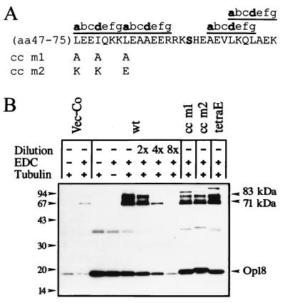
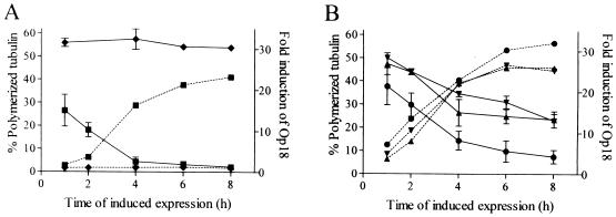
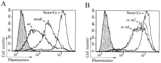
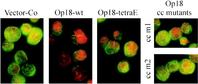
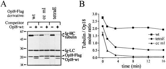
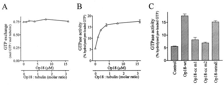
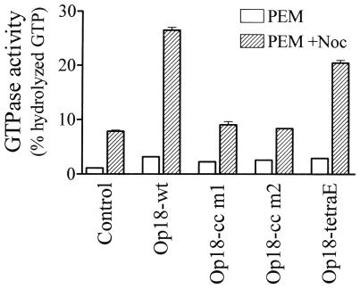
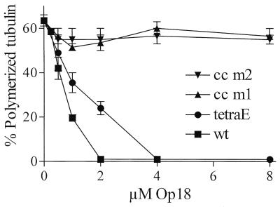
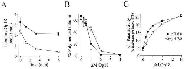
Oncoprotein 18/stathmin (Op18) is a recently identified phosphorylation-responsive regulator of the microtubule (MT) system. It was originally proposed that Op18 specifically regulates dynamic properties of MTs by associating with tubulin, but it has subsequently been proposed that Op18 acts simply by sequestering of tubulin heterodimers. We have dissected the mechanistic action of Op18 by generation of two distinct classes of mutants. One class has interruptions of the heptad repeats of a potential coiled-coil region of Op18, and the other involves substitution at all four phosphorylation sites with negatively charged Glu residues. Both types of mutation result in Op18 proteins with a limited decrease in tubulin complex formation. However, the MT-destabilizing activities of the coiled-coil mutants are more severely reduced in transfected leukemia cells than those of the Glu-substituted Op18 derivative, providing evidence for tubulin-directed regulatory activities distinct from tubulin complex formation. Analysis of Op18-mediated regulation of tubulin GTPase activity and taxol-promoted tubulin polymerization showed that while wild-type and Glu-substituted Op18 derivatives are active, the coiled-coil mutants are essentially inactive. This suggests that Op18-tubulin contact involves structural motifs that deliver a signal of regulatory importance to the MT system.


Figures

FIG. 1
FIG. 1
Cross-linking of tubulin to wt and mutated Op18 in crude cell extracts of transfected K562 cells. (A) The locations of mutations in Op18-cc m1 (L47A, I50A, and L54A) and Op18-cc m2 (L47K, I50K, and L54E) in the putative coiled-coil region are depicted. The Ser-63 phosphorylation site is indicated in bold. (B) K562 cells were transfected with pMEP4 (Vec-Co), pMEP-Op18-wt (wt), pMEP-Op18-cc m1 (cc m1), pMEP-Op18-cc m2 (cc m2), or pMEP-Op18-S16,25,38,63E (tetraE), and cell lines were selected as described in Materials and Methods. Cells were treated with Cd2+ (0.1 μM) for 7 h to induce expression from the hMTIIa promoter and subsequently lysed. In each case, crude cell extract was mixed with bovine tubulin and subjected to the EDC cross-linking as described in Materials and Methods. After separation with SDS-PAGE, complexes were revealed by rabbit anti-Op18:33-149. For semi-quantification of complex formation, the sample of cells expressing Op18-wt was diluted twofold (2×), fourfold (4×), and eightfold (8×) as indicated. The positions of the 71- and 83-kDa Op18-tubulin complexes are indicated. Data are representative for two experiments.
FIG. 2
FIG. 2
Alterations in MT polymerization status in response to overexpressed wt and mutated Op18. K562 cells were transfected with (A) pMEP4 (⧫) or Op18-wt (■) and with (B) Op18-cc m1 (▴), Op18-cc m2 (▾), or Op18-tetraE (●) as described in the legend for Fig. 1. Transfected cell lines were induced with 0.1 μM Cd2+ for the indicated times, and the fraction of polymerized tubulin was determined by the extraction protocol described in Materials and Methods. The mean of two independent determinations is shown. In parallel the induced levels of ectopic Op18 were determined by quantitative Western blot analysis (dashed lines), and data are presented as fold induction over that by endogenous Op18. Data are representative for two independent transfection experiments.
FIG. 3
FIG. 3
Flow cytometric analysis of MTs in cells overexpressing wt and mutated Op18. Transfected K562 cells were induced with 0.1 μM Cd2+ for 5.5 h, extracted with MT-stabilizing buffer, fixed, and stained with anti-α-tubulin. Open graphs depict α-tubulin-specific fluorescence of cells transfected with the indicated pMEP4-Op18 constructs, and shaded graphs show control staining in the absence of anti-α-tubulin but in the presence of fluorescein-conjugated rabbit anti-mouse immunoglobulin. To facilitate comparison, the histograms depicting control staining and cells expressing vector control (pMEP4) are shown in both panel A and B. The median fluorescence signals were as follows: control staining, 19; vector control (Vector-Co), 1,275; Op18-wt, 95; Op18-cc m1, 567; Op18-cc m2, 692; and Op18-tetraE, 311.
FIG. 4
FIG. 4
Immunodetection of MTs in cells induced to overexpress Op18. K562 cells, transfected with the indicated pMEP4-based constructs, were induced with 0.1 μM Cd2+ for 5.5 h and double stained with anti-α-tubulin (green) and propidium iodide (DNA staining; red). Representative interphase cells analyzed by epifluorescence (1,000-fold original magnification) are shown.
FIG. 5
FIG. 5
Characterization of in vitro tubulin binding to wt and mutated Op18. (A) Tubulin (10 μM in PEM, pH 6.8) was mixed with the indicated Flag-epitope-tagged Op18 derivative coupled to M2 beads (Op18-F/M2 beads). To control for nonspecific binding, competitor Op18-wt (50 μM), which does not bind to M2 beads, was added where indicated (+). After a 10-min incubation at 37°C, bead-bound material was pelleted through a sucrose cushion and separated by SDS-PAGE. Proteins were detected by Coomassie blue staining, and the positions of the immunoglobulin (Ig-) heavy (HC) and light (LC) chain derived from the M2 antibody, as well as tubulin, Op18-F (Op18-Flag), and competitor Op18, are indicated. (B) Tubulin sufficient for saturated binding (20 μM in PEM, pH 6.8) was allowed to bind for 15 min at 37°C to Op18-wt-F (■), Op18-cc m1-F (▴), or Op18-tetraE-F (●) coupled to M2 beads. Beads were thereafter either pelleted through a sucrose cushion (t = 0) or diluted 40-fold, and dissociation of tubulin was monitored at 37°C by pelleting of beads at the indicated time points. The molar ratio of tubulin associated with Op18 was determined as described in Materials and Methods, and the contribution of nonspecific binding (about 8%) was subtracted from the presented data. Data are representative for at least two independent experiments.
FIG. 6
FIG. 6
Tubulin-directed activities of wt and mutated Op18. (A) Tubulin (5 μM) was incubated with a graded concentration of Op18-wt on ice for 30 min in the presence of [γ-32P]GTP. GTP exchange was calculated by determination of tubulin-associated [γ-32P]GTP as described in Materials and Methods. (B) [γ-32P]GTP was allowed to bind to tubulin in the presence of graded concentrations of Op18 as described for panel A. Unbound [γ-32P]GTP was thereafter removed on a desalting column, and [γ-32P]GTP-loaded tubulin and Op18 were incubated at 37°C. The mean of duplicate determinations of hydrolyzed GTP, after 40 min of incubation, is shown. (C) Modulation of tubulin GTPase activity by 16 μM concentration of the indicated Op18 derivative was determined as described for panel B. Data are representative for at least two independent experiments.
FIG. 7
FIG. 7
Modulation of nocodazole-stimulated tubulin GTPase activity by wt and mutated Op18. Tubulin (5 μM) was incubated at 37°C in the presence of [γ-32P]GTP in the absence (open bars) or presence (striped bars) of nocodazole (33 μM). The indicated Op18 derivative (16 μM) was also included in the reaction mixtures. The means of duplicate determinations of hydrolyzed GTP, after 120 min of incubation, are shown. Data are representative for at least two independent experiments.
FIG. 8
FIG. 8
Op18-mediated inhibition of taxol-driven MT polymerization. Tubulin was incubated with taxol (5 μM) in the presence of graded concentrations of the indicated Op18 derivatives for 30 min at 37°C. Polymerized tubulin was sedimented by centrifugation and quantitated by using the bicinchoninic acid protein assay. Incubation of tubulin with taxol on ice resulted in sedimentation of less than 2 to 3% of all tubulin protein. The means of duplicate determinations are shown. Data are representative for at least two independent experiments.
FIG. 9
FIG. 9
pH dependent modulation of Op18-tubulin interactions and tubulin-directed activities. (A) Tubulin (20 μM) was allowed to bind for 15 min at 37°C to Op18-wt-F-coupled M2 beads in PEM buffer at pH 6.8 (■) or pH 7.5 (□). Tubulin binding at t = 0 and tubulin dissociation were determined at each pH as described for Fig. 5. (B) Op18-mediated inhibition of taxol-driven MT polymerization in PEM buffer at pH 6.8 (■) and pH 7.5 (□) was determined as described for Fig. 8. (C) Op18-stimulated tubulin GTPase activity at pH 6.8 (■) and pH 7.5 (□) was determined as described for Fig. 6.

Similar articles

Cited by

References

    1. Austin S, Dixon R. The prokaryotic enhancer binding protein NTRC has an ATPase activity which is phosphorylation and DNA dependent. EMBO J. 1992;11:2219–2228. - PMC - PubMed
    1. Belmont L D, Mitchison T J. Identification of a protein that interacts with tubulin dimers and increases the catastrophe rate of microtubules. Cell. 1996;84:623–631. - PubMed
    1. Boron W F. Intracellular pH regulation in epithelial cells. Annu Rev Physiol. 1986;48:377–388. - PubMed
    1. Brattsand G, Roos G, Marklund U, Ueda H, Landberg G, Nanberg E, Sideras P, Gullberg M. Quantitative analysis of the expression and regulation of an activation-regulated phosphoprotein (oncoprotein 18) in normal and neoplastic cells. Leukemia. 1993;7:569–579. - PubMed
    1. Correia J J. Effects of antimitotic agents on tubulin-nucleotide interactions. Pharmacol Ther. 1991;52:127–147. - PubMed

Publication types

LinkOut - more resources