. 1999 Mar;19(3):2251-64.
doi: 10.1128/MCB.19.3.2251.

Progesterone inhibits estrogen-induced cyclin D1 and cdk4 nuclear translocation, cyclin E- and cyclin A-cdk2 kinase activation, and cell proliferation in uterine epithelial cells in mice

Affiliations

Progesterone inhibits estrogen-induced cyclin D1 and cdk4 nuclear translocation, cyclin E- and cyclin A-cdk2 kinase activation, and cell proliferation in uterine epithelial cells in mice

W Tong et al. Mol Cell Biol. 1999 Mar.

Abstract

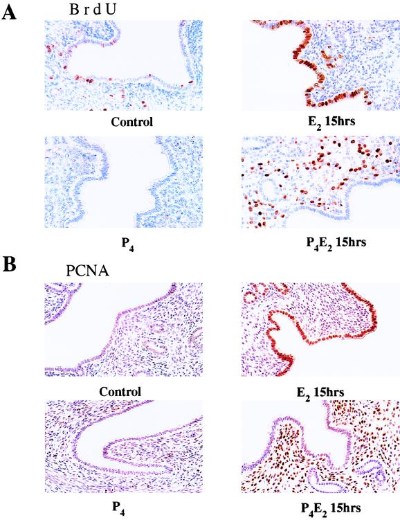
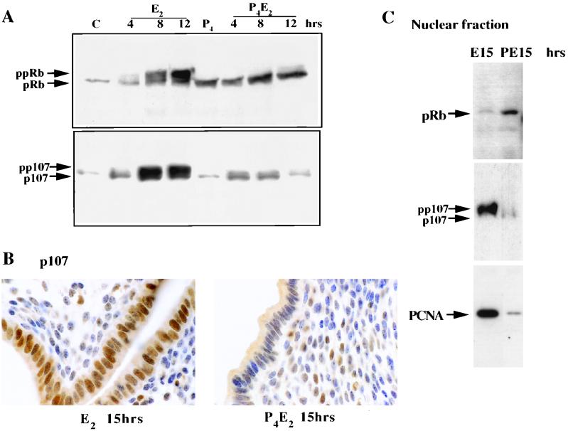
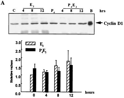
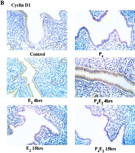
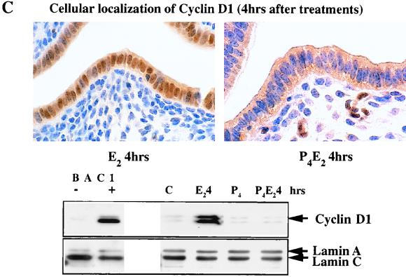
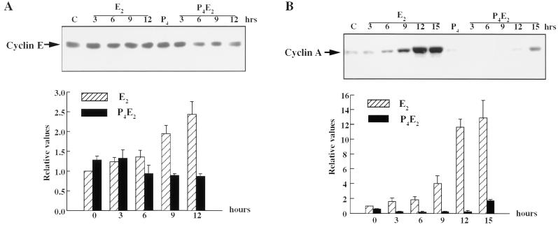
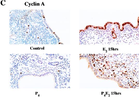
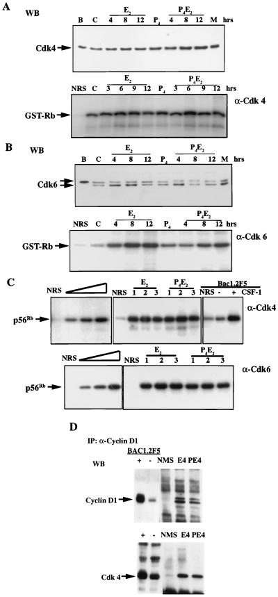
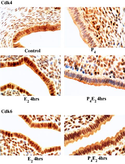
The response of the uterine epithelium to female sex steroid hormones provides an excellent model to study cell proliferation in vivo since both stimulation and inhibition of cell proliferation can be studied. Thus, when administered to ovariectomized adult mice 17beta-estradiol (E2) stimulates a synchronized wave of DNA synthesis and cell division in the epithelial cells, while pretreatment with progesterone (P4) completely inhibits this E2-induced cell proliferation. Using a simple method to isolate the uterine epithelium with high purity, we have shown that E2 treatment induces a relocalization of cyclin D1 and, to a lesser extent, cdk4 from the cytoplasm into the nucleus and results in the orderly activation of cyclin E- and cyclin A-cdk2 kinases and hyperphosphorylation of pRb and p107. P4 pretreatment did not alter overall levels of cyclin D1, cdk4, or cdk6 nor their associated kinase activities but instead inhibited the E2-induced nuclear localization of cyclin D1 to below the control level and, to a lesser extent, nuclear cdk4 levels, with a consequent inhibition of pRb and p107 phosphorylation. In addition, it abrogated E2-induced cyclin E-cdk2 activation by dephosphorylation of cdk2, followed by inhibition of cyclin A expression and consequently of cyclin A-cdk2 kinase activity and further inhibition of phosphorylation of pRb and p107. P4 is used therapeutically to oppose the effect of E2 during hormone replacement therapy and in the treatment of uterine adenocarcinoma. This study showing a novel mechanism of cell cycle inhibition by P4 may provide the basis for the development of new antiestrogens.


Figures

FIG. 1
FIG. 1
Cell proliferation in response to E2 and P4 treatment in the mouse uterus. Shown are the results of immunohistochemistry studies of transverse sections of uteri from ovariectomized mice given BrdU intraperitoneally 2 h before killing and 15 h after the following hormone treatments: control (no treatment), 50 ng of E2 (E2 15hrs), 1 mg of P4 for 4 days (P4), and 1 mg of P4 for 4 days and 50 ng of E2 on the fourth day (P4E2 15hrs). (A) Bouin’s fixed uteri immunostained for BrdU; (B) PLPG-fixed uteri immunostained for PCNA. In the immunohistochemistry studies, the figures shown are representative of five mice analyzed per group (magnification, ×400).
FIG. 2
FIG. 2
Phosphorylation status and localization of pRb and p107 following E2 or P4E2 treatment. (A) Uterine epithelial lysates from the same numbers of cells were prepared from control (C) mice at the indicated times after E2 or P4E2 treatment and analyzed for pRb (top) and p107 (bottom) proteins by Western blotting. The hypophosphorylated (pRb) and hyperphosphorylated (ppRb) bands are indicated, as are p107 and its phosphorylated form (pp107). (B) Immunohistochemical localization of p107 following 15 h of E2 and P4E2 treatment. Note the strong nuclear localization following E2 treatment (magnification, ca. ×900). (C) Western blot showing nuclear localization of pRb preferentially in the epithelial nuclear fraction from P4E2 at 15 h (PE15) compared to the fraction from E2 at 15 h (E15) and that of p107 preferentially in the nuclear fraction from E2 at 15 h compared to the fraction from P4E2 at 15 h. PCNA immunodetection acts as a control for the E2 effect.
FIG. 3
FIG. 3
Levels and localization of cyclin D1 in mouse uterus influenced by E2 and P4. (A) Western blotting of cyclin D1 with equivalent amounts of protein from total epithelial cell lysates with the DCS-6 mouse monoclonal anti-cyclin D1 antibody. The histogram indicates densitometric determination of cyclin D1 expression for four independent determinations (means ± standard errors of the means) (lane B, BAC1.2F5 cell lysates; lane C, untreated control epithelial cell lysates). (B) Localization of cyclin D1 determined by immunohistochemistry of transverse sections of uteri from ovariectomized mice after the following hormone treatments: control (no treatment), 50 ng of E2 (E2 4hrs), 50 ng of E2 (E2 15hrs); 1 mg of P4 for 4 days (P4), 1 mg of P4 for 4 days and 50 ng of E2 on the fourth day (P4E2 4hrs), and the treatment designated P4E2 15hrs. (magnification, ×400). (C) Immunohistochemistry of cyclin D1 in transverse sections of mouse uteri after the treatments designated E2 4hrs and P4E2 4hrs (magnification, ca. ×800). The lower portion shows concentrations of cyclin D1 in the nuclear fraction of the epithelial cells determined by Western blotting with the Ab-3 rabbit polyclonal anti-cyclin D1 antibody. It should be noted that this polyclonal antibody recognizes the upper D1 band with greater sensitivity than that of the mouse monoclonal antibody used in panel A. As loading controls, the expression levels of lamins A and C (constitutively expressed) are shown. (BAC1 −, BAC1.2F5 cells without CSF-1 stimulation; BAC1 +, BAC1.2F5 cells 6 h after CSF-1 stimulation).
FIG. 3
FIG. 3
Levels and localization of cyclin D1 in mouse uterus influenced by E2 and P4. (A) Western blotting of cyclin D1 with equivalent amounts of protein from total epithelial cell lysates with the DCS-6 mouse monoclonal anti-cyclin D1 antibody. The histogram indicates densitometric determination of cyclin D1 expression for four independent determinations (means ± standard errors of the means) (lane B, BAC1.2F5 cell lysates; lane C, untreated control epithelial cell lysates). (B) Localization of cyclin D1 determined by immunohistochemistry of transverse sections of uteri from ovariectomized mice after the following hormone treatments: control (no treatment), 50 ng of E2 (E2 4hrs), 50 ng of E2 (E2 15hrs); 1 mg of P4 for 4 days (P4), 1 mg of P4 for 4 days and 50 ng of E2 on the fourth day (P4E2 4hrs), and the treatment designated P4E2 15hrs. (magnification, ×400). (C) Immunohistochemistry of cyclin D1 in transverse sections of mouse uteri after the treatments designated E2 4hrs and P4E2 4hrs (magnification, ca. ×800). The lower portion shows concentrations of cyclin D1 in the nuclear fraction of the epithelial cells determined by Western blotting with the Ab-3 rabbit polyclonal anti-cyclin D1 antibody. It should be noted that this polyclonal antibody recognizes the upper D1 band with greater sensitivity than that of the mouse monoclonal antibody used in panel A. As loading controls, the expression levels of lamins A and C (constitutively expressed) are shown. (BAC1 −, BAC1.2F5 cells without CSF-1 stimulation; BAC1 +, BAC1.2F5 cells 6 h after CSF-1 stimulation).
FIG. 4
FIG. 4
E2 and P4 effects on the levels of cyclin E and cyclin A in mouse uterine epithelial cells. (A and B) Uterine epithelial cell extracts, prepared at the indicated times, were analyzed for cyclin E (A) and cyclin A (B) protein expression by Western blotting. Lanes C, epithelial cell lysates from untreated control mice. Relative values are shown for three to five determinations per time point in the bar figures underneath (means ± standard errors of the means). (C) Immunohistochemistry with anti-cyclin A antibodies of uterine transverse sections from ovariectomized mice after the hormone treatments, as described for Fig. 1 (magnification, ×400).
FIG. 4
FIG. 4
E2 and P4 effects on the levels of cyclin E and cyclin A in mouse uterine epithelial cells. (A and B) Uterine epithelial cell extracts, prepared at the indicated times, were analyzed for cyclin E (A) and cyclin A (B) protein expression by Western blotting. Lanes C, epithelial cell lysates from untreated control mice. Relative values are shown for three to five determinations per time point in the bar figures underneath (means ± standard errors of the means). (C) Immunohistochemistry with anti-cyclin A antibodies of uterine transverse sections from ovariectomized mice after the hormone treatments, as described for Fig. 1 (magnification, ×400).
FIG. 5
FIG. 5
Levels and localization of p27Kip1 in the mouse uterus influenced by E2 and P4. (A) Western blotting of uterine epithelial cell lysates at different time points following E2 and P4E2 treatments with anti-p27 antibodies. Lane C, epithelial cell lysates from untreated control mice. (B) Densitometric quantitation of p27Kip1 concentrations; three to five independent determinations per time point (means ± standard errors of the means). (C) Immunohistochemical determination of p27Kip1 distribution in the uterine epithelium (magnification, ×400).
FIG. 6
FIG. 6
Suppression of E2-induced cyclin E- and cyclin A-cdk2-associated kinase activities by P4. Cyclin E (A)-, cyclin A (B)-, or cdk2 (C [top])-associated kinase activities were assessed with histone H1 as a substrate. The total cell lysates were analyzed for cdk2 protein by Western blotting (C [bottom]). Thr 160-P indicates the threonine-160-phosphorylated active form of cdk2, and Thr 160 indicates the nonphosphorylated form. Also shown are cyclin E- and cyclin A-cdk2 kinase activities in extracts from uterine epithelial cells of p27Kip1 nullizygous mice (p27KO) 12 h after E2 or P4E2 treatment (D). Details are as described for panels A to C. Abbreviations: NRS, normal rabbit immunoglobulin G; WT, wild type; C, control; HH1, histone H1; WB, Western blot.
FIG. 7
FIG. 7
Concentrations and kinase activities of cdk4 and cdk6 following E2 or P4E2 treatment. (A) cdk4 levels and associated kinase activities remain unchanged following different hormonal treatments as determined by Western blotting (WB) (top) and IP kinase assay with GST-Rb (769) as substrate (bottom). (B) cdk6 levels and associated kinase activities are comparable between E2 and P4E2 treatments as determined by Western blotting (WB) (top) and IP kinase assay with GST-Rb (769) as substrate (bottom). (Abbreviations: B, BAC1.2F5; M, MEF; C, control; NRS, normal rabbit immunoglobulin G). (C) (upper panel) Left, titration of kinase activity with increasing concentrations of uterine epithelial cell lysate. Middle, three independent experiments (1 to 3) with cdk4 activities 4 h after E2 and P4E2 treatment with recombinant truncated Rb (p56Rb) as substrate. Total epithelial cell lysates with protein concentrations in the middle of the linear portion of the titration were used in the experiments. Right, BAC1.2F5 cells either unstimulated or stimulated with CSF-1 for 9 h were used as positive controls. (Lower panel) As described for the upper panel, except that cdk6 kinase activities were determined. (D) Coimmunoprecipitation of cdk4 with cyclin D1, at 4 h following E2 and P4E2 treatments. NMS, normal mouse immunoglobulin G; WB, Western blot.
FIG. 8
FIG. 8
Immunostaining of uterine transverse sections for cdk4 (0 and 4 h) and cdk6 (4 h) after E2 and P4E2 treatments. Magnification, ×1,000.

Similar articles

Cited by

References

    1. Altucci L, Addeo R, Cicatiello L, Dauvois S, Parker M G, Truss M, Beato M, Sica V, Bresciani F, Weisz A. 17β-Estradiol induces cyclin D1 gene transcription, p36D1-p34cdk4 complex activation and p105Rb phosphorylation during mitogenic stimulation of G1-arrested human breast cancer cells. Oncogene. 1996;12:2315–2324. - PubMed
    1. Altucci L, Addeo R, Cicatiello L, Germano D, Pacilio C, Battista T, Cancemi M, Petrizzi V B, Bresciani F, Weisz A. Estrogen induces early and timed activation of cyclin-dependent kinases 4, 5, and 6 and increases cyclin messenger ribonucleic acid expression in rat uterus. Endocrinology. 1997;138:978–984. - PubMed
    1. Baldin V, Lukas J, Marcote M, Pagano M, Draetta G. Cyclin D1 is a nuclear protein required for cell cycle progression in G1. Genes Dev. 1993;7:812–821. - PubMed
    1. Castles C G, Fuqua S A W. Alterations within the estrogen receptor in breast cancer. In: Pasqualini J R, Katzenellenbogen B S, editors. Hormonal-dependent cancer. New York, N.Y: Marcel Dekker, Inc.; 1996. pp. 81–105.
    1. Cheng M, Diehl J A, Fero M, Olivier P, Randel E, Roberts J M, Sherr C J, Roussel M F. Fourteenth Annual Meeting on Oncogenes. (Abstract.) 1998. Cdk inhibitors p27Kip1 and p21Cip1 are required for assembly of active cyclin D-dependent kinases in mouse fibroblasts; p. 39.

MeSH terms

Substances