. 1999 Mar;19(3):2278-88.
doi: 10.1128/MCB.19.3.2278.

Identification of a novel family of targets of PYK2 related to Drosophila retinal degeneration B (rdgB) protein

Affiliations

Identification of a novel family of targets of PYK2 related to Drosophila retinal degeneration B (rdgB) protein

S Lev et al. Mol Cell Biol. 1999 Mar.

Abstract

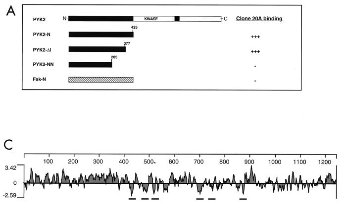
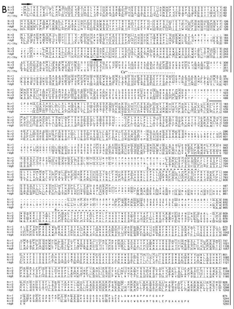
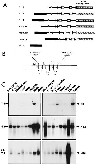
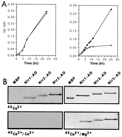
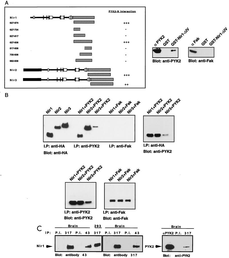
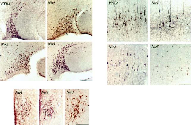
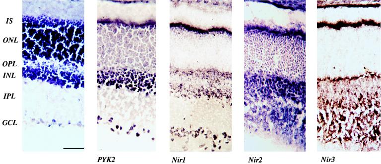
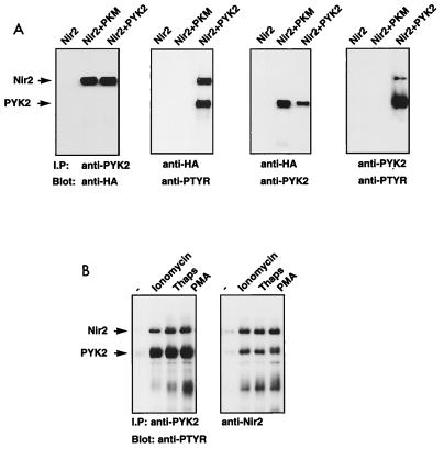
The protein tyrosine kinase PYK2 has been implicated in signaling pathways activated by G-protein-coupled receptors, intracellular calcium, and stress signals. Here we describe the molecular cloning and characterization of a novel family of PYK2-binding proteins designated Nirs (PYK2 N-terminal domain-interacting receptors). The three Nir proteins (Nir1, Nir2, and Nir3) bind to the amino-terminal domain of PYK2 via a conserved sequence motif located in the carboxy terminus. The primary structures of Nirs reveal six putative transmembrane domains, a region homologous to phosphatidylinositol (PI) transfer protein, and an acidic domain. The Nir proteins are the human homologues of the Drosophila retinal degeneration B protein (rdgB), a protein implicated in the visual transduction pathway in flies. We demonstrate that Nirs are calcium-binding proteins that exhibit PI transfer activity in vivo. Activation of PYK2 by agents that elevate intracellular calcium or by phorbol ester induce tyrosine phosphorylation of Nirs. Moreover, PYK2 and Nirs exhibit similar expression patterns in several regions of the brain and retina. In addition, PYK2-Nir complexes are detected in lysates prepared from cultured cells or from brain tissues. Finally, the Nir1-encoding gene is located at human chromosome 17p13.1, in proximity to a locus responsible for several human retinal diseases. We propose that the Nir and rdgB proteins represent a new family of evolutionarily conserved PYK2-binding proteins that play a role in the control of calcium and phosphoinositide metabolism downstream of G-protein-coupled receptors.


Figures

FIG. 1
FIG. 1
Cloning and amino acid sequences of Nir1, Nir2, and Nir3. (A) Identification of PYK2-binding proteins using the yeast two-hybrid system. PYK2-N is shown along with heterologous baits that were used to determine binding specificity toward clone 20A. Yeast two-hybrid interactions were determined by induction of the reporter genes for HIS3 and β-Gal. (B) Comparison of amino acid sequences of human Nir1, Nir2, and Nir3; Drosophila rdgB; and human PI transfer protein α. The amino acid sequences are shown in single-letter code, and the numbers represent the positions of amino acid residues. Amino acids present in two or more sequences are boxed. Boundaries of the PI transfer domain (first 270 amino acids at the amino terminus) and the PYK2-binding domain (last 350 amino acids at the carboxy terminus) are marked by arrows and vertical lines to the right. The six putative transmembrane domains are overlined. The hydrophobicity of transmembrane domains 1 to 4 is stronger (solid lines) than the hydrophobicity of transmembrane domains 5 and 6 (dashed lines). The putative calcium-binding site is marked with a dotted line. Three Xs were incorporated into the Drosophila rdgB sequence; two represent amino acid deletions, whereas the third X located the position of a predicted frame shift in the published sequence that extends the rdgB-Nir homology. (C) Hydropathy profile of Nir2. The profile was calculated with a window size of 15 amino acids (18).
FIG. 1
FIG. 1
Cloning and amino acid sequences of Nir1, Nir2, and Nir3. (A) Identification of PYK2-binding proteins using the yeast two-hybrid system. PYK2-N is shown along with heterologous baits that were used to determine binding specificity toward clone 20A. Yeast two-hybrid interactions were determined by induction of the reporter genes for HIS3 and β-Gal. (B) Comparison of amino acid sequences of human Nir1, Nir2, and Nir3; Drosophila rdgB; and human PI transfer protein α. The amino acid sequences are shown in single-letter code, and the numbers represent the positions of amino acid residues. Amino acids present in two or more sequences are boxed. Boundaries of the PI transfer domain (first 270 amino acids at the amino terminus) and the PYK2-binding domain (last 350 amino acids at the carboxy terminus) are marked by arrows and vertical lines to the right. The six putative transmembrane domains are overlined. The hydrophobicity of transmembrane domains 1 to 4 is stronger (solid lines) than the hydrophobicity of transmembrane domains 5 and 6 (dashed lines). The putative calcium-binding site is marked with a dotted line. Three Xs were incorporated into the Drosophila rdgB sequence; two represent amino acid deletions, whereas the third X located the position of a predicted frame shift in the published sequence that extends the rdgB-Nir homology. (C) Hydropathy profile of Nir2. The profile was calculated with a window size of 15 amino acids (18).
FIG. 2
FIG. 2
Structure of Nir proteins and mRNA distribution. (A) Schematic diagram of the primary structures of Nir1, Nir2, and Nir3. The amino-terminal PI transfer domain (black), the acidic domain (white), the three conserved subdomains (gray), and the carboxy-terminal PYK2-binding domain (striped) are represented. The six putative transmembrane domains are shown as vertical lines. (B) Model of the structure of Nir proteins. The amino-terminal PI transfer domain, the acidic calcium-binding domain, and the carboxy-terminal PYK2-binding domain are located on one face of the membrane. The six transmembrane domains are numbered 1 to 6. The loops between the transmembrane domains are designated I, II, III, and IV. (C) Northern blot analysis of Nir mRNA expression in various human tissues. Human multiple-tissue Northern blots were hybridized with radiolabeled probes for Nir1 (top), Nir2 (middle), and Nir3 (bottom). Specific probes are described in Materials and Methods. Marker sizes are shown in kilobases at the left.
FIG. 3
FIG. 3
Functional analysis of the PI transfer and acidic domains of Nirs. (A) Rescue of the growth lesion of a sec14ts mutant by a Nir PI transfer domain. Growth curves of a sec14ts mutant transformed either with a yeast expression vector (○) or with an expression vector containing the PI transfer domain of Nir3 (●) at 25 and 35°C. Growth was determined by measuring the optical density at 600 nm (OD 600) of a liquid culture at different time points as indicated. An identical growth curve was observed at 25°C, while Nir3 rescued the growth lesion of the sec14ts mutant at 39°C. (B) The acidic domain (AD) of Nir proteins binds calcium. The acidic domains of Nir1, Nir2, and Nir3 were expressed in Escherichia coli in the form of MBP fusion proteins. Following affinity purification, the recombinant acidic domains and MBP alone were resolved on a sodium dodecyl sulfate–12% polyacrylamide gel and either stained with Coomassie brilliant blue (upper right) or transferred to a nitrocellulose filter and incubated with 45Ca2+ as described in Materials and Methods in the absence (upper left) or presence of 10 mM CaCl2 (lower left) or MgCl2 (lower right).
FIG. 4
FIG. 4
Interaction between PYK2 and Nirs in yeast, in cultured cells, and in brain tissue. (A) Determination of the PYK2-binding domain of Nir proteins. The binding region responsible for binding of Nir1 to PYK2-N was determined by assaying growth on selection medium lacking histidine by a β-Gal assay (left) and by an in vitro binding assay (right). The diagram shows the sequences of Nir1 that were fused to the GAL-4 activation domain. The numbers indicate the amino acid residues of Nir1 that were included in each fusion protein. The interaction of the C-terminal domains of Nir2 and Nir3 with PYK2-N is shown at the bottom. An in vitro assay of PYK2 binding to an immobilized GST fusion protein expressing the Nir1-ΔIV cDNA (amino acids 627 to 936) is shown at the right. Lysates of cells expressing either PYK2 or FAK were incubated with immobilized GST or GST–Nir1-ΔIV for 2 h at 4°C. Following extensive washing, binding of PYK2 or FAK to the recombinant proteins was detected by immunoblotting with anti-PYK2 or anti-FAK antibodies as indicated. (B) Association between PYK2 and Nir proteins in vivo. Lysates from 293T cells transfected with Nirs-HA alone, Nirs-HA cotransfected with PYK2, or Nirs-HA cotransfected with FAK were immunoprecipitated with anti-HA, anti-PYK2, or anti-FAK antibodies as indicated. Immunoprecipitates (I.P) were subjected to immunoblotting with anti-HA, anti-PYK2, or anti-FAK antibodies as indicated. PYK2 was detected in Nir immunoprecipitates (left), and Nir proteins were detected in PYK2 immunoprecipitates (right). Expression levels of PYK2 and FAK in cells coexpressing Nir proteins are shown at the bottom. (C) Association of PYK2 with Nir1 in brain tissue. Adult rat brain was homogenized as described in Materials and Methods. The homogenate was subjected to immunoprecipitation with different antibodies as indicated: P.I. (preimmune serum) anti-Nir1 antibodies, antibody 317 raised against a synthetic peptide corresponding to the last 10 amino acids, antibody 43 raised against a GST fusion protein containing amino acids 260 to 380 of Nir1, or anti-PYK2 antibodies. The Nir1 protein was also immunoprecipitated from 293 cells transfected with a Nir1 expression vector as indicated. Antibodies against Nir1 recognized a single protein that migrates in sodium dodecyl sulfate gels with an apparent molecular mass of 120 kDa (two left panels). This protein was coimmunoprecipitated with anti-PYK2 antibodies (right panel).
FIG. 5
FIG. 5
Immunohistochemical analysis of PYK2, Nir1, Nir2, or Nir3 distribution in the rat brain. The left panel shows the localization of PYK2, Nir1, Nir2, or Nir3 in the supraoptic nucleus (scale bar, 130 μm). The right panel shows staining in pyramidal layer V (scale bar, 95 μm), and the bottom panel depicts staining of the three Nir proteins in the middle preoptic area (scale bar, 100 μM).
FIG. 6
FIG. 6
Distribution of the PYK2 and Nir proteins in the rat retina. (A) Hematoxylin staining of a vertical cryostat section of a rat retina showing different layers of the retina. (B) PYK2 immunoreactivity in the retina. Strong staining for PYK2 is detected throughout the inner nuclear layer (INL) and in the ganglion cell layer (GCL); specific staining is also observed in the outer nuclear layer (ONL). (C) Nir1 immunoreactivity in the retina. Strong staining is detected in the inner segment (IS), in the outer plexiform layer (OPL), in specific cells within the inner nuclear layer, and in the ganglion cell layer. (D) Nir2 immunostaining is seen in the inner segment, outer nuclear layer, and inner plexiform layer (IPL), and a moderate expression level is seen in the inner nuclear layer and the outer plexiform layer. (E) Strong immunostaining of Nir3 is detected in the inner segment and the inner and outer plexiform layers (scale bar, 50 μM).
FIG. 7
FIG. 7
Tyrosine phosphorylation of Nir2 in response to PYK2 activation. (A) Human 293T cells were transfected with expression vectors for Nir2-HA, PKM, or PYK2. Lysates from transfected cells were subjected to immunoprecipitation (I.P) with anti-HA or anti-PYK2 antibodies as indicated. Tyrosine phosphorylation was determined by immunoblotting with antiphosphotyrosine antibodies (anti-PTYR). (B) Quiescent HL60 cells were stimulated with ionomycin (6 μM), thapsigargin (Thaps; 2 μM), or phorbol myristate acetate (PMA; 1.6 μM) for 10 min at 37°C. PYK2 and Nir2 were immunoprecipitated from lysates of unstimulated (−) and stimulated cells. The samples were resolved by sodium dodecyl sulfate-polyacrylamide gel electrophoresis, transferred to a nitrocellulose filter, and immunoblotted with anti-P-Tyr antibodies.

References

    1. Aikawa Y, Hara H, Watanabe T. Molecular cloning and characterization of mammalian homologues of the Drosophila retinal degeneration B gene. Biochem Biophys Res Commun. 1997;236:559–564. - PubMed
    1. Astier A, Avraham H, Manie S N, Groopman J, Canty T, Avraham S, Freedman A S. The related adhesion focal tyrosine kinase is tyrosine-phosphorylated after beta1-integrin stimulation in B cells and binds to p130cas. J Biol Chem. 1997;272:228–232. - PubMed
    1. Avraham S, London R, Fu Y, Ota S, Hiregowdara D, Li J, Jiang S, Pasztor L M, White R A, Groopman J E, et al. Identification and characterization of a novel related adhesion focal tyrosine kinase (RAFTK) from megakaryocytes and brain. J Biol Chem. 1995;270:27742–27751. - PubMed
    1. Balciuniene J, Johansson K, Sandgren O, Wachtmeister L, Holmgren G, Forsman K. A gene for autosomal dominant progressive cone dystrophy (CORD5) maps to chromosome 17p12-p13. Genomics. 1995;30:281–286. - PubMed
    1. Banfi S, Borsani G, Rossi E, Bernard L, Guffanti A, Rubboli F, Marchitiello A, Giglio S, Coluccia E, Zollo M, Zuffardi O, Ballabio A. Identification and mapping of human cDNAs homologous to Drosophila mutant genes through EST database searching. Nat Genet. 1996;13:167–174. - PubMed

MeSH terms