. 1999 Mar;19(3):2338-50.
doi: 10.1128/MCB.19.3.2338.

Identification of a new Pyk2 target protein with Arf-GAP activity

Affiliations

Identification of a new Pyk2 target protein with Arf-GAP activity

J Andreev et al. Mol Cell Biol. 1999 Mar.

Abstract

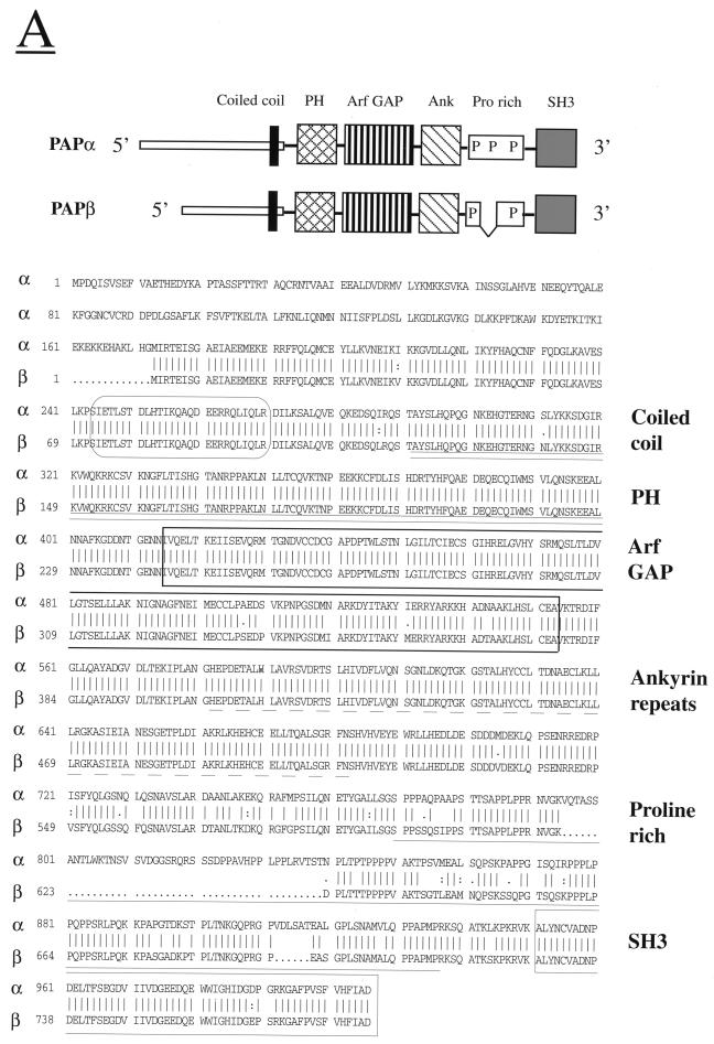
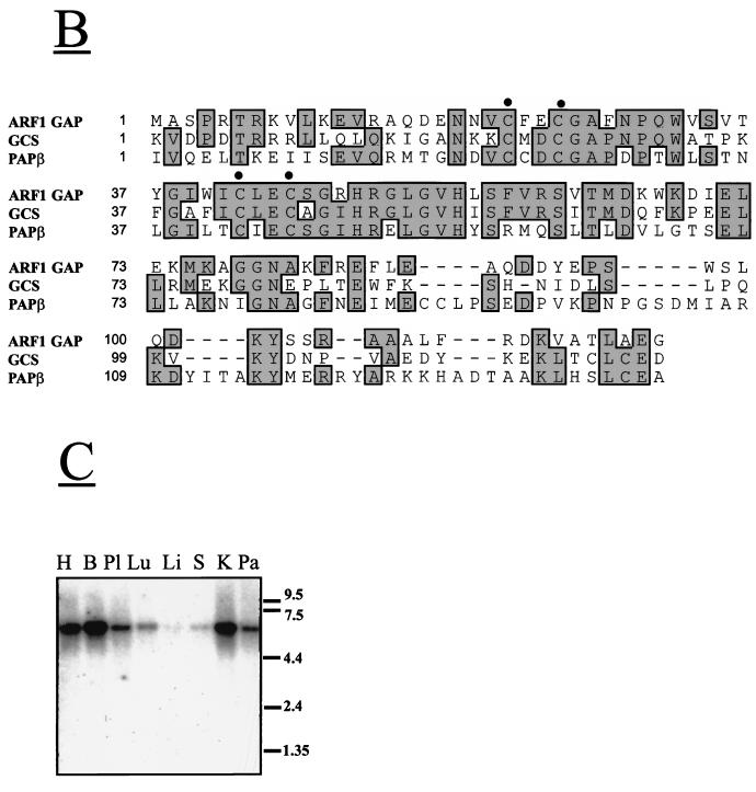
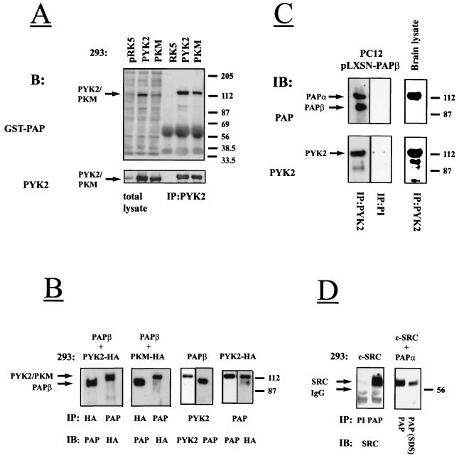
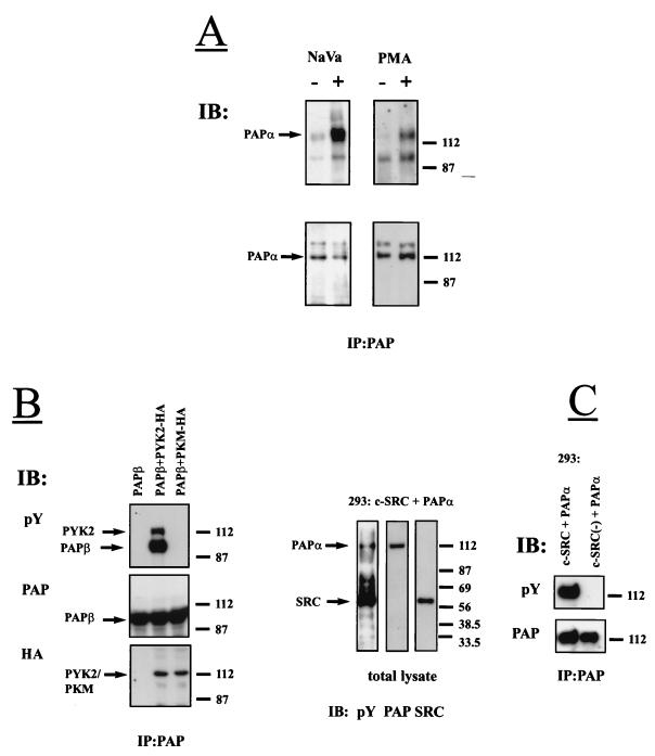
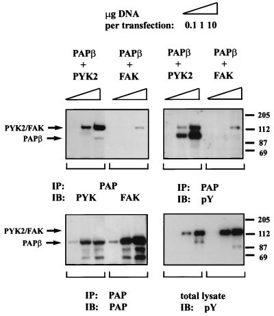
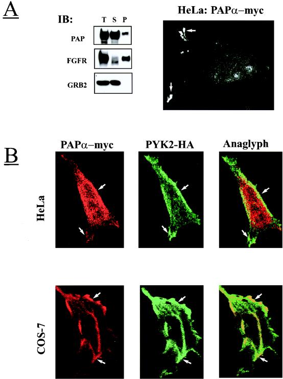
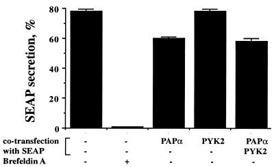
Protein tyrosine kinase Pyk2 is activated by a variety of G-protein-coupled receptors and by extracellular signals that elevate intracellular Ca2+ concentration. We have identified a new Pyk2 binding protein designated Pap. Pap is a multidomain protein composed of an N-terminal alpha-helical region with a coiled-coil motif, followed by a pleckstrin homology domain, an Arf-GAP domain, an ankyrin homology region, a proline-rich region, and a C-terminal SH3 domain. We demonstrate that Pap forms a stable complex with Pyk2 and that activation of Pyk2 leads to tyrosine phosphorylation of Pap in living cells. Immunofluorescence experiments demonstrate that Pap is localized in the Golgi apparatus and at the plasma membrane, where it is colocalized with Pyk2. In addition, in vitro recombinant Pap exhibits strong GTPase-activating protein (GAP) activity towards the small GTPases Arf1 and Arf5 and weak activity towards Arf6. Addition of recombinant Pap protein to Golgi preparations prevented Arf-dependent generation of post-Golgi vesicles in vitro. Moreover, overexpression of Pap in cultured cells reduced the constitutive secretion of a marker protein. We propose that Pap functions as a GAP for Arf and that Pyk2 may be involved in regulation of vesicular transport through its interaction with Pap.


Figures

FIG. 1
FIG. 1
Primary structure of Pap isoforms and tissue expression pattern. (A) Schematic diagram and amino acid sequences of Papα (human) and Papβ (murine). The amino acid sequences are shown in single-letter code. The numbers represent positions of the amino acid residues. Predicted coiled-coil region (CoilScan program, GCG package) is enclosed in a rectangle with rounded corners. The PH domain is underlined with a double line. The Arf GAP domain is boxed. The ankyrin homology region is underlined with a dashed line. The proline-rich region is underlined with a solid line. The SH3 domain is boxed. (B) Comparison of Arf GAP domains of murine Pap, Arf1 GAP, and GCS1. Multiple sequence alignment of the regions containing the zinc finger motif (CXXCX16CXXC, where X is any amino acid) of Arf1 GAP (residues 1 to 119), GCS1 (residues 5 to 122), and murine Pap (residues 243 to 381). Identical residues are framed and shadowed. The positions of four conserved cysteins of zinc finger motif are marked by dots. (C) Northern blot analysis of Pap mRNA expression in various human tissues. Human multiple tissues for Northern blotting were hybridized with radiolabeled probe for Pap as described in Materials and Methods. Size markers in kilobases are shown on the right of the figure. Abbreviations: H, heart; B, brain; Pl, placenta; Lu, lungs; Li, liver; S, spleen; K, kidney; Pa, pancreas.
FIG. 1
FIG. 1
Primary structure of Pap isoforms and tissue expression pattern. (A) Schematic diagram and amino acid sequences of Papα (human) and Papβ (murine). The amino acid sequences are shown in single-letter code. The numbers represent positions of the amino acid residues. Predicted coiled-coil region (CoilScan program, GCG package) is enclosed in a rectangle with rounded corners. The PH domain is underlined with a double line. The Arf GAP domain is boxed. The ankyrin homology region is underlined with a dashed line. The proline-rich region is underlined with a solid line. The SH3 domain is boxed. (B) Comparison of Arf GAP domains of murine Pap, Arf1 GAP, and GCS1. Multiple sequence alignment of the regions containing the zinc finger motif (CXXCX16CXXC, where X is any amino acid) of Arf1 GAP (residues 1 to 119), GCS1 (residues 5 to 122), and murine Pap (residues 243 to 381). Identical residues are framed and shadowed. The positions of four conserved cysteins of zinc finger motif are marked by dots. (C) Northern blot analysis of Pap mRNA expression in various human tissues. Human multiple tissues for Northern blotting were hybridized with radiolabeled probe for Pap as described in Materials and Methods. Size markers in kilobases are shown on the right of the figure. Abbreviations: H, heart; B, brain; Pl, placenta; Lu, lungs; Li, liver; S, spleen; K, kidney; Pa, pancreas.
FIG. 2
FIG. 2
Interaction between Pyk2 and Pap in vitro, in cultured cells, and in brain tissue. (A) Lysates from 293 cells transfected with expression vectors for Pyk2 or PKM or with vector alone were subjected to SDS-PAGE immediately (total lysate) or after immunoprecipitation (IP) with anti-Pyk2 antibodies, then transferred to nitrocellulose filters, and blotted with GST fusion protein containing the proline-rich region and the SH3 domain of Pap (3 μg/ml) and anti-GST monoclonal antibodies (1:1,000) (upper panel). The same filter was reprobed with anti-Pyk2 antibodies (lower panel). About 0.5 mg of total protein was used per immunoprecipitation experiment. Endogenous Pyk2 protein could not be detected with anti-Pyk2 antibodies under these conditions. Arrows mark the Pyk2 and PKM. Positions of standard protein markers (in kilodaltons) are indicated on the right. (B) Lysates from 293 cells transfected with expression vectors for Pyk2-HA and Papβ, with expression vectors for PKM-HA and Papβ, or with Papβ and Pyk2-HA alone were immunoprecipitated (IP) with anti-HA, anti-Pap, or anti-Pyk2 antibodies. Immunoprecipitates were subjected to immunoblotting (IB) with anti-HA, anti-Pyk2, or anti-Pap antibodies. About 5 mg of total protein was used per immunoprecipitation experiment. Arrows mark the Pyk2/PKM or Papβ. Positions of standard protein markers (in kilodaltons) are indicated on the right. (C) Lysates of PC12 cells infected with Papβ virus were immunoprecipitated with anti-Pyk2 antibodies and preimmune serum followed by immunoblotting (IB) with anti-Pap (left upper panels) or anti-Pyk2 (left lower panels) antibodies. Neither Pyk2 nor Pap were detected in this experiment with a preimmune serum (PI). Arrows mark the Papα or Papβ. Adult mouse brain homogenate was subjected to immunoprecipitation (IP) with anti-Pyk2 antibodies and immunoblotting (IB) with anti-Pap antibodies (right upper panel) or anti-Pyk2 antibodies (right lower panel). (D) Lysates from 293 cells transfected with expression vector for Src or with expression vectors for Papα and Src were immunoprecipitated (IP) with either preimmune (PI) or anti-Pap antibodies (PAP). Immunoprecipitations were performed either in lysis buffer or in lysis buffer supplemented with 1% Nonidet P-40, 0.5% deoxycholate, and 0.1% SDS instead of 1% Triton X-100 (SDS). Immunoprecipitates were subjected to immunoblotting (IB) with anti-Src antibodies. The arrows mark Src or the immunoglobulin G (IgG) heavy chain. Positions of standard protein markers (in kilodaltons) are indicated on the right.
FIG. 3
FIG. 3
Tyrosine phosphorylation of Pap by Pyk2 and by Src kinases. (A) PC12 cells were stimulated with the mixture of H2O2 and Na3VO4 (1 mM) for 20 min or with PMA (2 μM) for 10 min at 37°C. Pap was immunoprecipitated from unstimulated (−) or stimulated (+) cells, immunoblotted (IB) with anti-pY antibodies (upper panels), and reprobed with anti-Pap antibodies (lower panels). The arrows mark Papα. Positions of standard protein markers (in kilodaltons) are indicated on the right. Apart from endogenous Papα (112 kDa), anti-Pap antibodies precipitated a band with an apparent molecular size of 140 kDa (lower panels), which may represent an additional uncharacterized PAP isoform expressed in PC12 cells. (B) Lysates from 293 cells transfected with expression vectors for Papβ and Pyk2-HA, expression vectors for Papβ and PKM-HA, or expression vectors for Papβ alone were subjected to immunoprecipitation with anti-Pap antibodies and immunoblotting with anti-pY antibodies. The same filters were reprobed with anti-Pap and anti-HA antibodies. The arrows mark the Pyk2/PKM or Papβ. Positions of standard protein markers (in kilodaltons) are indicated on the right. (C) Lysates from 293 cells transfected with expression vectors for Papα and Src or expression vectors for Papα and Src(−) (kinase-negative mutant of Src) were either separated by SDS-PAGE, immunoblotted with anti-pY antibodies, and reprobed with anti-Pap or anti-Src antibodies or else subjected to immunoprecipitation with anti-Pap antibodies and immunoblotting with anti-pY and anti-Pap antibodies. The arrows mark the Papα or Src. Positions of standard protein markers (in kilodaltons) are indicated on the right.
FIG. 4
FIG. 4
Pap is tyrosine phosphorylated by Pyk2 but not by FAK. Human 293 cells were transfected with different amounts of expression vectors for Papβ and Pyk2 or expression vectors for Papβ and FAK. Lysates from these cells were subjected to SDS-PAGE immediately (total lysate) or after immunoprecipitation (IP) with anti-Pap antibodies and then processed for immunoblotting with anti-pY antibodies (right lower and upper panels). The right upper filter was reprobed with anti-Pyk2 or anti-FAK antibodies (left upper panel) and anti-Pap antibodies (left lower panel). The tyrosine phosphorylated 112-kDa proteins detected in the total cell lysate (right lower panel) correspond to Pyk2 and FAK, as determined by reprobing the same filter with anti-Pyk2 or anti-FAK antibodies (data not shown). The arrows mark the Pyk2, FAK, or Papβ. Positions of standard protein markers (in kilodaltons) are indicated on the right.
FIG. 5
FIG. 5
Recombinant Pap exhibits Arf GAP activity in vitro. (A) A single round of GTP hydrolysis on myristoylated Arf1 (open circles), myristoylated Arf5 (solid circles), myristoylated Arf6 (squares), and unmodified Arl2 (triangles) was measured in the presence of crude phosphoinositides (1 mg/ml) as a source of PtdIns(4,5)P2 and the indicated concentrations of recombinant Pap. GAP activity is expressed as the percentage of initially bound GTP hydrolyzed in 4 min. Myristoylated Arf1 was indistinguishable from nonmyristoylated Arf1 (data not shown). (B) A single round of GTP hydrolysis on nonmyristoylated Arf1 was measured in the presence of 1.5 nM recombinant Pap, and the indicated phospholipids are as described in Materials and Methods. None, no added phospholipid; PIP2, 90 μM phosphatidylinositol 4,5-bisphosphate; PA, 750 μM phosphatidic acid; PI, 720 μM phosphatidylinositol; PS, 720 μM phosphatidylserine; PC, 720 μM phosphatidylcholine. Error bars indicate the standard deviation.
FIG. 6
FIG. 6
Subcellular localization of Pap at the cell surface and in the Golgi complex. (A) 293 cells overexpressing Papα, fibroblast growth factor receptor (FGFR1, transmembrane protein), or Grb2 (cytosolic protein) were subjected to subcellular fractionation as described in Materials and Methods. Total (T), soluble (S), and particulate (P) fractions were separated by SDS-PAGE and immunoblotted (IB) with anti-Pap, anti-FGFR1, or anti-Grb2 antibodies. HeLa cells were transiently transfected with expression vector for Papα-myc. After 48 h, the cells were fixed, permeabilized, labeled with anti-myc antibodies, stained with fluorescein-conjugated anti-myc antibodies, and then examined with a confocal microscope. The arrows mark the Papα localization in the plasma membrane protrusions. (B) HeLa and COS-7 cells were transiently transfected with Papα-myc and Pyk2-HA expression vectors. After 48 h, the cells were fixed, permeabilized, and double labeled with anti-HA monoclonal antibodies and anti-Pap polyclonal antibodies, followed by staining with fluorescein-conjugated anti-mouse IgG antibodies and rhodamine-conjugated anti-rabbit IgG antibodies, and then examined by confocal fluorescence microscopy. The images were superimposed (anaglyph) to detect the areas of overlapping localization. The arrows mark the regions of Pap and Pyk2 colocalization at the plasma membrane. (C) COS-7 cells were transiently transfected with expression vectors for Papα-myc. After 48 h, cells were fixed, permeabilized, labeled with anti-myc, anti-mannosidase II, or anti-β-Cop antibodies. The cells were then stained with fluorescein-conjugated anti-mouse IgG antibodies and with rhodamine-conjugated anti-rabbit IgG antibodies and examined by confocal fluorescence microscopy. The images were superimposed (anaglyph) to detect the areas of overlapping localization. The arrows indicate the regions of Pap and mannosidase II or β-Cop colocalization in the perinuclear area.
FIG. 6
FIG. 6
Subcellular localization of Pap at the cell surface and in the Golgi complex. (A) 293 cells overexpressing Papα, fibroblast growth factor receptor (FGFR1, transmembrane protein), or Grb2 (cytosolic protein) were subjected to subcellular fractionation as described in Materials and Methods. Total (T), soluble (S), and particulate (P) fractions were separated by SDS-PAGE and immunoblotted (IB) with anti-Pap, anti-FGFR1, or anti-Grb2 antibodies. HeLa cells were transiently transfected with expression vector for Papα-myc. After 48 h, the cells were fixed, permeabilized, labeled with anti-myc antibodies, stained with fluorescein-conjugated anti-myc antibodies, and then examined with a confocal microscope. The arrows mark the Papα localization in the plasma membrane protrusions. (B) HeLa and COS-7 cells were transiently transfected with Papα-myc and Pyk2-HA expression vectors. After 48 h, the cells were fixed, permeabilized, and double labeled with anti-HA monoclonal antibodies and anti-Pap polyclonal antibodies, followed by staining with fluorescein-conjugated anti-mouse IgG antibodies and rhodamine-conjugated anti-rabbit IgG antibodies, and then examined by confocal fluorescence microscopy. The images were superimposed (anaglyph) to detect the areas of overlapping localization. The arrows mark the regions of Pap and Pyk2 colocalization at the plasma membrane. (C) COS-7 cells were transiently transfected with expression vectors for Papα-myc. After 48 h, cells were fixed, permeabilized, labeled with anti-myc, anti-mannosidase II, or anti-β-Cop antibodies. The cells were then stained with fluorescein-conjugated anti-mouse IgG antibodies and with rhodamine-conjugated anti-rabbit IgG antibodies and examined by confocal fluorescence microscopy. The images were superimposed (anaglyph) to detect the areas of overlapping localization. The arrows indicate the regions of Pap and mannosidase II or β-Cop colocalization in the perinuclear area.
FIG. 7
FIG. 7
Inhibition of generation of post-Golgi vesicles by Pap. (A) Assay mixtures containing Golgi fractions with 35S-labeled sialylated VSV-G proteins, liver cytosol proteins, and ATP were incubated at 37°C for 1 h with the indicated concentrations of recombinant Pap, and the reactions were stopped by chilling on ice. The radioactivity recovered in the supernatant after removal of the residual Golgi membranes is expressed as a percentage of the initial radioactivity in the Golgi (release of VSV-G [%]). Assay mixtures were incubated at 37°C for 1 h with (solid circles) or without (open circles) recombinant Pap (0.25 mg/ml), with liver cytosol, with (B) or without (C) ATP (1 mM), or with GMP-PNP (100 μM) (D). After incubation, the mixtures were chilled on ice, and the released vesicles were separated in a sucrose density gradient as described in Materials and Methods. The radioactivity distribution in the gradient fractions, loading zone (S), and resuspended pellet (P) is expressed as a percentage of the total VSV-G radioactivity recovered in the gradient. Uncoated vesicles (fractions 2 to 5) sedimented more slowly than coated ones (fractions 5 to 11). Points represent the averages from three independent experiments with two different Golgi and cytosolic protein preparations. Error bars represent the standard-deviation values.
FIG. 8
FIG. 8
Inhibition of SEAP secretion in 293 cells by overexpression of Pap. 293 cells were cotransfected with expression vectors for SEAP or with expression vectors for SEAP together with expression vector(s) for Pap or Pyk2 or both. Brefeldin A (5 μg/ml) was added to the medium for the entire duration of the assay (see Materials and Methods). The graphs depict the amount of SEAP released into the medium as a percentage of the total SEAP expressed. All experiments were done three times in triplicate. Error bars represent standard deviation values.

References

    1. Aoe T, Cukierman E, Lee A, Cassel D, Peters P J, Hsu V W. The KDEL receptor, ERD2, regulates intracellular traffic by recruiting a GTPase-activating protein for Arf1. EMBO J. 1997;16:7305–7316. - PMC - PubMed
    1. Aronheim A. Improved efficiency Sos recruitment system: expression of the mammalian GAP reduces isolation of Ras GTPase false positives. Nucleic Acids Res. 1997;25:3373–3374. - PMC - PubMed
    1. Aronheim A, Zandi E, Hennemann H, Elledge S J, Karin M. Isolation of an AP-1 repressor by a novel method for detecting protein-protein interactions. Mol Cell Biol. 1997;17:3094–3102. - PMC - PubMed
    1. Astier A, Manie S N, Avraham H, Hirai H, Law S F, Zhang Y, Golemis E A, Fu Y, Druker B J, Haghayeghi N, Freedman A S, Avraham S. The related focal adhesion tyrosine kinase differentially phosphorylates p130Cas and the Cas-like protein, p105HEF1. J Biol Chem. 1997;272:19719–19724. - PubMed
    1. Austin C D, Shields D. Formation of nascent secretory vesicles from the trans-Golgi network of endocrine cells is inhibited by tyrosine kinase and phosphatase inhibitors. J Cell Biol. 1996;135:1471–1483. - PMC - PubMed

MeSH terms