

Cellular reservoirs for coronavirus infection of the brain in beta2-microglobulin knockout mice
- PMID: 10023135
- PMCID: PMC7179536
- DOI: 10.1159/000028054
Cellular reservoirs for coronavirus infection of the brain in beta2-microglobulin knockout mice
Abstract
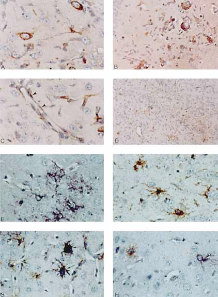
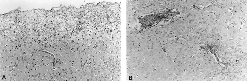
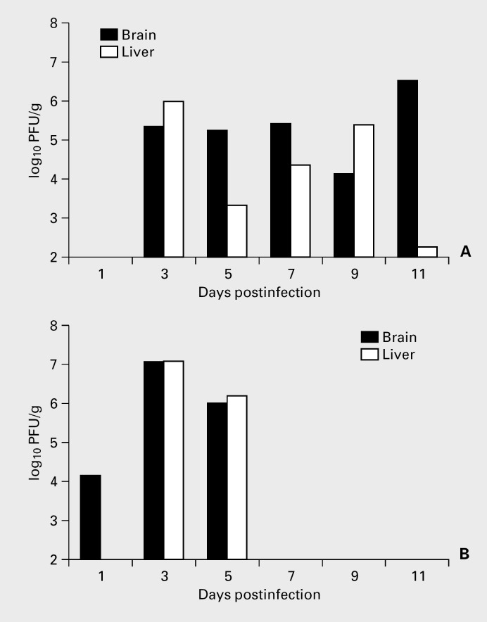
Mouse hepatitis virus (MHV) A59 infection which causes acute encephalitis, hepatitis, and chronic demyelination, is one of the experimental models for multiple sclerosis. Previous studies showed that lethal infection of beta2-microglobulin 'knockout' (beta2M(-/-)) mice required 500-fold less virus and viral clearance was delayed as compared to infection of immunocompetent C57Bl/6 (B6) mice. To investigate the mechanism of the increased susceptibility of beta2M(-/-) mice to MHV-A59, we studied organ pathology and the distribution of viral antigen and RNA during acute and chronic infection. A59-infected beta2M(-/-) mice were more susceptible to acute encephalitis and hepatitis, but did not have increased susceptibility to demyelination. Viral antigen and RNA distribution in the brain was increased in microglia, lymphocytes, and small vessel endothelial cells while the distribution in neurons and glia was similar in beta2M(-/-) mice and B6 mice. Acute hepatitis and thymus cortical hypoplasia in beta2M(-/-) mice were delayed in onset but pathologic changes in these organs were similar to those in B6 mice. The low rate of demyelination in beta2M(-/-) mice was consistent with the low dose of the virus given. A less neurotropic virus MHV-2, caused increased parenchymal inflammation in beta2M(-/-) mice, but without demyelination. Thus, CD8+ cells were important for viral clearance from endothelial cells, microglia and inflammatory cells, but not from neuronal and glial cells. In addition, CD8+ cells played a role in preventing the spread of encephalitis.
Figures
Similar articles
-
Mouse hepatitis virus A59-induced demyelination can occur in the absence of CD8+ T cells.Microb Pathog. 1995 Mar;18(3):211-21. doi: 10.1016/s0882-4010(95)90058-6. Microb Pathog. 1995. PMID: 7565015 Free PMC article.
-
Mouse hepatitis virus type-2 infection in mice: an experimental model system of acute meningitis and hepatitis.Exp Mol Pathol. 2001 Aug;71(1):1-12. doi: 10.1006/exmp.2001.2378. Exp Mol Pathol. 2001. PMID: 11502093
-
Sequence analysis of the S gene of recombinant MHV-2/A59 coronaviruses reveals three candidate mutations associated with demyelination and hepatitis.J Neurovirol. 2001 Oct;7(5):432-6. doi: 10.1080/135502801753170282. J Neurovirol. 2001. PMID: 11582515 Free PMC article.
-
Pathogenesis of mouse hepatitis virus-induced demyelination.J Neurovirol. 1996 Dec;2(6):361-76. doi: 10.3109/13550289609146902. J Neurovirol. 1996. PMID: 8972418 Review.
-
Pathogenesis of murine coronavirus in the central nervous system.J Neuroimmune Pharmacol. 2010 Sep;5(3):336-54. doi: 10.1007/s11481-010-9202-2. Epub 2010 Apr 6. J Neuroimmune Pharmacol. 2010. PMID: 20369302 Free PMC article. Review.
Cited by
-
Mouse hepatitis virus 3 binding to macrophages correlates with resistance to experimental infection.Scand J Immunol. 2005 Jul;62 Suppl 1(Suppl 1):95-9. doi: 10.1111/j.1365-3083.2005.01616.x. Scand J Immunol. 2005. PMID: 15953191 Free PMC article.
-
Connecting Neuroinflammation and Neurodegeneration in Multiple Sclerosis: Are Oligodendrocyte Precursor Cells a Nexus of Disease?Front Cell Neurosci. 2021 Jun 21;15:654284. doi: 10.3389/fncel.2021.654284. eCollection 2021. Front Cell Neurosci. 2021. PMID: 34234647 Free PMC article. Review.
-
Brain Invasion by Mouse Hepatitis Virus Depends on Impairment of Tight Junctions and Beta Interferon Production in Brain Microvascular Endothelial Cells.J Virol. 2015 Oct;89(19):9896-908. doi: 10.1128/JVI.01501-15. Epub 2015 Jul 22. J Virol. 2015. PMID: 26202229 Free PMC article.
-
Virus demyelination.J Neurovirol. 2003 Apr;9(2):148-64. doi: 10.1080/13550280390194046. J Neurovirol. 2003. PMID: 12707846 Free PMC article. Review.
-
Murine hepatitis virus--a model for virus-induced CNS demyelination.J Neurovirol. 2002 Apr;8(2):76-85. doi: 10.1080/13550280290049534. J Neurovirol. 2002. PMID: 11935460 Free PMC article. Review.
References
-
- McIntosh K. Coronaviruses: A comparative review. Curr Top Microbiol Immunol. 1974;63:80–129.
-
- Wege H, Siddell S, ter Meulen V. The biology and pathogenesis of coronaviruses. Adv Virol Immunol. 1982;99:165–200. - PubMed
-
- Lavi E, Weiss SR, Coronaviruses . In: Clinical and Molecular Aspects of Neurotropic Viral Infections. Gilden DH, Lipton HL, editors. Boston, Kluwer: Dev Med Virol; 1989. pp. pp 101–139.
-
- Weiner LP. Pathogenesis of demyelination induced by a mouse hepatitis virus (JHM virus) Arch Neurol. 1973;28:298–303. - PubMed
-
- Stohlman SA, Weiner LP. Chronic central nervous system demyelination in mice after JHM virus infection. Neurology. 1981;31:38–44. - PubMed
Publication types
MeSH terms
Substances
LinkOut - more resources
Full Text Sources
Molecular Biology Databases
Research Materials
