

doi: 10.1126/science.283.5405.1180.
Regulation of neurotrophin-3 expression by epithelial-mesenchymal interactions: the role of Wnt factors
Affiliations
- PMID: 10024246
- PMCID: PMC2710127
- DOI: 10.1126/science.283.5405.1180
Item in Clipboard
Regulation of neurotrophin-3 expression by epithelial-mesenchymal interactions: the role of Wnt factors
Science.
.
Abstract
Neurotrophins regulate survival, axonal growth, and target innervation of sensory and other neurons. Neurotrophin-3 (NT-3) is expressed specifically in cells adjacent to extending axons of dorsal root ganglia neurons, and its absence results in loss of most of these neurons before their axons reach their targets. However, axons are not required for NT-3 expression in limbs; instead, local signals from ectoderm induce NT-3 expression in adjacent mesenchyme. Wnt factors expressed in limb ectoderm induce NT-3 in the underlying mesenchyme. Thus, epithelial-mesenchymal interactions mediated by Wnt factors control NT-3 expression and may regulate axonal growth and guidance.
Figures
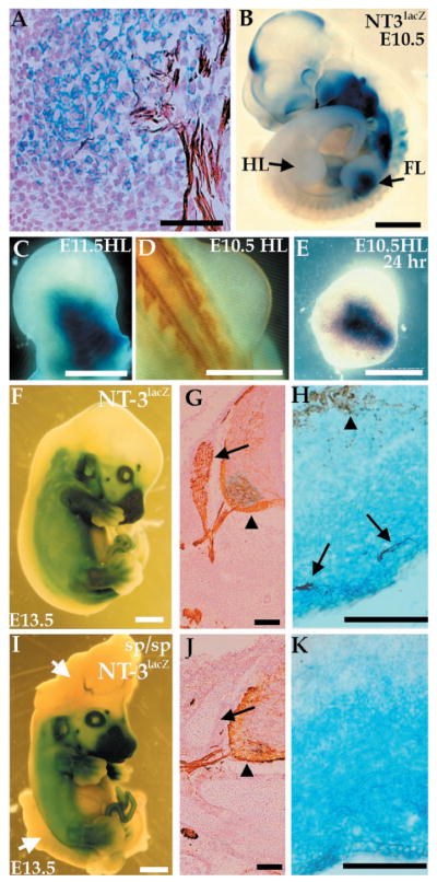
Expression of NT-3 in the limb does not depend on innervation. Expression of NT-3 was detected by a lacZ reporter gene stained with X-Gal (blue), and axons were visualized with a neurofilament antibody (NF, brown). (A) E11.5 NT-3lacZ embryo section shows colocalization of X-Gal staining with ends of sensory neuron projections. (B) E10.5 NT-3lacZ heterozygous embryo shows lacZ expression in forelimb (FL) but not hindlimb (HL). (C) E11.5 hindlimb stained for lacZ expression. (D) Dorsal view of an E10.5 embryo stained with NF. No axons are present in the hindlimb at this time. (E) E10.5 hindlimb cultured for 24 hours and stained for lacZ expression. (F to K) Expression of lacZ or NF (or both) in E13.5 NT-3lacZ embryos in a wild-type (F to H) or splotch background (I to K). Embryos were stained for lacZ expression (F and I). The splotch embryo exhibits severe defects in the cranial and lumbosacral region (arrows, I). No significant difference in lacZ expression in wild-type and mutant embryos is detected. (G and J) NF staining of caudal neural tube sections. DRGs (arrows) are absent in splotch mutants, although motor neurons (arrowheads) are present. (H and K) lacZ/NF costaining of the distal hindlimb. Sensory (arrows) but not motor (arrowhead) axons correlate with NT3 expression as detected by lacZ expression (H). (K) NT-3 expression is normal in the absence of NF staining in splotch mice. Bars, 50 μm (A, H, and K); 1 mm (B, F, and I); 0.5 mm (C to E); 100 μm (G and J).
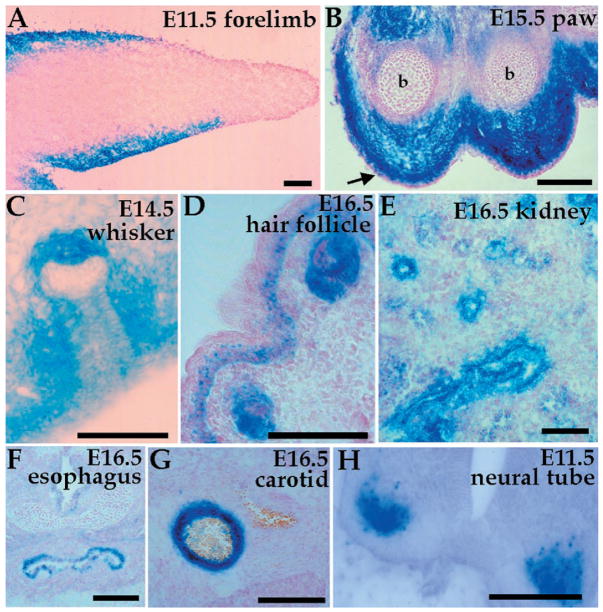
NT-3 is expressed in many sites of epithelial-mesenchymal interactions during embryogenesis. X-Gal staining of NT-3lacZ heterozygous embryo sections shows expression in (A) E11.5 forelimb mesenchyme, (B) E15.5 forelimb paw [note the absence of expression in ectoderm (arrow) and bone (b)], (C) E14.5 whisker pad, (D) E16.5 hair follicle and skin dermis, (E) E16.5 epithelial cells of the metanephric kidney, (F) E16.5 upper esophagus, (G) E16.5 carotid artery, and (H) motor neurons of E11.5 neural tube. Bars, 100 μm.
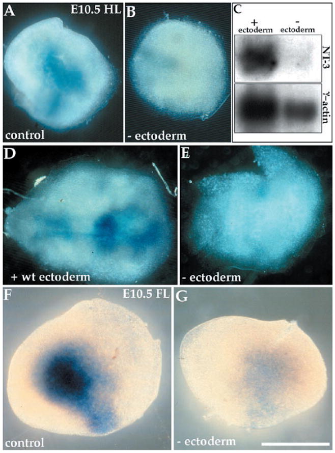
Ectoderm induces NT-3 expression in limb mesenchyme. Limbs were isolated from E10.5 NT-3lacZ heterozygotes and cultured in vitro. (A) An intact hindlimb expresses NT-3, as assessed by X-Gal staining, but (B) a hindlimb lacking ectoderm does not. (C) A Northern blot containing mRNA isolated from limbs cultured with or without ectoderm (10 limbs per lane), probed for NT-3 and γ-actin. (D) NT-3lacZ hindlimb mesenchyme reconstituted with wild-type ectoderm exhibits lacZ expression, but (E) a hindlimb lacking ectoderm does not. (F) Forelimb shows strong lacZ expression, (G) whereas a forelimb lacking ectoderm does not. All limbs were cultured for 24 hours with the exception of (D) and (E), which were cultured for 48 hours. Bar, 0.5 mm.
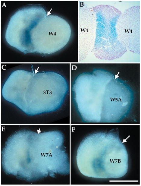
Wnt proteins are sufficient to induce NT-3 expression. Mesenchymal hindlimb tissue (without ectoderm) isolated from E10.5 NT-3lacZ heterozygous embryos was cocultured with aggregates of NIH 3T3–derived cell lines for 24 hours, after which they were stained with X-Gal. (A) Wnt-4–expressing NIH 3T3 cells induce NT-3 in limb mesenchyme. (B) A Wnt-4/limb/Wnt-4 explant stained and sectioned shows two blue stripes at the junction of the two tissues. (C) NIH 3T3 cells or (D) Wnt-5a– expressing cells do not induce NT-3, whereas (E) Wnt-7a– or (F) Wnt-7b–expressing cells induce NT-3 to a certain extent. Arrows point to junctions of limb mesenchyme and cell aggregates. Bar, 0.5 mm.
References
-
- Reichardt LF, Fariñas I. In: Molecular Approaches to Neural Development. Cowan MW, Jessell TM, Zipurski L, editors. Oxford Univ. Press; New York: 1997. pp. 220–263.
-
- Elshamy WM, Ernfors P. ibid. 1996;16:963. - PubMed
-
- Verdi JM, et al. ibid. :515.
-
- Song HJ, Ming GL, Poo MM. Nature. 1997;388:275. - PubMed
Publication types
MeSH terms
Substances
Grants and funding
LinkOut - more resources
Full Text Sources
Research Materials
