. 1999 Mar 1;19(5):1566-76.
doi: 10.1523/JNEUROSCI.19-05-01566.1999.

Developmental synaptic changes increase the range of integrative capabilities of an identified excitatory neocortical connection

Affiliations

Developmental synaptic changes increase the range of integrative capabilities of an identified excitatory neocortical connection

M C Angulo et al. J Neurosci. .

Abstract

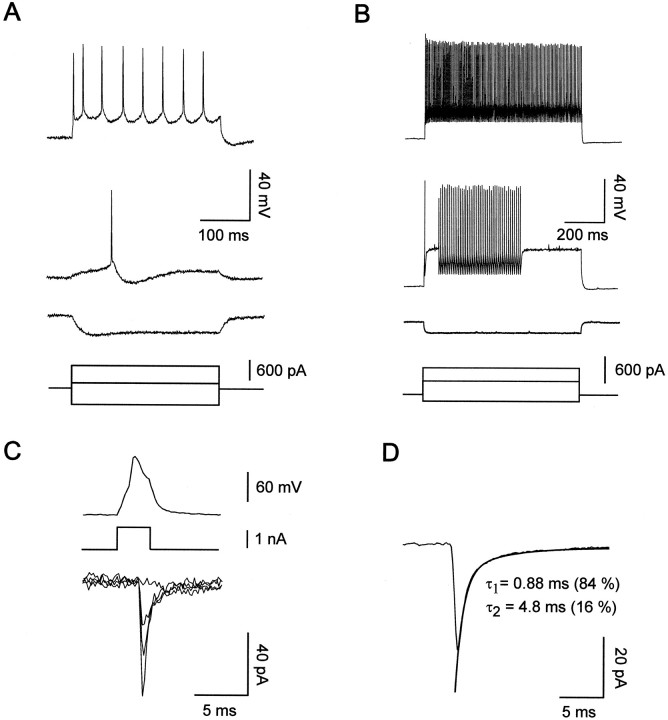
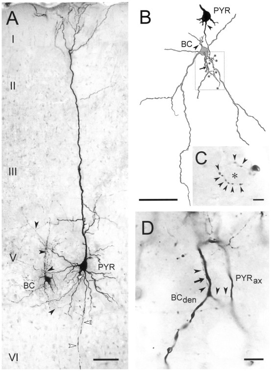
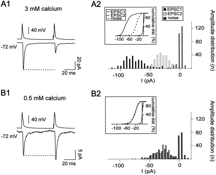
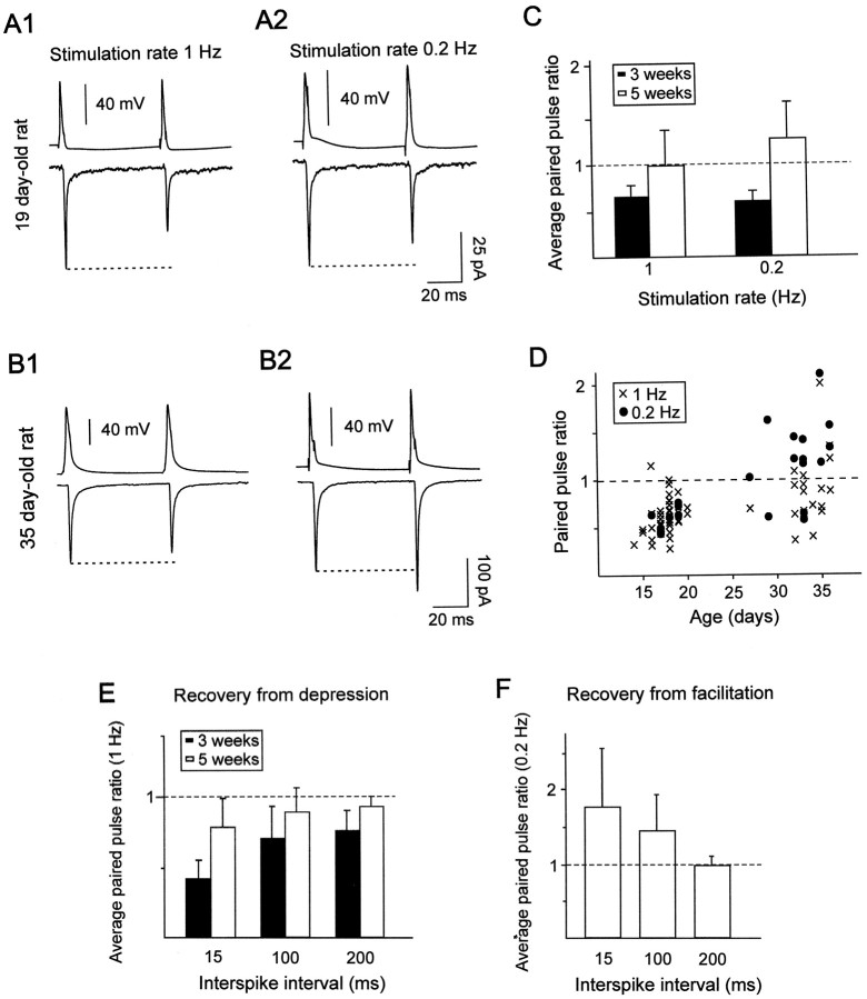
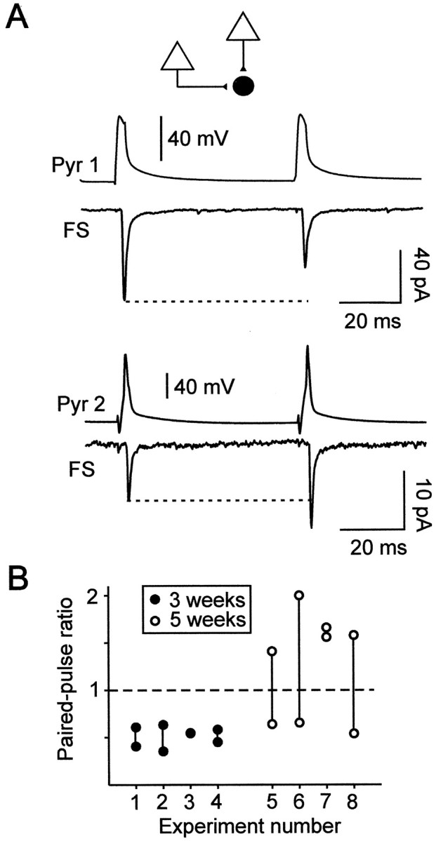
Excitatory synaptic transmission between pyramidal cells and fast-spiking (FS) interneurons of layer V of the motor cortex was investigated in acute slices by using paired recordings at 30 degrees C combined with morphological analysis. The presynaptic and postsynaptic properties at these identified central synapses were compared between 3- and 5-week-old rats. At these two postnatal developmental stages, unitary EPSCs were mediated by the activation of AMPA receptors with fast kinetics at a holding potential of -72 mV. The amplitude distribution analysis of the EPSCs indicates that, at both stages, pyramidal-FS connections consisted of multiple functional release sites. The apparent quantal size obtained by decreasing the external calcium ([Ca2+]e) varied from 11 to 29 pA near resting membrane potential. In young rats, pairs of presynaptic action potentials elicited unitary synaptic responses that displayed paired-pulse depression at all tested frequencies. In older animals, inputs from different pyramidal cells onto the same FS interneuron had different paired-pulse response characteristics and, at most of these connections, a switch from depression to facilitation occurred when decreasing the rate of presynaptic stimulation. The balance between facilitation and depression endows pyramidal-FS connections from 5-week-old animals with wide integrative capabilities and confers unique functional properties to each synapse.


Figures

Fig. 1.
Fig. 1.
Electrophysiological characterization of connected pyramidal cells and FS interneurons from 18-d-old rats.A, Current-clamp recordings of a pyramidal cell during injection of current pulses with a sharp intracellular electrode. In response to near-threshold current pulses, the pyramidal cell emitted a single action potential with a slow AHP (middle voltage trace). Note the slow action potentials and the accommodation of the firing during larger depolarizing current pulses (top voltage trace) and the time-dependent rectification in response to hyperpolarizing current pulses (bottom voltage trace). Recordings are from the pyramidal cell shown in Figure2A. B, Current-clamp recordings of a FS interneuron during injection of current pulses with a patch pipette. In response to near-threshold current pulses (middle voltage trace), this FS cell emitted a single fast action potential with a large fast AHP followed by a silent period and a late discharge of action potentials. Application of a larger depolarizing current (top voltage trace) induced a continuous discharge at high frequency. Note the low input resistance of the FS neuron as seen in response to hyperpolarizing current pulses (bottom voltage trace). Recordings are from the FS cell shown in Figure 2BD.C, Unitary EPSCs at a pyramidal–FS connection. Thetop trace illustrates an action potential elicited in the pyramidal neuron by a brief depolarizing current pulse (middle voltage trace). The bottom tracesare four superimposed current responses recorded in the FS interneuron at a holding potential of −72 mV during the stimulation of the pyramidal neuron. Note an apparent transmission failure and the large variation of the EPSC amplitudes. Recordings are from the connection illustrated in Figure 2BD.D, Kinetics of unitary EPSCs at the same pyramidal–FS connection as in C. A biexponential fit of the decay of the EPSC was superimposed to the average of 203 postsynaptic responses, excluding failures. The numbers inparentheses represent the relative amplitude of the first (τ1) and the second (τ2) exponential.
Fig. 2.
Fig. 2.
Morphological characterization of pyramidal–FS connections from 18-d-old rats. A, Photomontage of a typical pyramidal cell (PYR) to a putative basket cell (BC) connection as recorded and stained with biocytin in the present study. Both cells have large somata located in layer V of motor cortex and possess a characteristic morphology for the respective cell class. White arrowheads mark the axon of the pyramidal cell, black arrowheads those of the interneuron. Roman numerals denominate cortical laminae.B, Camera lucida drawing showing the contact formed by another pair, also from layer V. The soma of the pyramidal cell and the skeleton of the axon until it contacts a secondary dendrite of the large multipolar putative basket cell (arrow) is traced. Axonal branch points that have not been reconstructed further are marked with an asterisk. Arrowheadsindicate the axon initial segments of both cells. Stippled frame marks area shown as micrograph in D.C, Basket-like terminal formation of the axon of the interneuron shown in B. Arrowheads point to individual boutons presumably contacting the encircled soma (asterisk). D, High-power micrograph showing the pyramidal cell axon (PYRax), specifically the course of the recurrent collateral (arrowheads) that forms a delicate contact (arrow) with the putative basket cell dendrite (BCden). Scale bars: A,B, 100 μm; C, D, 10 μm.
Fig. 3.
Fig. 3.
Multiple quanta of transmitter released at a single pyramidal–FS connection from a 16-d-old rat. A1,B1, The bottom traces show the mean amplitude, including failures, of the postsynaptic currents (EPSC1 and EPSC2) recorded in a FS cell in response to pairs of action potentials (top traces) elicited in a presynaptic pyramidal cell with an interspike interval of 50 msec at a stimulation rate of 1 Hz in 3 mm (A1) and 0.5 mm(B1) of [Ca2+]e. A marked PPD (ratio of 0.37) was observed in 3 mm[Ca2+]e (A1), whereas a PPF (ratio of 1.2) was obtained when reducing [Ca2+]e to 0.5 mm(B1). The response probabilities of EPSC1 and EPSC2 were, respectively, 0.99 and 0.88 in 3 mm[Ca2+]e and 0.52 and 0.6 in 0.5 mm [Ca2+]e. Traces are averages of 270 and 330 responses for A1 andB1, respectively. A2, B2, Amplitude distributions of EPSC1 and EPSC2, excluding failures, obtained in 3 mm [Ca2+]e(A2) and 0.5 mm[Ca2+]e (B2). The histograms and cumulative plots (A2,inset) show that the distribution of EPSC1 and EPSC2 were significantly different in 3 mm[Ca2+]e, suggesting that multiple quanta were released at this connection. This was confirmed by the reduction in amplitude of both responses in 0.5 mm[Ca2+]e (B2,inset). In low [Ca2+]e, the amplitude distribution of EPSC1 and EPSC2 were identical suggesting that, under these conditions of low release probability, a single quantum was released when there was a response (B2). An apparent quantal size of −20 pA could be estimated from the current corresponding to 50% of the cumulative distribution (B2, inset).
Fig. 4.
Fig. 4.
Paired-pulse response characteristics at pyramidal–FS connections at two different developmental stages.A, B, Paired-pulse responses elicited with a presynaptic interspike interval of 50 msec (top traces) at stimulation rates of 1 (A1,B1) and 0.2 Hz (A2, B2) in FS interneurons from 19- and 35-d-old rats. The bottom traces correspond to the mean amplitudes, including failures, of the postsynaptic currents. For the connection in A, the response probabilities of EPSC1 and EPSC2 were, respectively, 0.96 and 0.93 at a stimulation rate of 1 Hz (A1) and 1 and 0.98 at a stimulation rate of 0.2 Hz (A2). For the connection in B, the response probabilities of EPSC1 and EPSC2 were, respectively, 0.99 and 1 at a stimulation rate of 1 Hz (B1) and 1 at a stimulation rate of 0.2 Hz (B2). Note that in A the connection showed a PPD at stimulation rates of 1 Hz (ratio of 0.64) and 0.2 Hz (ratio of 0.73), whereas in B the connection showed a PPD at a stimulation rate of 1 Hz (ratio of 0.89) and a PPF at 0.2 Hz (ratio of 1.2). Traces are averages from 69 to 131 responses.C, Average of the paired-pulse ratios at connections from 3-week-old rats (black bars) and 5-week-old rats (white bars). Only pairs tested at both stimulation rates were included. There was a statistical difference between the ratios obtained at 1 and 0.2 Hz for pairs from 5-week-old rats (p < 0.001; n = 11) but not for pairs from younger animals (n = 8). Note that the PPD was more marked at connections from younger animals.D, Plot of the paired-pulse ratio of all individual connections as a function of the age of the preparation at stimulation rates of 1 Hz (crosses; n = 57) and 0.2 Hz (circles; n = 23). Note the large variability of responses at pairs from 5-week-old animals.E, Recovery from depression. Average paired-pulse ratios at connections from 3-week-old (n = 5) and 5-week-old (n = 6) rats obtained at presynaptic interspike intervals of 15, 100, and 200 msec at a stimulation rate of 1 Hz. F, Recovery from facilitation. Average paired-pulse ratios at connections from 5-week-old rats (n = 5) obtained at presynaptic interspike intervals of 15, 100, and 200 msec at a stimulation rate of 0.2 Hz.
Fig. 5.
Fig. 5.
Paired-pulse responses elicited by two different presynaptic pyramidal cells onto single postsynaptic FS interneurons.A, Paired-pulse responses of a FS interneuron elicited by two presynaptic pyramidal cells recorded sequentially in a slice from a 33-d-old rat. A PPD (ratio of 0.64; traces are averages of 50 responses) was observed at the first connection (Pyr 1-FS) at which the response probabilities of EPSC1 and EPSC2 were 0.94 and 0.86, respectively. A PPF (ratio of 1.4; traces are averages of 76 responses) was observed at the second connection (Pyr 2-FS) with response probabilities of 0.7 and 0.87 for EPSC1 and EPSC2, respectively.B, Comparison of the paired-pulse ratios of two different presynaptic inputs onto single FS interneurons.Vertical lines link the two data points corresponding to the paired-pulse ratios of the two connections studied for each of the eight FS cells recorded in 3-week-old (n = 4) and 5-week-old (n = 4) animals. Note that PPD was obtained at all young connections, whereas different inputs onto three of four single FS cells from 5-week-old rats could display either PPD or PPF. It is noteworthy that, at older connections, the presence of PPD or PPF was independent of the order in which the sequential connections were obtained.

References

    1. Angulo MC, Lambolez B, Audinat E, Hestrin S, Rossier J. Subunit composition, kinetic, and permeation properties of AMPA receptors in single neocortical nonpyramidal cells. J Neurosci. 1997;17:6685–6696. - PMC - PubMed
    1. Bolshakov VY, Siegelbaum SA. Regulation of hippocampal transmitter release during development and long-term potentiation. Science. 1995;269:1730–1734. - PubMed
    1. Buhl EH, Tamas G, Szilagyi T, Stricker C, Paulsen O, Somogyi P. Effect, number and location of synapses made by single pyramidal cells onto aspiny interneurones of cat visual cortex. J Physiol (Lond) 1997;500:689–713. - PMC - PubMed
    1. Buzsaki G. Functions for interneuronal nets in the hippocampus. Can J Physiol Pharmacol. 1997;75:508–515. - PubMed
    1. Cauli B, Audinat E, Lambolez B, Angulo M-C, Ropert N, Tsuzuki K, Hestrin S, Rossier J. Molecular and physiological diversity of cortical nonpyramidal cells. J Neurosci. 1997;17:3894–3906. - PMC - PubMed

Publication types

MeSH terms

LinkOut - more resources