. 1999 Mar 1;19(5):1577-85.
doi: 10.1523/JNEUROSCI.19-05-01577.1999.

Molecular dynamics of the sodium channel pore vary with gating: interactions between P-segment motions and inactivation

Affiliations

Molecular dynamics of the sodium channel pore vary with gating: interactions between P-segment motions and inactivation

J P Bénitah et al. J Neurosci. .

Abstract

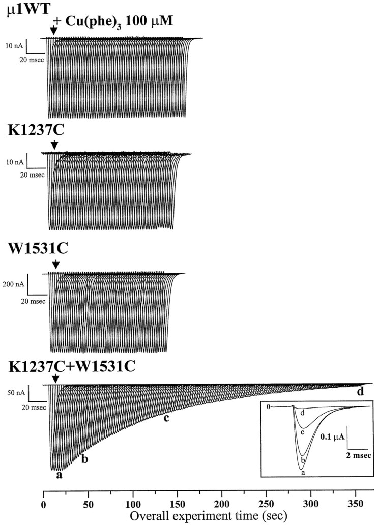
Disulfide trapping studies have revealed that the pore-lining (P) segments of voltage-dependent sodium channels undergo sizable motions on a subsecond time scale. Such motions of the pore may be necessary for selective ion translocation. Although traditionally viewed as separable properties, gating and permeation are now known to interact extensively in various classes of channels. We have investigated the interaction of pore motions and voltage-dependent gating in micro1 sodium channels engineered to contain two cysteines within the P segments. Rates of catalyzed internal disulfide formation (kSS) were measured in K1237C+W1531C mutant channels expressed in oocytes. During repetitive voltage-clamp depolarizations, increasing the pulse duration had biphasic effects on the kSS, which first increased to a maximum at 200 msec and then decreased with longer depolarizations. This result suggested that occupancy of an intermediate inactivation state (IM) facilitates pore motions. Consistent with the known antagonism between alkali metals and a component of slow inactivation, kSS varied inversely with external [Na+]o. We examined the converse relationship, namely the effect of pore flexibility on gating, by measuring recovery from inactivation in Y401C+E758C (YC/EC) channels. Under oxidative conditions, recovery from inactivation was slower than in a reduced environment in which the spontaneous YC/EC cross-link is disrupted. The most prominent effects were slowing of a component with intermediate recovery kinetics, with diminution of its relative amplitude. We conclude that occupancy of an intermediate inactivation state facilitates motions of the P segments; conversely, flexibility of the P segments alters an intermediate component of inactivation.


Figures

Fig. 1.
Fig. 1.
Effect of external application of Cu(phe)3 on wild-type and mutant μ1 Na current. Whole-cell Na currents were recorded from Xenopusoocytes coexpressing wild-type or mutant α subunits and the rat brain β1 subunit. The α subunits were (fromtop to bottom) μ1 wild-type, K1237C, W1531C, and the double-mutant K1237C+W1531C. Na currents were elicited by 50 msec pulses from −100 to −30 mV at 0.5 Hz. The calibration bar represents the time base at which the individual current records are displayed. The total experimental time in seconds is shown on the abscissa. The arrowhead indicates the time at which the redox catalyst Cu(phe)3 (100 μm) was applied. Currents from the wild-type and each of the single mutants were not modified by application of Cu(phe)3, whereas the current through the double-mutant K1237C+W1531C was progressively inhibited. Inset, Selected currents recorded from the double-mutant K1237C+W1531C during depolarizing voltage steps in the absence (a) and after 40 sec (b), 2 min (c), and 6 min (d) of exposure to 100 μmCu(phe)3.
Fig. 2.
Fig. 2.
Disulfide formation time course for the K1237C+W1531C channels. The normalized current amplitude of the superimposed current traces of the bottom panel of Figure 1 is plotted as function of the duration of redox catalyst Cu(phe)3exposure (plus symbols). A function describing the single-component reaction scheme (see Results) was fitted to the data: 1 − I/Imax = 1 − exp(−kss(t + {exp(−t/tp)} − 1). The fitted rate parameters kss for this reaction is 9.13 × 10⁻³ · sec−1 · molecule−1after correction for the rate of bath exchange (tp, 9.9 sec in this case) (Bénitah et al., 1997a).
Fig. 3.
Fig. 3.
Effect of pulse duration on the rate of Cu(phe)3 catalysis. Representative examples are shown for Cu(phe)3 inhibition of the peak K1237C+W1531C Na current as a function of pulse duration. Whole-cell currents were elicited in 96 mm [Na+]o using 0.05 Hz pulse trains with individual depolarizations from −100 to −40 mV lasting 50, 200, or 5000 msec (from top tobottom). The total time of the experiment is shown on the x-axis; the time base of the individual currents is indicated by the calibration bar. The arrowheadindicates the time of application of the redox catalyst.
Fig. 4.
Fig. 4.
Cu(phe)3 catalyzed disulfide bond formation in the K1237C+W1531C double mutant has a biphasic dependence on pulse duration. The disulfide formation rates (kss) plotted as a function of the pulse duration for oocytes expressing the K1237C+W1531C double mutant. The Na currents were elicited by depolarizing pulses at a rate of 0.05 Hz and durations ranging from 3 msec to 5 sec. We detect no cumulative of inactivation at this slow stimulation frequency for even the longest (5 sec) depolarizations. Currents were measured before and after addition of 100 μm Cu(phe)3, and the reaction rate constants were determined as described for Figure 2. The plotted values (means ± SEM) represent measurements from at least four different oocytes for each data point. *, p < 0.02; **, p < 0.002.
Fig. 5.
Fig. 5.
Rate of development of intermediate and slow-inactivation for the double-mutant K1237C+W1531C. As shown in the voltage-clamp protocol (top), the duration of the depolarizing prepulse (−40 mV) was varied, and the extent of recovery after 20 msec at −100 mV was assessed using a 50 msec test pulse to −40 mV. The 20 msec recovery interval removed the most rapidly recovering inactivation component (IF) from consideration. Plotted is the fractional recovery from inactivation as a function of the prepulse duration. Data were collected from seven oocytes. The dotted line shows a nonlinear fit to the mean data using the function:y = A1exp(−t1) + A2exp(−t2). The least squares error was minimized when A1 = 0.2, τ1 = 66 msec, A2 = 0.8, and τ2= 5212 msec.
Fig. 6.
Fig. 6.
External Na+ concentration modulates the rate of inactivation and disulfide bond formation.A, Representative examples of the time course of the inhibition of K1237C+W1531C by 100 mm Cu(phe)3obtained from oocytes bathed in either (▵) 48 mm[Na+]o, (○) 96 mm[Na+]o, or (⋄) 140 mm [Na+]o. Sorbitol was used as a substitute for Na+ to maintain constant osmolarity. The currents were elicited by repetitive 50 msec pulses from −100 to −35 mV (0.5 Hz). Plotted are the whole-cell Na current amplitudes normalized to the value measured before the application of Cu(phe)3 (100 μm). Summary data forkss as a function of extracellular [Na+]o are given in Table 1.B, Rates of the development of inactivation of wild-type μ1 channels coexpressed with the β1 subunit. The protocol is identical to that shown in Figure 5 for the KC/WC mutant. Reducing the [Na+]o enhances the rate of development of slow inactivation. In 96 mm[Na+]o (▪) there is little development of intermediate inactivation (< 3%). In contrast, in 10 mm [Na+]o (○) a 1 sec prepulse inactivates 35% of the current.
Fig. 7.
Fig. 7.
Redox state modulates the rate of recovery from inactivation for the double mutant Y401C+E758C. A, B, A standard two-pulse protocol was used to compare recovery from inactivation of Y401C+E758C channels in oxidized control solution (left panels) and after addition of 1 mm DTT (right panels) in the same oocyte. Oocytes were held at −100 mV. After a 50 msec (A) or 1 sec (B) conditioning pulse to −35 mV (the peak of the IV relationship), a pulse to −100 mV ranging from 1 msec to 1000 msec was followed by a 50 msec test depolarization. C, Fractional recovery from inactivation as a function of time at −100 mV is plotted for the double mutant Y401C+E758C in oxidized control solution (solid symbols) and after paired addition of 1 mm DTT (open symbols). Data sets obtained using either a 50 msec (squares) or a 1 sec (circles) conditioning pulse are shown. A three-exponential function of the form y = A1exp(−t1) + A2exp(−t2) + A3exp(−t3) was fitted to the individual data sets (fitted parameters are in Table 2). Fitted parameters to the mean data for a 50 msec prepulse (control vs DTT) were τ1 = 2.6 versus 2.4 msec, A1 = 0.84 versus 0.80; τ2 = 40.0 versus 40.9 msec, A2 = 0.11 versus 0.12; τ3 = 1565 versus 1315 msec, A3 = 0.05 versus 0.08. For the 1 sec prepulse, fitted parameters to the mean data (control vs DTT) were τ1 = 2.6 versus 3.3 msec, A1 = 0.51 versus 0.62; τ2 = 100.2 versus 49.8 msec, A2 = 0.23 versus 0.33; τ3 = 2660 versus 3449 msec, A3 = 0.16 versus 0.15.
Fig. 8.
Fig. 8.
Occupancy of an intermediate inactivated state facilitates disulfide formation in the outer pore. The diagram illustrates sequential occupancy of three distinct inactivated states (IF,IM, andIS) as the length of the depolarization is extended. Time constants for entry intoIM and IS are based on the biexponential fit to the data in Figure 5, and the time constant for development of fast inactivation (IF) was estimated from the literature (Nuss et al., 1995). The scheme suggests that when theIM state is occupied, the 1237 and 1531 cysteines are spatially optimized in the outer pore for disulfide formation.

References

    1. Balser JR, Nuss HB, Chiamvimonvat N, Pérez-García MT, Marban E, Tomaselli GF. External pore residue mediates slow inactivation in μ1 rat skeletal muscle sodium channels. J Physiol (Lond) 1996;494.2:431–442. - PMC - PubMed
    1. Bénitah J-P, Tomaselli GF, Marban E. Adjacent pore-lining residues within sodium channels identified by paired cysteine mutagenesis. Proc Natl Acad Sci USA. 1996;93:7392–7396. - PMC - PubMed
    1. Bénitah J-P, Ranjan R, Yamagishi T, Janecki M, Tomaselli GF, Marban E. Molecular motions within the pore of voltage-dependent sodium channels. Biophys J. 1997a;73:603–613. - PMC - PubMed
    1. Bénitah J-P, Balser JR, Marban E, Tomaselli GF. Proton inhibition of sodium channels: mechanism of gating shifts and reduced conductance. J Membr Biol. 1997b;155:121–131. - PubMed
    1. Careaga CL, Falke JJ. Thermal motions of surfacea-helces in the d-galactose chemosensory receptor: detection by disulfide trapping. J Mol Biol. 1992;226:1219–1235. - PMC - PubMed

Publication types

LinkOut - more resources