. 1999 Mar;67(3):1086-92.
doi: 10.1128/IAI.67.3.1086-1092.1999.

The staphylococcal transferrin-binding protein is a cell wall glyceraldehyde-3-phosphate dehydrogenase

Affiliations

The staphylococcal transferrin-binding protein is a cell wall glyceraldehyde-3-phosphate dehydrogenase

B Modun et al. Infect Immun. 1999 Mar.

Abstract

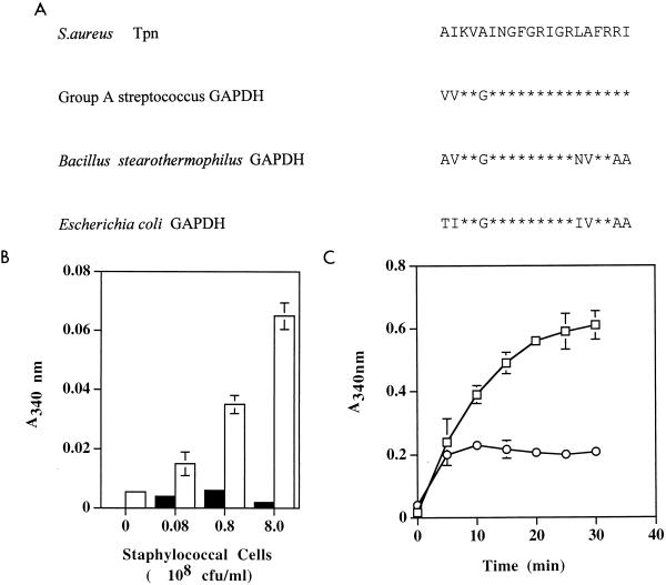
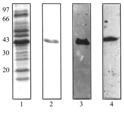
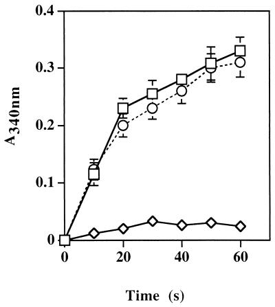
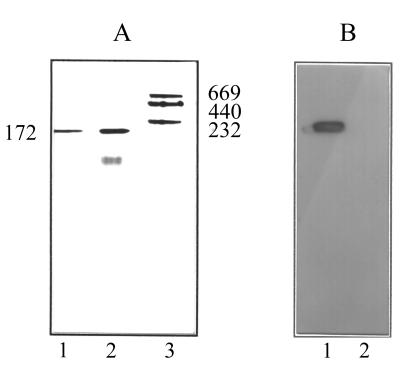
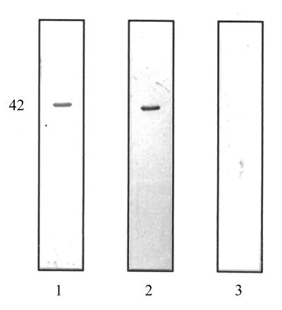
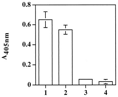
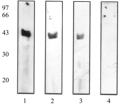
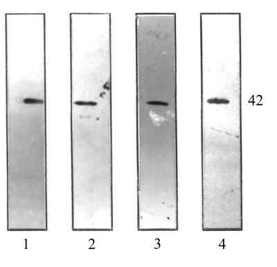
Staphylococcus aureus and Staphylococcus epidermidis possess a 42-kDa cell wall transferrin-binding protein (Tpn) which is involved in the acquisition of transferrin-bound iron. To characterize this protein further, cell wall fractions were subjected to two-dimensional sodium dodecyl sulfate (SDS)-polyacrylamide gel electrophoresis blotted, and the N-terminus of Tpn was sequenced. Comparison of the first 20 amino acid residues of Tpn with the protein databases revealed a high degree of homology to the glycolytic enzyme glyceraldehyde-3-phosphate dehydrogenase (GAPDH). Analysis of staphylococcal cell wall fractions for GAPDH activity confirmed the presence of a functional enzyme which, like Tpn, is regulated by the availability of iron in the growth medium. To determine whether Tpn is responsible for this GAPDH activity, it was affinity purified with NAD+ agarose. Both S. epidermidis and S. aureus Tpn catalyzed the conversion of glyceraldehyde-3-phosphate to 1,3-diphosphoglycerate. In contrast, Staphylococcus saprophyticus, which lacks a Tpn, has no cell wall-associated GAPDH activity. Native polyacrylamide gel electrophoresis of the affinity-purified Tpn revealed that it was present in the cell wall as a tetramer, consistent with the structures of all known cytoplasmic GAPDHs. Furthermore, the affinity-purified Tpn retained its ability to bind human transferrin both in its native tetrameric and SDS-denatured monomeric forms. Apart from interacting with human transferrin, Tpn, in common with the group A streptococcal cell wall GAPDH, binds human plasmin. Tpn-bound plasmin is enzymatically active and therefore may contribute to the ability of staphylococci to penetrate tissues during infections. These studies demonstrate that the staphylococcal transferrin receptor protein, Tpn, is a multifunctional cell wall GAPDH.


Figures

FIG. 1
FIG. 1
(A) Comparison of the N-terminal amino acid sequences of the S. aureus Tpn and the GAPDHs of group A streptococci, B. stearothermophilus, and Escherichia coli, whose SwissProt database accession numbers are P50467, P00362, and P06977 respectively. The S. epidermidis Tpn sequence is identical to that of S. aureus. ∗, identity to the S. aureus GAPDH. (B) GAPDH activity associated with whole S. aureus cells (cell concentrations ranging from 0 to 8 × 10⁸ CFU/ml) as determined by the conversion of NAD+ to NADH in the presence (open bars) or absence (solid bars) of G-3-P as described in Materials and Methods. (C) GAPDH activity associated with the cell wall fractions of S. aureus as measured following the catalytic conversion of G-3-P to 1,3-diphosphoglycerate and the formation of NADH from NAD+. Staphylococci grown to stationary phase in iron-replete RPMI 1640 (○) or iron-depleted RPMI 1640 (□) were harvested, washed in PBS, and resuspended to the same optical density at 600 nm prior to fractionation as described in Materials and Methods.
FIG. 2
FIG. 2
SDS-polyacrylamide gel (lanes 1 and 2) of S. aureus cell wall proteins (lane 1) and the NAD+-agarose affinity-purified S. aureus Tpn (lane 2). Lanes 3 and 4 show Western blots of the SDS-solubilized S. aureus cell wall proteins (lane 3) and affinity-purified S. aureus Tpn (lane 4) probed with a human transferrin-HRP conjugate. Molecular mass markers (in kilodaltons) are shown on the left-hand side.
FIG. 3
FIG. 3
GAPDH activity of the affinity-purified Tpn proteins from S. aureus (□) and S. epidermidis (○) as determined by the conversion of NAD+ to NADH. As a negative control, a cell wall extract from S. saprophyticus (◊), which lacks the transferrin-binding protein, was included.
FIG. 4
FIG. 4
Native polyacrylamide gel (A) of the affinity-purified S. aureus Tpn (lane 1), the B. stearothermophilus GAPDH (lane 2), and molecular mass markers ranging from 669 to 232 kDa (lane 3). Lanes 4 and 5 show Western blots of native Tpn and the B. stearothermophilus GAPDH, respectively, probed with a human transferrin-HRP conjugate.
FIG. 5
FIG. 5
Western blot of the affinity-purified S. aureus Tpn (lane 1) and S. epidermidis Tpn (lane 2) probed with biotinylated human plasmin and visualized with a streptavidin-HRP conjugate. As a negative control, Tpn was probed with streptavidin-HRP alone (lane 3).
FIG. 6
FIG. 6
Enzymatic activity of plasmin bound to the S. aureus (lane 1) and S. epidermidis (lane 2) affinity-purified Tpn. Activity is determined as the increase in A405 following the release of paranitroanilide from the synthetic substrate N-p-tosyl-Gly-Pro-Lys-p-paranitroanilide. As negative controls, the experiment was repeated in the absence of either plasmin (lane 3) or the purified Tpn (lane 4).
FIG. 7
FIG. 7
Western strip blot competition assay to show the inhibition of binding of HRP-conjugated human transferrin to the S. aureus Tpn by human plasmin. Western blots containing the purified Tpn were incubated with a mixture of HRP-conjugated human transferrin (0.18 nM) and a range of concentrations of human plasmin (lane 1, 0 μM; lane 2, 3.75 μM; lane 3, 7.5 μM; lane 4, 11.25 μM).
FIG. 8
FIG. 8
Western strip blot competition assay to show the inhibition of binding of biotinylated human plasmin to the S. aureus Tpn by human transferrin. Western blots containing the purified Tpn were incubated with a mixture of biotinylated human plasmin (0.18 nM) and a range of concentrations of human transferrin (lane 1, 0 μM; lane 2, 3.75 μM; lane 3, 7.5 μM; lane 4, 11.25 μM).

References

    1. Arnold H, Pette D. Bundling of glycolytic enzymes to structure proteins of the muscle. Eur J Biochem. 1968;6:163–171. - PubMed
    1. Arnold H, Pette D. Binding of aldolase and triosephosphate dehydrogenase to F-actin and modification of catalytic properties of aldolase. Eur J Biochem. 1970;15:360–366. - PubMed
    1. Cockayne A, Hill P J, Powell N B L, Bishop K, Sims C, Williams P. Molecular cloning of a 32-kilodalton lipoprotein component of a novel iron-regulated Staphylococcus epidermidis ABC transporter. Infect Immun. 1998;66:3767–3774. - PMC - PubMed
    1. Cornelissen C N, Sparling P F. Iron piracy: acquisition of transferrin-bound iron by bacterial pathogens. Mol Microbiol. 1994;14:843–850. - PubMed
    1. Cornelissen C N, Anderson J E, Sparling P F. Energy-dependent changes in the gonococcal transferrin receptor. Mol Microbiol. 1997;26:25–35. - PubMed

Publication types

LinkOut - more resources