. 1999 Mar;67(3):1139-48.
doi: 10.1128/IAI.67.3.1139-1148.1999.

Vibrio parahaemolyticus thermostable direct hemolysin modulates cytoskeletal organization and calcium homeostasis in intestinal cultured cells

Affiliations

Vibrio parahaemolyticus thermostable direct hemolysin modulates cytoskeletal organization and calcium homeostasis in intestinal cultured cells

A Fabbri et al. Infect Immun. 1999 Mar.

Abstract

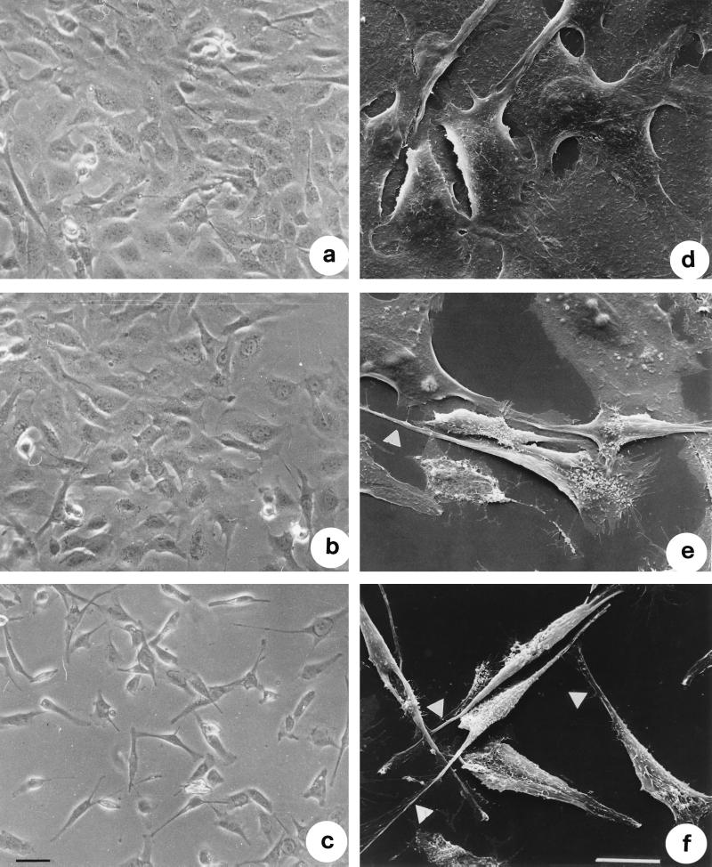
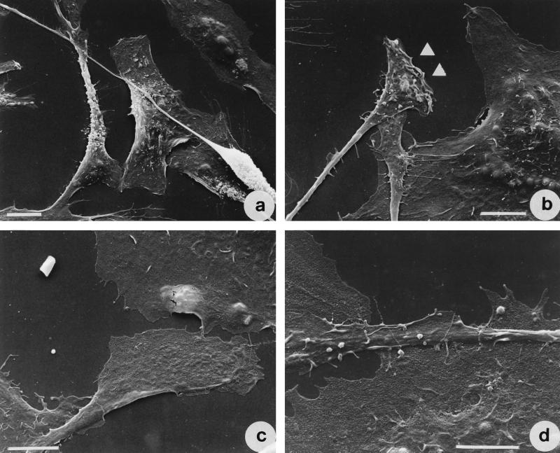
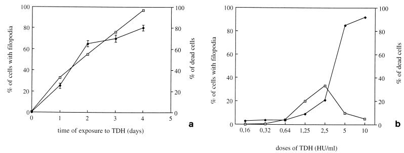
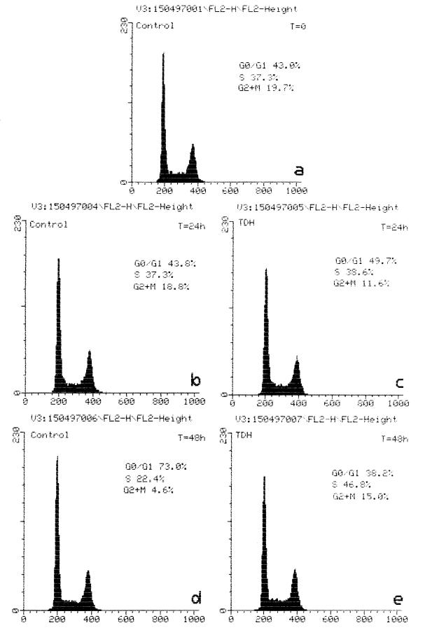
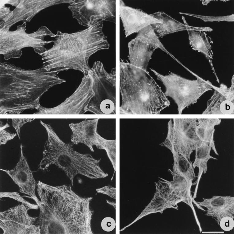
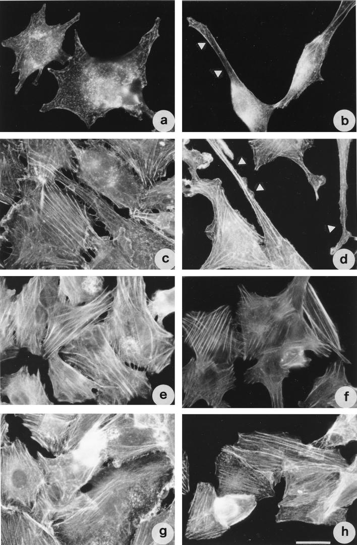
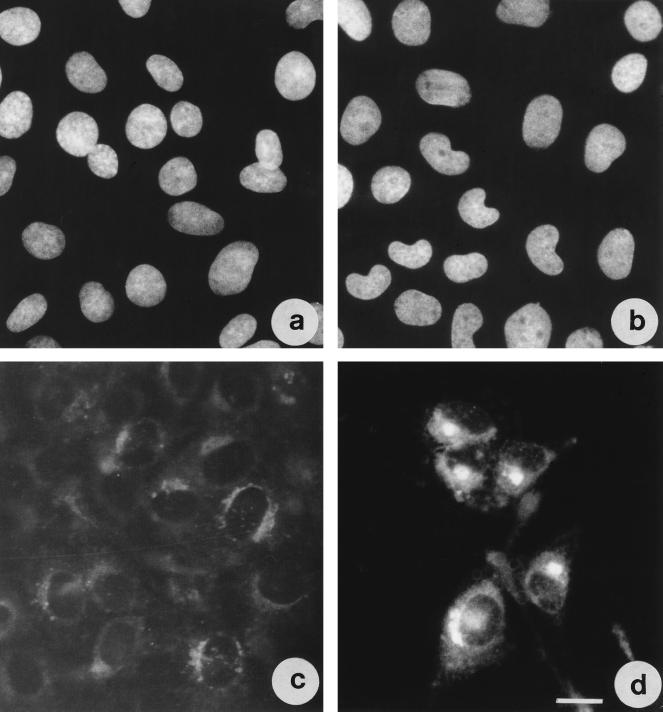
Vibrio parahaemolyticus is a marine bacterium known to be the leading cause of seafood gastroenteritis worldwide. A 46-kDa homodimer protein secreted by this microorganism, the thermostable direct hemolysin (TDH), is considered a major virulence factor involved in bacterial pathogenesis since a high percentage of strains of clinical origin are positive for TDH production. TDH is a pore-forming toxin, and its most extensively studied effect is the ability to cause hemolysis of erythrocytes from different mammalian species. Moreover, TDH induces in a variety of cells cytotoxic effects consisting mainly of cell degeneration which often leads to loss of viability. In this work, we examined the cellular changes induced by TDH in monolayers of IEC-6 cells (derived from the rat crypt small intestine), which represent a useful cell model for studying toxins from enteric bacteria. In experimental conditions allowing cell survival, TDH induces a rapid transient increase in intracellular calcium as well as a significant though reversible decreased rate of progression through the cell cycle. The morphological changes seem to be dependent on the organization of the microtubular network, which appears to be the preferential cytoskeletal element involved in the cellular response to the toxin.


Figures

FIG. 1
FIG. 1
SDS-PAGE of TDH on a 12% acrylamide gel stained with Coomassie brilliant blue. A single band of about 23 kDa is detected, confirming the purity of the toxin used.
FIG. 2
FIG. 2
TDH influences calcium homeostasis in IEC-6 cells. (a) Cells treated with 2.5 HU of TDH per ml; (b) cells exposed to 1 mM EGTA for 20 min before TDH treatment; (c) cells exposed to TDH for 30 min and then washed. Graphs show the first 40 min of the experiments. After the rapid increase in intracellular calcium level, the decrease occurs between 5 and 25 min (a and c). The y axis represents the ratio 340/380. The negative deflection at about 5 min represents an artifact due to the closure of the shutter to expose the dish and check whether the TDH was added correctly. The rise in cytosolic Ca2+ level is transient, totally reversible, and prevented by EGTA. ↓, TDH addition; ↑, washing.
FIG. 3
FIG. 3
TDH induces morphological effects in IEC-6 cells. Shown are phase-contrast images of control cells (a) and cells treated with 2.5 HU of TDH per ml for 2 (b) and 18 (c) h and scanning electron micrographs of control cells (d) and cells exposed to 2.5 HU of TDH per ml for 2 (e) and 18 (f) h all at the same magnification. Note the extrusion of thin filopodia from the cell body of cells exposed to the hemolysin (arrows). Bars represent 10 μm.
FIG. 4
FIG. 4
Scanning electron micrographs of IEC-6 cells treated with 2.5 HU of TDH per ml for 18 h. Note the small lamellipodia at the end of filopodia (arrows). Bar represents 10 μm.
FIG. 5
FIG. 5
TDH-induced effects in IEC-6 cells are dose and time dependent. (a) Percentages of cells with filopodia and of dead cells after various times of exposure to 2.5 HU of TDH per ml; (b) dose dependence of TDH-induced cellular effects (filopodium formation and cell death), detectable after 18 h of exposure to the toxin. The results reported as percentages (± standard deviations) of cells with filopodia (⊡) or of dead cells (⧫) (as detected by trypan blue) with respect to the total number of cells counted, are from three different experiments in each of which at least 500 cells (randomly chosen) were counted.
FIG. 6
FIG. 6
TDH influences the cell cycle of IEC-6 cells. Histograms show DNA content of control cells (a, b, and d) and cells treated with 2.5 HU of TDH per ml (c and e) for the time periods indicated. Note the augmented percentage of cells in S phase after 2 days of exposure to TDH.
FIG. 7
FIG. 7
TDH provokes changes in actin and tubulin cytoskeletal networks in IEC-6 cells. Shown are fluorescence micrographs of cells stained for detection of F-actin (a and b) and tubulin (c and d). (a and c) Control cells; (b and d) cells exposed to 2.5 HU of TDH per ml for 18 h. Both cytoskeletal elements are present in the filopodia. Bar represents 10 μm.
FIG. 8
FIG. 8
Microtubule perturbants inhibit TDH-induced morphological changes in IEC-6 cells as viewed by fluorescence detection of F-actin. Cells were treated with cytochalasin D (a and b), jasplakinolide (c and d), vinblastine (e and f), or taxol (g and h). Cells in panel b, d, f, and h were subsequently exposed to TDH for 18 h. Note that only the microtubule-perturbing drugs are able to inhibit the formation of filopodia. Arrows indicate filopodia. Bar represents 10 μm.
FIG. 9
FIG. 9
Nuclear shape and Golgi apparatus positioning are altered in TDH-treated IEC-6 cells. Shown are fluorescence micrographs of cells stained with Hoechst 33258 (a and b) or with NBD C6-ceramide (c and d) for Golgi detection. (a and c) Control cells; (b and d) cells exposed to TDH for 18 h. Bar represents 10 μm.

References

    1. Abrami L, Fivaz M, Glauser P E, Parton R G, van der Goot F G. A pore-forming toxin interacts with a GPI-anchored protein and causes vacuolation of the endoplasmic reticulum. J Cell Biol. 1998;140:525–540. - PMC - PubMed
    1. Boquet P, Munro P, Fiorentini C, Just I. Toxins from anaerobic bacteria: specificity and molecular mechanisms of action. Curr Opin Microbiol. 1998;1:66–74. - PubMed
    1. Budd M, Senderowicz A, Sausville E, Duncan K, Korn E. Jasplakinolide, a cytotoxic natural product, induces action polymerization and competitively inhibits the binding of phalloidin to F-actin. J Biol Chem. 1994;269:14869–14871. - PubMed
    1. Cherwonogrodzky J W, Clark A G. The purification of the Kanagawa haemolysin from Vibrio parahaemolyticus. FEMS Microbiol Lett. 1982;15:175. . (Abstract.)
    1. Darzynkiewicz Z, Juan G, Li X, Gorczyca W, Murakami T, Traganos F. Cytometry in cell necrobiology: analysis of apoptosis and accidental cell death (necrosis) Cytometry. 1997;27:1–20. - PubMed

Publication types

LinkOut - more resources