. 1999 Mar;67(3):1227-37.
doi: 10.1128/IAI.67.3.1227-1237.1999.

Identification of a cytolethal distending toxin gene locus and features of a virulence-associated region in Actinobacillus actinomycetemcomitans

Affiliations

Identification of a cytolethal distending toxin gene locus and features of a virulence-associated region in Actinobacillus actinomycetemcomitans

M P Mayer et al. Infect Immun. 1999 Mar.

Abstract

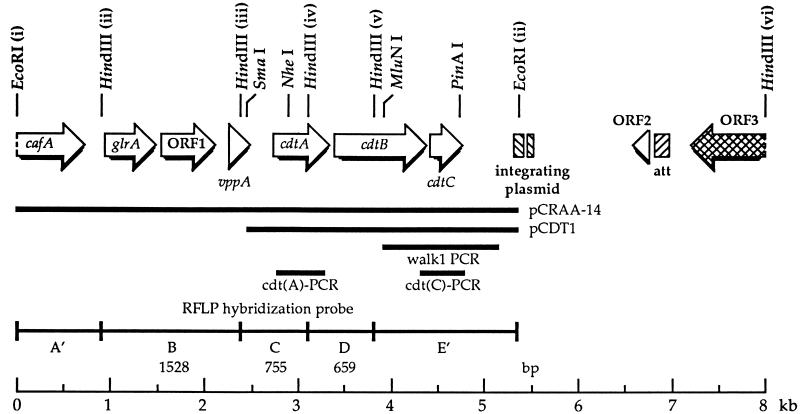
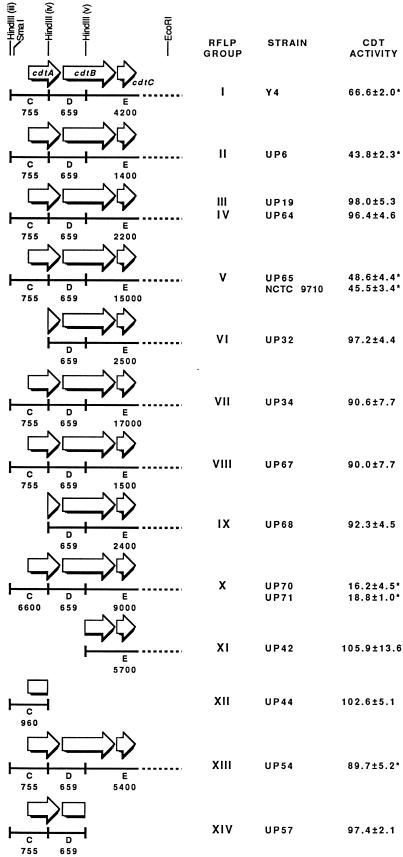
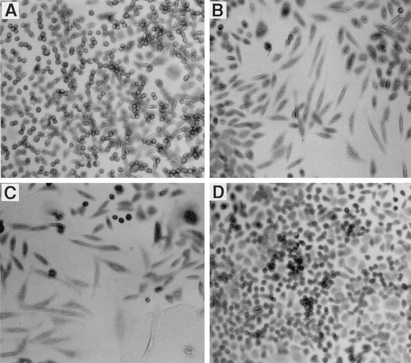
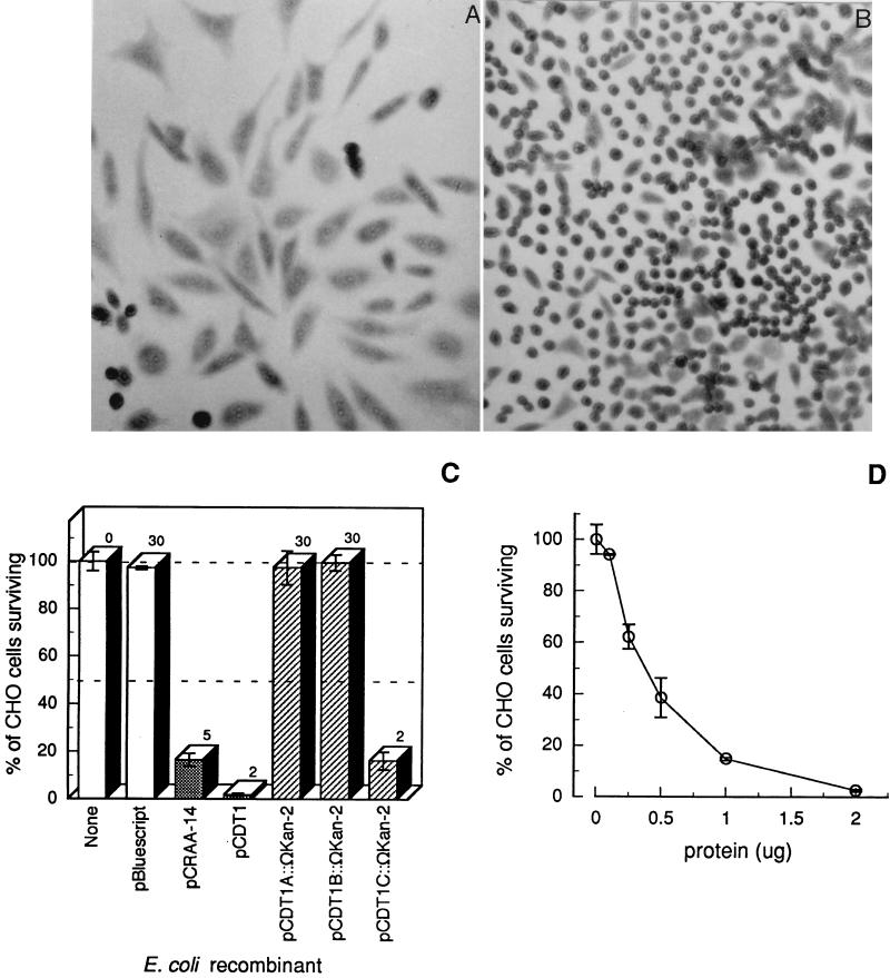
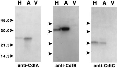
A genetic locus for a cytolethal distending toxin (CDT) was identified in a polymorphic region of the chromosome of Actinobacillus actinomycetemcomitans, a predominant oral pathogen. The locus was comprised of three open reading frames (ORFs) that had significant amino acid sequence similarity and more than 90% sequence identity to the cdtABC genes of some pathogenic Escherichia coli strains and Haemophilus ducreyi, respectively. Sonic extracts from recombinant E. coli, containing the A. actinomycetemcomitans ORFs, caused the distension and killing of Chinese hamster ovary cells characteristic of a CDT. Monoclonal antibodies made reactive with the CdtA, CdtB, and CdtC proteins of H. ducreyi recognized the corresponding gene products from the recombinant strain. CDT-like activities were no longer expressed by the recombinant strain when an OmegaKan-2 interposon was inserted into the cdtA and cdtB genes. Expression of the CDT-like activities in A. actinomycetemcomitans was strain specific. Naturally occurring expression-negative strains had large deletions within the region of the cdt locus. The cdtABC genes were flanked by an ORF (virulence plasmid protein), a partial ORF (integrase), and DNA sequences (bacteriophage integration site) characteristic of virulence-associated regions. These results provide evidence for a functional CDT in a human oral pathogen.


Figures

FIG. 1
FIG. 1
Genetic organization of the RFLP probe region from the A. actinomycetemcomitans Y4 chromosome. ORFs and the direction of transcription are designated by the large arrows. Selected restriction endonuclease sites are shown. The HindIII fragments A to E define the various RFLP groups (15). A′ and E′ signify that these end fragments extend to the next upstream and downstream HindIII sites, i and vi, respectively, on the chromosome. The numbers represent the lengths of the HindIII fragments, in base pairs. The hatched boxes indicate sequence identity with an H. influenzae integrating plasmid and bacteriophage HP1 lysogenic insertion site (19, 58). The solid lines show the regions contained in the plasmid constructs pCRAA-14 and pCDT1 and the positions of the DNA probes used for sequence walking (walk1-PCR) and RFLP variant analysis [cdt(A)-PCR and cdt(C)-PCR].
FIG. 2
FIG. 2
Relationship between the lysogenic integration site and tRNA gene sequences of H. influenzae and the homologous sequence from A. actinomycetemcomitans. Bases in lowercase differ from the attP sequence. The relative location of the A. actinomycetemcomitans sequence is shown in Fig. 1. The attB site is within the tRNA genes for lysine and leucine. Only a portion of the chromosome of H. influenzae Rd001 and the left arm of bacteriophage HP1 are shown. attP, bacteriophage HP1 attachment site sequence (19, 58); attB, H. influenzae Rd001 attachment site sequence (19, 20); Aa, A. actinomycetemcomitans sequence marked “att” in Fig. 1.
FIG. 3
FIG. 3
Comparison of the cytotoxic effects of sonic extracts from representative members of the RFLP groups of A. actinomycetemcomitans on CHO cells. CHO cells (3 × 10³/well) were seeded in 6-well tissue culture plates. Sonic extract (30 μg of protein, except for strains UP6 [14 μg] and UP57 [56 μg]) from each RFLP group variant was added at zero time. The attached cells were stained and counted after 6 days of growth. CFU were expressed as percentages of surviving cells relative to CFU of untreated CHO cell controls. Deletions in the cdt locus were identified by RFLP analyses with the 4.7-kb hybridization probe (15) and Southern blotting with cdtAB and cdtC DNA probes made by PCR (see Materials and Methods). The letters under the solid lines correspond to the RFLP HindIII DNA fragments defined in Fig. 1. Only the region of the cdt locus is shown. The numbers under the solid lines represent the sizes, in base pairs, of these DNA fragments as identified in each genetic variant. RFLP group III and IV variants differ in relation to the size of HindIII DNA fragment B (data not shown). Two genetic variants from RFLP groups V and X were examined. Values marked with an asterisk were statistically significant (P ≤ 0.05).
FIG. 4
FIG. 4
Microscopic examination of CHO cells following treatment with sonic extracts from A. actinomycetemcomitans. CHO cells (3 × 10³/well) and serial dilutions of the fractions were added to 96-well microtiter plates. The cells were stained and photographed after 3 days of growth. Shown are untreated CHO cells (A) and CHO cells treated with extract from strain Y4 (2.0 μg of protein/well) (B), an RFLP group II variant (UP6; 350 ng of protein/well) (C), and an RFLP group XIV variant (UP57; 18.7 μg of protein/well) (D).
FIG. 5
FIG. 5
Expression of CDT-like activity by recombinant E. coli strains containing the cdt ORFs. The CHO assay was performed as described in the legend to Fig. 4. (A) CHO cells treated with 169 ng of protein/well of sonic extract from E. coli DH5α(pCDT1). (B) CHO cells treated with 12 μg of protein/well of sonic extract from E. coli DH5α(pCDT1A::ΩKan-2). (C) Quantitative estimation of cytotoxicity with sonic extracts from E. coli DH5α[pBluescript II SK(+)] (30 μg of protein/well), E. coli DH5α(pCRAA-14) (5 μg of protein/well), E. coli DH5α(pCDT1) (2 μg of protein/well), E. coli DH5α(pCDT1A::ΩKan-2) (30 μg of protein/well), E. coli DH5α(pCDT1B::ΩKan-2) (30 μg of protein/well), and E. coli DH5α(pCDT1C::ΩKan-2) (2 μg of protein/well). (D) Dose-response curve. Various amounts of sonic extract (in micrograms) from E. coli DH5α(pCDT1) were added to 300 CHO cells/well and processed as described in the legend to Fig. 3. Error bars indicate standard deviations.
FIG. 6
FIG. 6
Immunoblot-based detection of recombinant Cdt proteins with monoclonal antibodies raised against the H. ducreyi Cdt proteins. Proteins present in whole cell lysates from E. coli DH5α(pPCDT3) (lanes H), E. coli DH5α(pCDT1) (lanes A), and E. coli DH5α(pBluescript II SK) (lanes V) were incubated with monoclonal antibodies reactive with the CdtA, CdtB, and CdtC proteins of H. ducreyi. The positions of molecular size markers are shown by the arrows on the left side of each blot. The sizes of the markers are shown in kilodaltons and were the same for each blot.

References

    1. Albert M J, Faruque S M, Faruque A S G, Bettelheim K A, Neogi P K B, Bhuiyan N A, Kaper J B. Controlled study of cytolethal distending toxin-producing Escherichia coli infections in Bangladeshi children. J Clin Microbiol. 1996;34:717–719. - PMC - PubMed
    1. Altschul S F, Gish W, Miller W, Myers E W, Lipman D J. Basic local alignment search tool. J Mol Biol. 1990;215:403–410. - PubMed
    1. Anderson J D, MacNab A J, Gransden W R, Damm S A M, Johnson W M, Lior H. Gastroenteritis and encephalopathy associated with a strain of Escherichia coli O55:K59:H4 that produced a cytolethal distending toxin. Pediatr Infect Dis J. 1987;6:1135–1136. - PubMed
    1. Aslund F, Ehn B, Miranda-Vizuete A, Pueyo C, Holmgren A. Two additional glutaredoxins exist in Escherichia coli: glutaredoxin 3 is a hydrogen donor for ribonucleotide reductase in a thioredoxin/glutaredoxin 1 double mutant. Proc Natl Acad Sci USA. 1994;91:9813–9817. - PMC - PubMed
    1. Baehni P C, Tsai C-C, McArthur W P, Hammond B F, Taichman N S. Interaction of inflammatory cells and oral microorganisms. VIII. Detection of leukotoxic activity of a plaque-derived gram-negative microorganism. Infect Immun. 1979;24:233–243. - PMC - PubMed

Publication types

Substances

Associated data

LinkOut - more resources