. 1999 Mar;67(3):1493-500.
doi: 10.1128/IAI.67.3.1493-1500.1999.

Probing the function of Bordetella bronchiseptica adenylate cyclase toxin by manipulating host immunity

Affiliations

Probing the function of Bordetella bronchiseptica adenylate cyclase toxin by manipulating host immunity

E T Harvill et al. Infect Immun. 1999 Mar.

Abstract

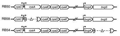
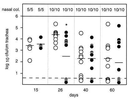
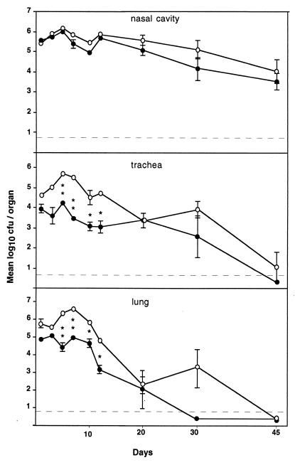
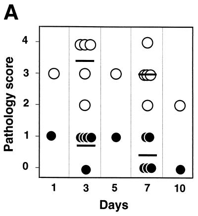
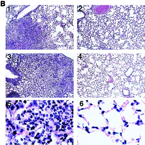
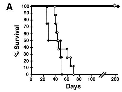
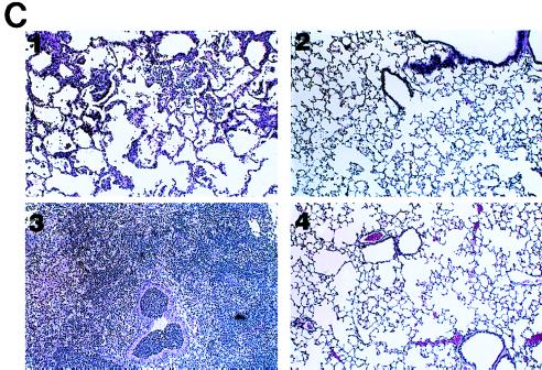
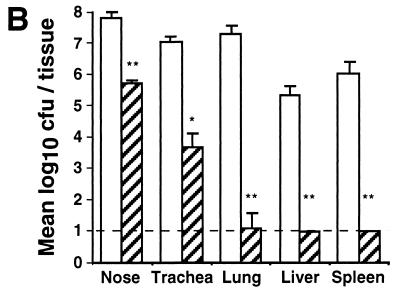
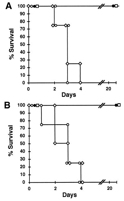
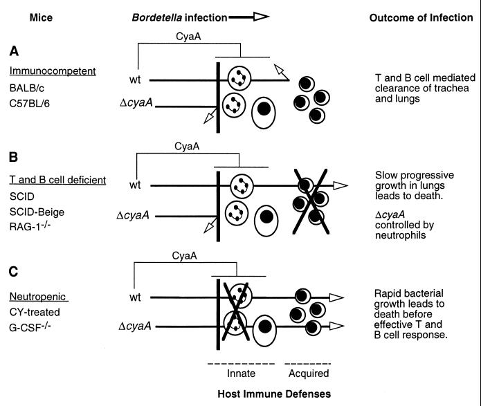
We have examined the role of adenylate cyclase-hemolysin (CyaA) by constructing an in-frame deletion in the Bordetella bronchiseptica cyaA structural gene and comparing wild-type and cyaA deletion strains in natural host infection models. Both the wild-type strain RB50 and its adenylate cyclase toxin deletion (DeltacyaA) derivative efficiently establish persistent infections in rabbits, rats, and mice following low-dose inoculation. In contrast, an inoculation protocol that seeds the lower respiratory tract revealed significant differences in bacterial numbers and in polymorphonuclear neutrophil recruitment in the lungs from days 5 to 12 postinoculation. We next explored the effects of disarming specific aspects of the immune system on the relative phenotypes of wild-type and DeltacyaA bacteria. SCID, SCID-beige, or RAG-1(-/-) mice succumbed to lethal systemic infection following high- or low-dose intranasal inoculation with the wild-type strain but not the DeltacyaA mutant. Mice rendered neutropenic by treatment with cyclophosphamide or by knockout mutation in the granulocyte colony-stimulating factor locus were highly susceptible to lethal infection by either wild-type or DeltacyaA strains. These results reveal the significant role played by neutrophils early in B. bronchiseptica infection and by acquired immunity at later time points and suggest that phagocytic cells are a primary in vivo target of the Bordetella adenylate cyclase toxin.


Figures

FIG. 1
FIG. 1
Genotypes of B. bronchiseptica strains. RB58 contains an in-frame deletion in the cyaA structural gene. RB54 contains an in-frame deletion in bvgS, resulting in the loss of expression of virulence-associated genes.
FIG. 2
FIG. 2
Cytotoxicity of wt and mutant B. bronchiseptica strains for J774 cells. Bacteria were incubated with J774 cells at an MOI of 10 for 4 h. Cytotoxicity was determined by the release of lactate dehydrogenase as measured with the Cytotox96 kit, and the means and standard errors are presented as percentages of the total lysis by detergent. All strains differ from one another (P < 0.001), except for the wt and ΔcyaA strains and the ΔbscNΔcyaA and Δbvg strains.
FIG. 3
FIG. 3
Time course of rat respiratory tract colonization by wt and ΔcyaA B. bronchiseptica. Wistar rats were inoculated intranasally with 50 μl of PBS containing 5 × 10⁵ CFU of either RB50 (wt; open circles) or RB58 (ΔcyaA; solid circles), and the numbers of bacteria present in the trachea were determined at the indicated times postinoculation. Each symbol represents a single animal, bars represent the means, and the dashed line represents the lower level of detection. Nasal colonization (col.) was determined by nasal swab and is presented as the number of B. bronchiseptica-colonized animals over the total number of rats. ∗, P < 0.01.
FIG. 4
FIG. 4
Time course of mouse respiratory tract colonization by wt and ΔcyaA B. bronchiseptica. Groups of three female, 4-week-old, BALB/c mice were inoculated intranasally with 50 μl of PBS containing 5 × 10⁵ CFU of either RB50 (wt; open circles) or RB58 (ΔcyaA; solid circles), and the numbers of CFU present in the nasal cavity, trachea, and lungs were determined at the indicated times postinoculation. ∗, P < 0.01. ∗∗, P < 0.001.
FIG. 5
FIG. 5
Mouse lung pathology induced by wt and ΔcyaA B. bronchiseptica. BALB/c mice were inoculated in parallel with those for Fig. 3. Histological sections of lung tissues were prepared as described in Materials and Methods. (A) Samples were submitted for observation in a blinded fashion. Upon examination, samples were given scores of 0 (no pathology), 1 (mild inflammation in ≤10% of the bronchioles and/or ≤10% of lung tissue), 2 (inflammation in 10 to 30% of bronchioles and/or mild inflammation in 10 to 30% of lung tissue), 3 (inflammation in >30% of bronchioles and mild to moderate inflammation in >30% of lung tissue), and 4 (inflammation in >50% of bronchioles and moderate to severe inflammation in >30% of lung tissue). Bars indicate the averages of five animals. Open circles represent animals infected with RB50 (wt), and solid circles represent animals infected with RB58 (ΔcyaA). (B) Representative sections of lungs infected with wt (panels 1 and 3) or ΔcyaA (panels 2 and 4) bacteria on day 3 (panels 1 and 2) or day 7 (panels 3 and 4). Magnification, ×100 (panels 1 to 4) and ×1,000 (panels 5 and 6).
FIG. 5
FIG. 5
Mouse lung pathology induced by wt and ΔcyaA B. bronchiseptica. BALB/c mice were inoculated in parallel with those for Fig. 3. Histological sections of lung tissues were prepared as described in Materials and Methods. (A) Samples were submitted for observation in a blinded fashion. Upon examination, samples were given scores of 0 (no pathology), 1 (mild inflammation in ≤10% of the bronchioles and/or ≤10% of lung tissue), 2 (inflammation in 10 to 30% of bronchioles and/or mild inflammation in 10 to 30% of lung tissue), 3 (inflammation in >30% of bronchioles and mild to moderate inflammation in >30% of lung tissue), and 4 (inflammation in >50% of bronchioles and moderate to severe inflammation in >30% of lung tissue). Bars indicate the averages of five animals. Open circles represent animals infected with RB50 (wt), and solid circles represent animals infected with RB58 (ΔcyaA). (B) Representative sections of lungs infected with wt (panels 1 and 3) or ΔcyaA (panels 2 and 4) bacteria on day 3 (panels 1 and 2) or day 7 (panels 3 and 4). Magnification, ×100 (panels 1 to 4) and ×1,000 (panels 5 and 6).
FIG. 6
FIG. 6
Infection of SCID-Beige mice with wt and ΔcyaA B. bronchiseptica. (A) Four-week-old SCID-Beige mice were inoculated with B. bronchiseptica, and percent survival over time is shown. Open symbols represent groups of eight animals inoculated with 103 CFU of either RB50 (wt; circles) or RB58 (ΔcyaA; diamonds) delivered in a 5-μl droplet to the external nares. Solid symbols represent groups of four mice inoculated with 5 × 10⁵ CFU of either RB50 (wt; circles) or RB58 (ΔcyaA; diamonds) delivered in a 50-μl volume to the respiratory tract via the nares. (B) Colonization of various tissues by wt and ΔcyaA B. bronchiseptica. Groups of three 4-week-old, female, SCID-Beige mice were inoculated with 1,000 CFU of either RB50 (wt; open bars) or RB58 (ΔcyaA; hatched bars) delivered in a 5-μl droplet to the external nares. Mice were sacrificed at 45 days postinoculation, and colonization levels in the nasal cavity, trachea, lungs, liver, and spleen were determined. Mean log10 CFU per organ or tissue section ± 1 standard error are shown. The dashed line represents the lower limit of detection. ∗, P < 0.01. ∗∗, P < 0.001. (C) Representative sections of SCID-Beige mouse lungs infected with wt (panels 1 and 3) or ΔcyaA (panels 2 and 4) bacteria on day 3 (panels 1 and 2) or day 7 (panels 3 and 4). Magnification, ×100.
FIG. 6
FIG. 6
Infection of SCID-Beige mice with wt and ΔcyaA B. bronchiseptica. (A) Four-week-old SCID-Beige mice were inoculated with B. bronchiseptica, and percent survival over time is shown. Open symbols represent groups of eight animals inoculated with 103 CFU of either RB50 (wt; circles) or RB58 (ΔcyaA; diamonds) delivered in a 5-μl droplet to the external nares. Solid symbols represent groups of four mice inoculated with 5 × 10⁵ CFU of either RB50 (wt; circles) or RB58 (ΔcyaA; diamonds) delivered in a 50-μl volume to the respiratory tract via the nares. (B) Colonization of various tissues by wt and ΔcyaA B. bronchiseptica. Groups of three 4-week-old, female, SCID-Beige mice were inoculated with 1,000 CFU of either RB50 (wt; open bars) or RB58 (ΔcyaA; hatched bars) delivered in a 5-μl droplet to the external nares. Mice were sacrificed at 45 days postinoculation, and colonization levels in the nasal cavity, trachea, lungs, liver, and spleen were determined. Mean log10 CFU per organ or tissue section ± 1 standard error are shown. The dashed line represents the lower limit of detection. ∗, P < 0.01. ∗∗, P < 0.001. (C) Representative sections of SCID-Beige mouse lungs infected with wt (panels 1 and 3) or ΔcyaA (panels 2 and 4) bacteria on day 3 (panels 1 and 2) or day 7 (panels 3 and 4). Magnification, ×100.
FIG. 7
FIG. 7
Comparison of wt and ΔcyaA B. bronchiseptica infections of neutropenic mice. Groups of four BALB/c mice rendered neutropenic by cyclophosphamide treatment (A) or G-CSF−/− mice (B) were inoculated with 5 × 10⁵ CFU of either RB50 (wt; open circles), RB58 (ΔcyaA; open diamonds), RB54 (ΔbvgS; solid circles), or PBS (open squares) delivered in a 50-μl volume. The percentages of animals (n = 4) surviving over time are indicated.
FIG. 8
FIG. 8
Model of the role of CyaA in infection of immunocompetent (A), T- and B-cell-deficient (B), and neutropenic (C) mice. See the text for description.

References

    1. Akerley B J, Cotter P A, Miller J F. Ectopic expression of the flagellar regulon alters development of the Bordetella-host interaction. Cell. 1995;80:611–620. - PubMed
    1. Appelberg R, Castro A G, Gomes S, Pedrosa J, Silva M T. Susceptibility of beige mice to Mycobacterium avium: role of neutrophils. Infect Immun. 1995;63:3381–3387. - PMC - PubMed
    1. Betsou F, Sismeiro O, Danchin A, Guiso N. Cloning and sequence of the Bordetella bronchiseptica adenylate cyclase-hemolysin-encoding gene: comparison with the Bordetella pertussis gene. Gene. 1995;162:165–166. - PubMed
    1. Confer D L, Eaton J W. Phagocyte impotence caused by an invasive bacterial adenylate cyclase. Science. 1982;217:948–950. - PubMed
    1. Cotter P A, Miller J F. BvgAS-mediated signal transduction: analysis of phase-locked regulatory mutants of Bordetella bronchiseptica in a rabbit model. Infect Immun. 1994;62:3381–3390. - PMC - PubMed

Publication types

LinkOut - more resources