

Oesophageal epithelial innervation in health and reflux oesophagitis
- PMID: 10026314
- PMCID: PMC1727420
- DOI: 10.1136/gut.44.3.317
Oesophageal epithelial innervation in health and reflux oesophagitis
Abstract
Background: The response of the oesophagus to refluxed gastric contents is likely to depend on intact neural mechanisms in the oesophageal mucosa. The epithelial innervation has not been systematically evaluated in health or reflux disease.
Aims: To study oesophageal epithelial innervation in controls, and also inflamed and non-inflamed mucosa in patients with reflux oesophagitis and healed oesophagitis.
Patients: Ten controls, nine patients with reflux oesophagitis, and five patients with healed oesophagitis.
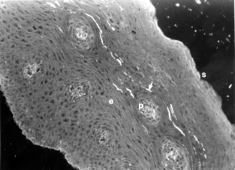
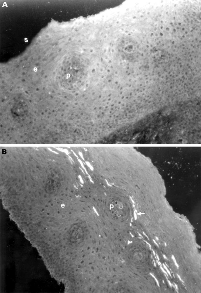
Methods: Oesophageal epithelial biopsy specimens were obtained at endoscopy. The distribution of the neuronal marker protein gene product 9.5 (PGP), and the neuropeptides calcitonin gene related peptide (CGRP), neuropeptide Y (NPY), substance P (SP), and vasoactive intestinal peptide (VIP) were investigated by immunohistochemistry. Density of innervation was assessed by the proportion of papillae in each oesophageal epithelial biopsy specimen containing immunoreactive fibres (found in the subepithelium and epithelial papillae, but not penetrating the epithelium).
Results: The proportion of papillae positive for PGP immunoreactive nerve fibres was significantly increased in inflamed tissue when compared with controls, and non-inflamed and healed tissue. There was also a significant increase in VIP immunoreactive fibres within epithelial papillae. Other neuropeptides showed no proportional changes in inflammation.
Conclusions: Epithelial biopsy specimens can be used to assess innervation in the oesophagus. The innervation of the oesophageal mucosa is not altered in non-inflamed tissue of patients with oesophagitis but alters in response to inflammation, where there is a selective increase (about three- to fourfold) in VIP containing nerves.
Figures
Similar articles
-
Increased capsaicin receptor TRPV1 nerve fibres in the inflamed human oesophagus.Eur J Gastroenterol Hepatol. 2004 Sep;16(9):897-902. doi: 10.1097/00042737-200409000-00014. Eur J Gastroenterol Hepatol. 2004. PMID: 15316415
-
Evaluation of Helicobacter pylori in reflux oesophagitis and Barrett's oesophagus.Gut. 1997 Jan;40(1):9-13. doi: 10.1136/gut.40.1.9. Gut. 1997. PMID: 9155568 Free PMC article.
-
Increased expression of epidermal growth factor receptors in basal cell hyperplasia of the oesophagus after acid reflux oesophagitis in rats.Aliment Pharmacol Ther. 2002 Apr;16 Suppl 2:52-8. doi: 10.1046/j.1365-2036.16.s2.29.x. Aliment Pharmacol Ther. 2002. PMID: 11966524
-
Eosinophilic oesophagitis versus reflux oesophagitis.Acta Gastroenterol Belg. 2011 Jun;74(2):323-9. Acta Gastroenterol Belg. 2011. PMID: 21861318 Review.
-
Oesophageal damage and defence in reflux oesophagitis: pathophysiological and cell biological mechanisms.Prog Histochem Cytochem. 1997;32(3-4):1-42. doi: 10.1016/s0079-6336(97)80005-8. Prog Histochem Cytochem. 1997. PMID: 9551487 Review. No abstract available.
Cited by
-
Gastroesophageal Reflux and Its Association With Atrial Fibrillation: A Traditional Review.Cureus. 2020 Sep 11;12(9):e10387. doi: 10.7759/cureus.10387. Cureus. 2020. PMID: 33062508 Free PMC article. Review.
-
Neuroepithelial control of mucosal inflammation in acute cystitis.Sci Rep. 2018 Jul 20;8(1):11015. doi: 10.1038/s41598-018-28634-0. Sci Rep. 2018. PMID: 30030504 Free PMC article.
-
Acid burn or cytokine sizzle in the pathogenesis of heartburn?J Gastroenterol Hepatol. 2013 Mar;28(3):385-7. doi: 10.1111/jgh.12103. J Gastroenterol Hepatol. 2013. PMID: 23441717 Free PMC article. No abstract available.
-
Distinct afferent innervation patterns within the human proximal and distal esophageal mucosa.Am J Physiol Gastrointest Liver Physiol. 2015 Mar 15;308(6):G525-31. doi: 10.1152/ajpgi.00175.2014. Epub 2015 Jan 8. Am J Physiol Gastrointest Liver Physiol. 2015. PMID: 25573174 Free PMC article.
-
The oesophagus as an immune organ.Nat Rev Gastroenterol Hepatol. 2025 Jun 18. doi: 10.1038/s41575-025-01086-4. Online ahead of print. Nat Rev Gastroenterol Hepatol. 2025. PMID: 40533621 Review.
References
MeSH terms
Substances
LinkOut - more resources
Full Text Sources
Research Materials
Miscellaneous