Comparative Study
. 1999 Mar;44(3):361-5.
doi: 10.1136/gut.44.3.361.

A blind comparison of the effectiveness of endoscopic ultrasonography and endoscopy in staging early gastric cancer

Affiliations
Comparative Study

A blind comparison of the effectiveness of endoscopic ultrasonography and endoscopy in staging early gastric cancer

H Yanai et al. Gut. 1999 Mar.

Abstract

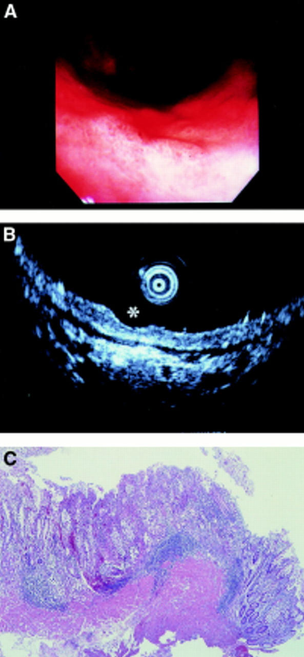
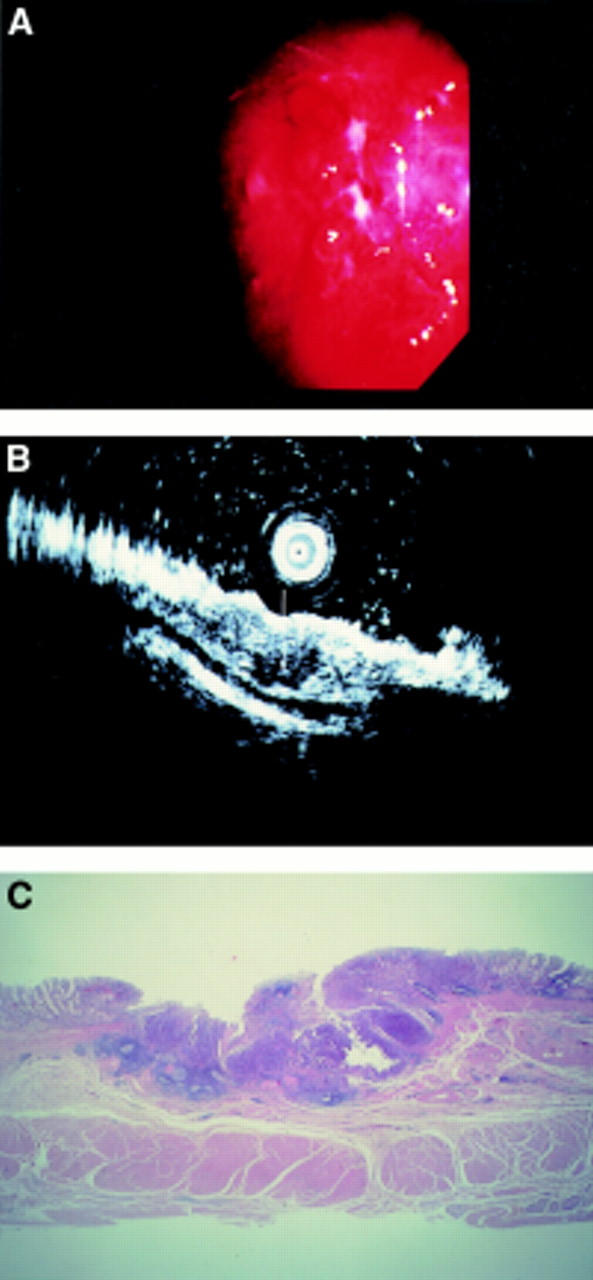
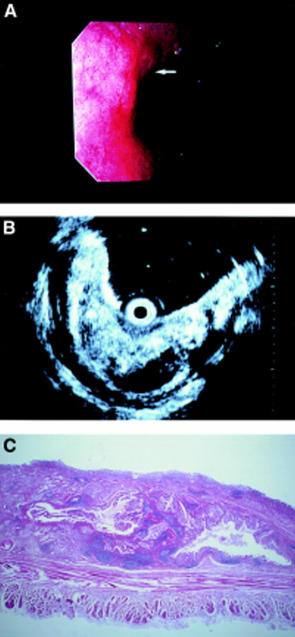
Background/aims: Endoscopic ultrasonography is expected to be useful for invasion depth staging of early gastric cancer. A prospective blind study of the staging characteristics of endoscopy and endoscopic ultrasonography for early gastric cancer was performed.

Methods: Findings of endoscopy and endoscopic ultrasonography using a 20 MHz thin ultrasound probe were independently reviewed and the results of 52 early gastric cancer lesions analysed.

Results: The overall accuracy rates in invasion depth staging of early gastric cancer were 63% for endoscopy and 71% for endoscopic ultrasonography. No statistically significant differences were observed in overall accuracy. Endoscopic ultrasonography tended to overstage, and lesions that were classified as mucosal cancer by endoscopic ultrasonography were very likely (95%) to be limited to the mucosa on histological examination. All 16 lesions staged as mucosal cancer independently but coincidentally by both methods were histologically limited to the mucosa.

Conclusions: Endoscopic ultrasonography is expected to compensate for the understaging of lesions with submucosal invasion that are endoscopically staged as mucosal cancer.


Figures

Figure 1
Figure 1
A superficial depressed type early gastric cancer located in the lower gastric body. Endoscopy (A) and endoscopic ultrasonography (B, asterisk) reviewers independently but coincidentally staged this as mucosal. Histological examination (C) of its endoscopically resected strip biopsy specimen also proved that it was mucosal cancer. In this case, both methods staged correctly.
Figure 2
Figure 2
A superficial depressed type early gastric cancer located in the middle gastric body. This case was understaged by endoscopy. The endoscopy reviewer observed that the surface of the depression was not so rough, and staged it as endoscopy-mucosal (A). In contrast, the endoscopic ultrasonography (EUS) reviewer observed hypoechoic tumorous invasion and irregular narrowing of the third layer (B, white arrow). He staged it as depth EUS-submucosal. Histological findings of a surgically resected specimen corresponded well to the EUS image analysis (C).
Figure 3
Figure 3
A superficial depressed type early gastric cancer located in the cardia. This was also understaged by endoscopy. As the lesser curvature of the cardia is a difficult location for a forward viewing type endoscope, it is likely that the endoscopy reviewer could not detect any apparent submucosal invasion (A, white arrow). In contrast, the endoscopic ultrasonography (EUS) picture provided a clear image of rough swelling and irregular cystic changes of the third layer (B). Histological examination (C) confirmed its submucosal invasion, which corresponded well to the EUS findings.

References

    1. Hepatogastroenterology. 1989 Apr;36(2):51-6 - PubMed
    1. Gastroenterology. 1989 Feb;96(2 Pt 1):433-41 - PubMed
    1. Gut. 1991 May;32(5):479-82 - PubMed
    1. Endoscopy. 1992 May;24 Suppl 1:297-303 - PubMed
    1. Endoscopy. 1992 May;24 Suppl 1:329-33 - PubMed

Publication types