. 1999 Feb;154(2):375-84.
doi: 10.1016/S0002-9440(10)65284-4.

Vascular endothelial growth factor (VEGF)-mediated angiogenesis is associated with enhanced endothelial cell survival and induction of Bcl-2 expression

Affiliations

Vascular endothelial growth factor (VEGF)-mediated angiogenesis is associated with enhanced endothelial cell survival and induction of Bcl-2 expression

J E Nör et al. Am J Pathol. 1999 Feb.

Abstract

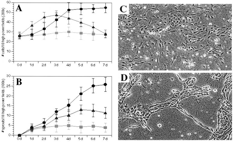
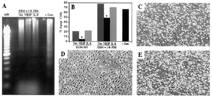
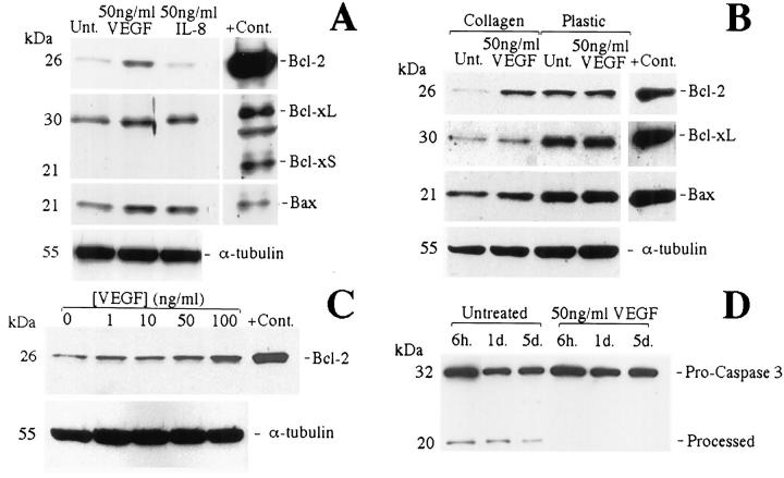
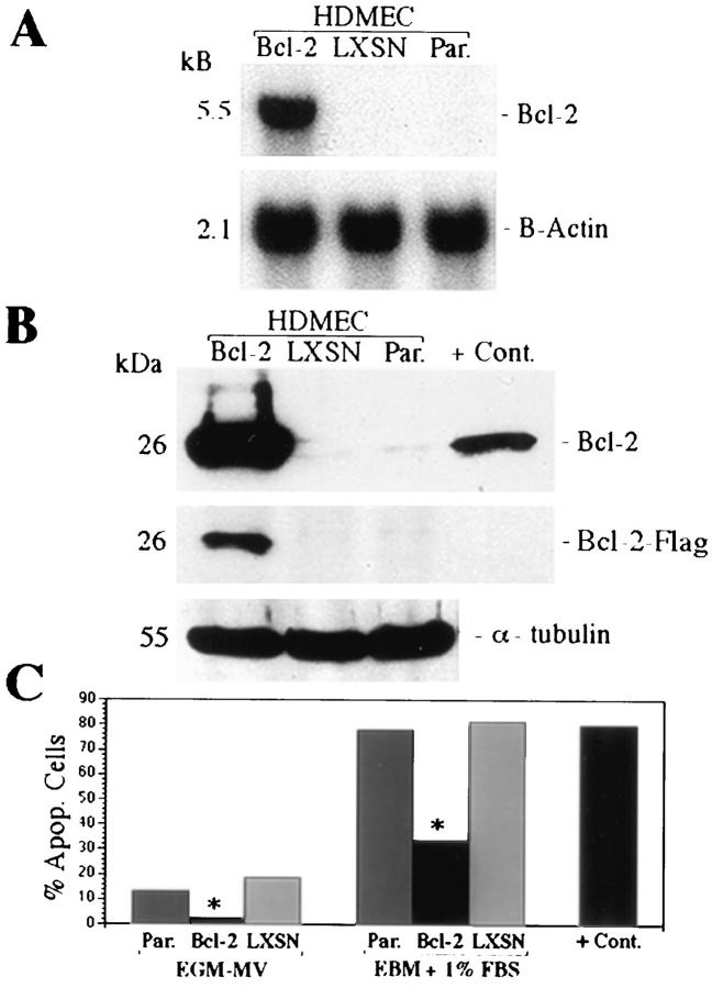
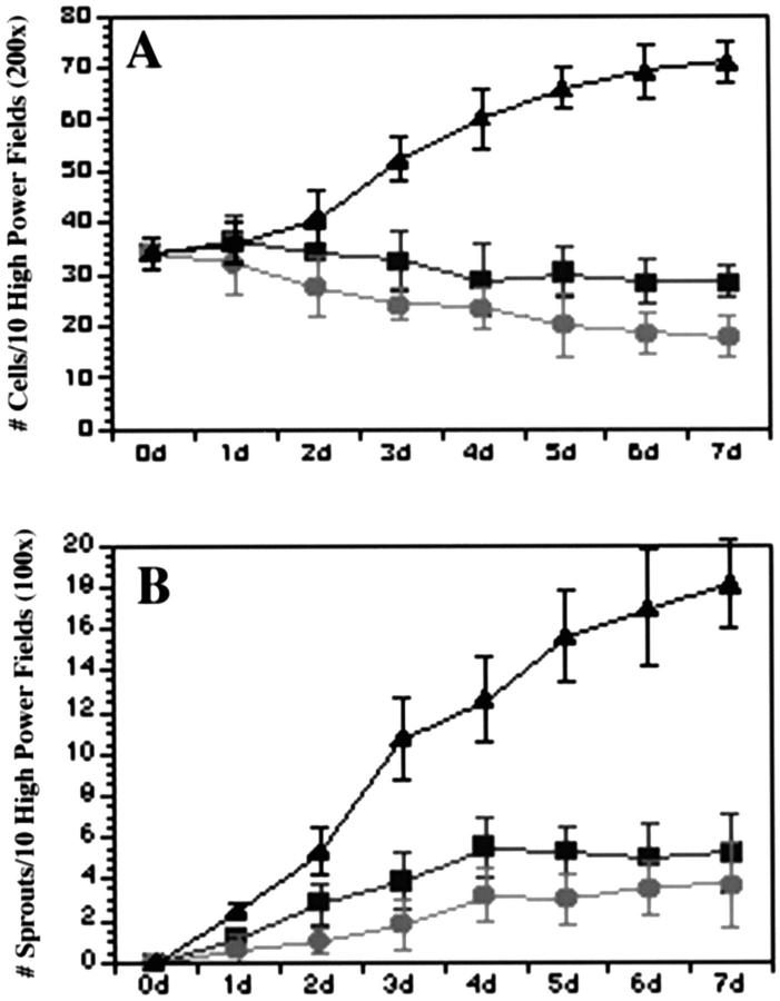
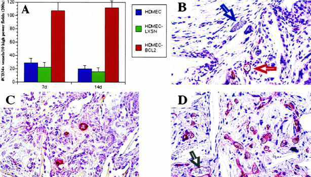
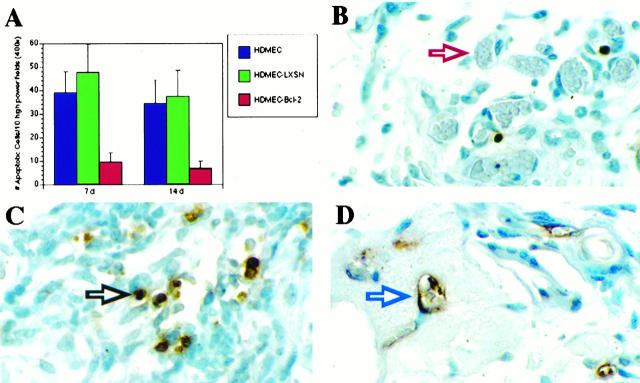
Vascular endothelial growth factor (VEGF) is an endothelial cell mitogen and permeability factor that is potently angiogenic in vivo. We report here studies that suggest that VEGF potentiates angiogenesis in vivo and prolongs the survival of human dermal microvascular endothelial cells (HDMECs) in vitro by inducing expression of the anti-apoptotic protein Bcl-2. Growth-factor-enriched and serum-deficient cultures of HDMECs grown on collagen type I gels with VEGF exhibited a 4-fold and a 1.6-fold reduction, respectively, in the proportion of apoptotic cells. Enhanced HDMEC survival was associated with a dose-dependent increase in Bcl-2 expression and a decrease in the expression of the processed forms of the cysteine protease caspase-3. Cultures of HDMECs transduced with and overexpressing Bcl-2 and deprived of growth factors showed enhanced protection from apoptosis and exhibited a twofold increase in cell number and a fourfold increase in the number of capillary-like sprouts. HDMECs overexpressing Bcl-2 when incorporated into polylactic acid sponges and implanted into SCID mice exhibited a sustained fivefold increase in the number of microvessels and a fourfold decrease in the number of apoptotic cells when examined 7 and 14 days later. These results suggest that the angiogenic activity attributed to VEGF may be due in part to its ability to enhance endothelial cell survival by inducing expression of Bcl-2.


Figures

Figure 1.
Figure 1.
Effect of VEGF on HDMEC proliferation and sprout formation in culture. VEGF was more potent than IL-8 and untreated controls in inducing HDMEC proliferation (A) and sprout formation (B to D). HDMECs were plated on type I collagen and cultured in complete EGM-MV in the presence of 50 ng/ml VEGF (•) or 50 ng/ml IL-8 (▴) or in the absence of additional cytokines (▪). C and D: Representative microscopic field (×200) of HDMECs seeded in collagen for 5 days in the absence of additional cytokines (C) or fed with EGM-MV supplemented with 50 ng/ml VEGF (D). At daily intervals, the number of cells and sprouts was counted in 10 random fields from three independent experiments.
Figure 2.
Figure 2.
Effect of VEGF and IL-8 on HDMEC apoptosis in culture. VEGF prevents DNA fragmentation, as shown in the DNA ladder assay (A) and in TUNEL assay followed by flow cytometry (B) and prevents cell detachment from collagen (C to E). DNA ladder assay (A) was performed with DNA extracted from HDMECs cultured 3 days on type I collagen and fed with EBM supplemented with 1% FBS in the presence of 50 ng/ml VEGF or 50 ng/ml IL-8 or untreated (ie, in the absence of additional cytokines). Flow cytometry (B) was performed with HDMECs cultured 3 days on type I collagen and fed either with complete EGM-MV (first three columns) or EBM supplemented with 1% FBS (second three columns) in the presence of 50 ng/ml VEGF, 50 ng/ml IL-8, or untreated. *Statistically different (P ≤ 0.01). The positive controls for apoptosis (A and B) were HDMECs cultured in suspension in 1.68% methylcellulose for 72 hours. Microscopic appearance of HDMECs cultured 3 days on type I collagen and fed with EBM supplemented with 1% FBS in absence of additional angiogenic factors (C) or in the presence of 50 ng/ml VEGF (D) or 50 ng/ml IL-8 (E).
Figure 3.
Figure 3.
VEGF induces Bcl-2 expression in HDMECs grown on collagen and prevents caspase-3 cleavage. Western blots of whole-cell lysates from HDMECs fed with complete EGM-MV and cultured 3 days on type I collagen in the presence of VEGF or IL-8 or untreated (A), cultured 3 days on type I collagen or plastic surface in the presence of VEGF or untreated (B), cultured 3 days on type I collagen in the presence of increasing concentrations of VEGF (C), or cultured on type I collagen in the presence of VEGF or untreated (D). Positive controls were whole-cell lysates from cells transduced with Bcl-2, Bcl-xL/S, or Bax.
Figure 4.
Figure 4.
Overexpression of Bcl-2 in endothelial cells increases survival. Northern (A) and Western (B) blot analyses of HDMECs stably transduced with Bcl-2 (HDMEC-Bcl-2), vector only (HDMEC-LXSN), or parental HDMEC (untransduced) were performed to confirm expression of Bcl-2. TUNEL assay followed by flow cytometry (C) was performed with cells cultured 3 days on type I collagen and fed either with complete EGM-MV (first three columns) or EBM supplemented with 1% FBS (second three columns). *Statistically different (P ≤ 0.01). Positive controls were HDMECs cultured in suspension in 1.68% methylcellulose for 72 hours (C).
Figure 5.
Figure 5.
Effect of Bcl-2 on endothelial cell proliferation and number of sprouts in culture. HDMECs overexpressing Bcl-2 show enhanced cell proliferation (A) and sprout formation (B). Endothelial cells transduced with Bcl-2 (▴), vector only (•), or parental (untransduced) cells (▪) were plated on type I collagen and cultured in complete EGM-MV. At daily intervals, the number of cells and sprouts was counted in 10 random fields from three independent experiments.
Figure 6.
Figure 6.
HDMECs overexpressing Bcl-2 show enhanced angiogenesis in SCID mice. HDMEC-Bcl-2, HDMEC-LXSN, or HDMEC were seeded in PLA sponges and implanted in SCID mice. After 7 or 14 days, the sponges were retrieved and stained with anti-CD34, and the number of CD34+ blood vessels was counted in 10 random fields from three independent sponges per time point and cell type (A). Photomicrograph from histological sections show anti-CD34 staining of HDMECs in the interior of the sponge (red arrow) and the absence of staining of mouse blood vessels (blue arrow) in the surrounding connective tissue (B), sponges seeded with HDMECs had fewer CD34+ blood vessels after 14 days (C), compared with sponges seeded with HDMEC-Bcl-2 after the same time period (D). Human blood vessels in the interior of the sponge showing blood-filled lumens (gray arrow). All photomicrographs were at ×200 magnification.
Figure 7.
Figure 7.
Apoptosis in sponges implanted in SCID mice. HDMEC-Bcl-2, HDMEC-LXSN, or HDMECs were seeded in PLA sponges and implanted subcutaneously in the back of the SCID mice. After 7 or 14 days, the sponges were retrieved, stained with the ApopTag peroxidase in situ kit. The number of TUNEL-positive cells was counted in 10 random fields from three independent sponges per time point and experimental condition (A). Photomicrographs from histological sections show sponges seeded with HDMEC-Bcl-2 had fewer TUNEL-positive cells (apoptotic cells) 14 days after implantation (B), compared with sponges seeded with HDMECs (C and D) after the same time period. The red arrow in B points to a TUNEL-negative microvessel populated by HDMEC-Bcl-2 cells. The black arrow in C points to a TUNEL-positive stromal cell in the sponge interior, and the blue arrow in D points to a TUNEL-positive microvessel populated by untransduced HDMECs. All photomicrographs were at ×1000 magnification.

References

    1. Ferrara N: Vascular endothelial growth factor. Eur J Cancer 1996, 32A:2413-2422 - PubMed
    1. Plate KH, Breiser G, Weich HA, Risau W: Vascular endothelial growth factor is a potential tumor angiogenesis factor in vivo. Nature 1992, 359:845-848 - PubMed
    1. Hashimoto M, Ohsawa M, Ohnishi A, Naka N, Hirota S, Kitamura Y, Aosaza K: Expression of vascular endothelial growth factor and its receptor mRNA in angiosarcoma. Lab Invest 1995, 73:859-863 - PubMed
    1. Benjamin LE, Keshet E: Conditional switching of vascular endothelial growth factor (VEGF) expression in tumors: induction of endothelial cell shedding and regression of hemangioblastoma-like vessels by VEGF withdrawal. Proc Natl Acad Sci USA 1997, 94:8761-8766 - PMC - PubMed
    1. Brown LF, Yeo KY, Berse B, Yeo TK, Senger DR, Dvorak HF, Van de Water L: Expression of vascular permeability factor (vascular endothelial growth factor) by epidermal keratinocytes during wound healing. J Exp Med 1992, 176:1375-1379 - PMC - PubMed

Publication types

MeSH terms

Substances