. 1999 Feb;154(2):385-94.
doi: 10.1016/S0002-9440(10)65285-6.

Angiosarcomas express mixed endothelial phenotypes of blood and lymphatic capillaries: podoplanin as a specific marker for lymphatic endothelium

Affiliations

Angiosarcomas express mixed endothelial phenotypes of blood and lymphatic capillaries: podoplanin as a specific marker for lymphatic endothelium

S Breiteneder-Geleff et al. Am J Pathol. 1999 Feb.

Abstract

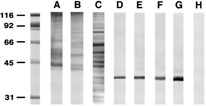
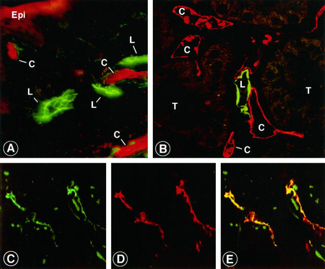
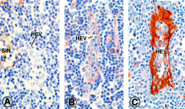
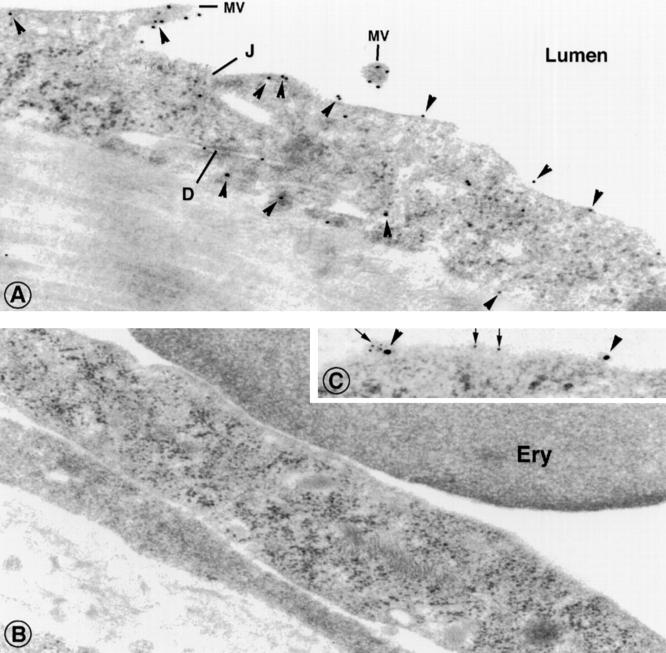
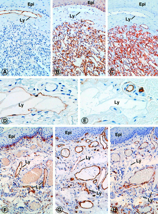
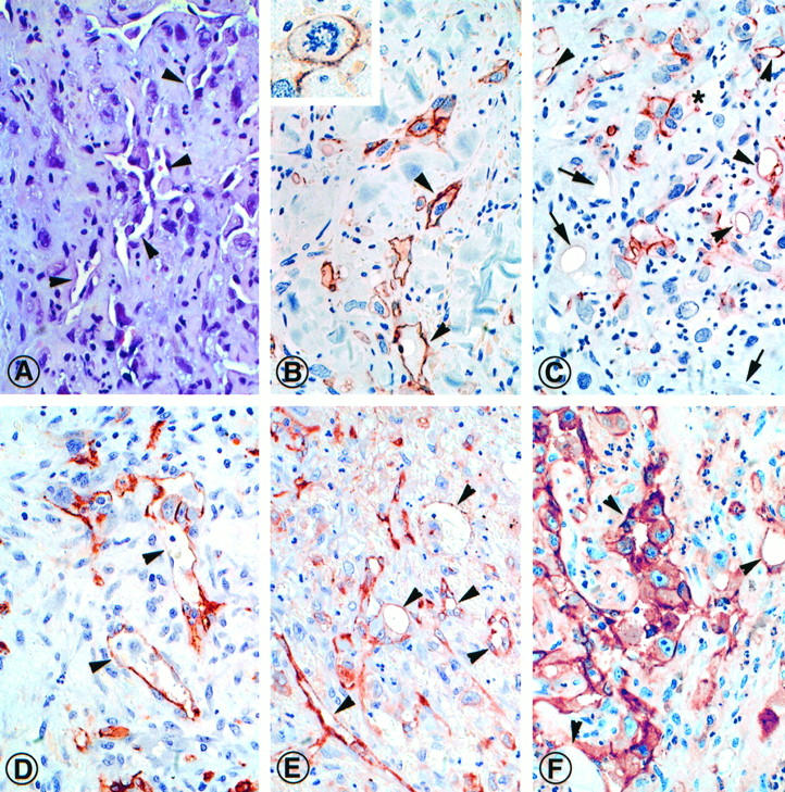
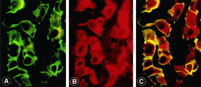
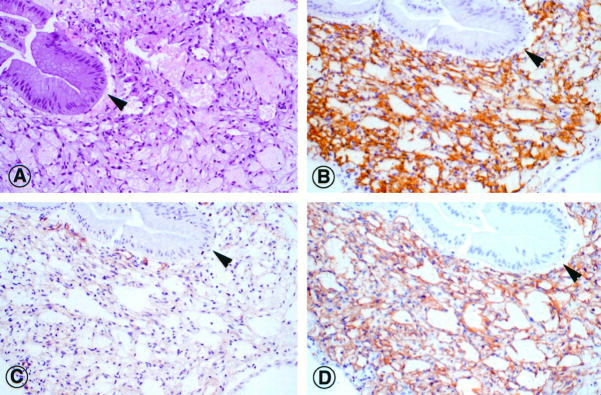
Angiosarcomas apparently derive from blood vessel endothelial cells; however, occasionally their histological features suggest mixed origin from blood and lymphatic endothelia. In the absence of specific positive markers for lymphatic endothelia the precise distinction between these components has not been possible. Here we provide evidence by light and electron microscopic immunohistochemistry that podoplanin, a approximately 38-kd membrane glycoprotein of podocytes, is specifically expressed in the endothelium of lymphatic capillaries, but not in the blood vasculature. In normal skin and kidney, podoplanin colocalized with vascular endothelial growth factor receptor-3, the only other lymphatic marker presently available. Complementary immunostaining of blood vessels was obtained with established endothelial markers (CD31, CD34, factor VIII-related antigen, and Ulex europaeus I lectin) as well as podocalyxin, another podocytic protein that is also localized in endothelia of blood vessels. Podoplanin specifically immunolabeled endothelia of benign tumorous lesions of undisputed lymphatic origin (lymphangiomas, hygromas) and was detected there as a 38-kd protein by immunoblotting. As paradigms of malignant vascular tumors, poorly differentiated (G3) common angiosarcomas (n = 8), epitheloid angiosarcomas (n = 3), and intestinal Kaposi's sarcomas (n = 5) were examined for their podoplanin content in relation to conventional endothelial markers. The relative number of tumor cells expressing podoplanin was estimated and, although the number of cases in this preliminary study was limited to 16, an apparent spectrum of podoplanin expression emerged that can be divided into a low-expression group in which 0-10% of tumor cells contained podoplanin, a moderate-expression group with 30-60% and a high-expression group with 70-100%. Ten of eleven angiosarcomas and all Kaposi's sarcomas showed mixed expression of both lymphatic and blood vascular endothelial phenotypes. By double labeling, most podoplanin-positive tumor cells coexpressed endothelial markers of blood vessels, whereas few tumor cells were positive for individual markers only. From these results we conclude that (1) podoplanin is a selective marker of lymphatic endothelium; (2) G3 angiosarcomas display a quantitative spectrum of podoplanin-expressing tumor cells; (3) in most angiosarcomas, a varying subset of tumor cells coexpresses podoplanin and endothelial markers of blood vessels; and (4) all endothelial cells of Kaposi's sarcomas expressed the lymphatic marker podoplanin.


Figures

Figure 1.
Figure 1.
Podoplanin is identified as a 38-kd protein in human tissues and cell lines by immunoblotting with an affinity-purified specific antibody raised in rabbit against a recombinant fusion protein containing the ectodomain. Tissues and cells were extracted with Na2CO3 (pH 11.5) to yield membrane protein fractions that were then used for immunoblotting. A-C: Membrane protein fractions stained with Coomassie Blue of lymphangioma (A), placenta (B), and lung (C). D-F: Immunoblots on these fractions revealed the 38-kd band specific for human podoplanin in lung (D), placenta (E), isolated glomeruli (F), and fetal lymphangioma (hygroma colli) (G). H: Podoplanin was not detected in membrane protein fractions of HUVECs.
Figure 2.
Figure 2.
Localization of podoplanin in lymphatic capillaries in cryostat sections of skin and kidney by indirect double immunofluorescence with affinity purified rabbit anti-podoplanin IgG. A and B: Normal human skin and kidney cortex labeled with rabbit anti-podoplanin IgG (detected by FITC-anti-rabbit IgG) and blood vessel endothelium-specific mouse monoclonal antibody PAL-E (detected by Texas Red-anti-mouse IgG). Lymphatic endothelia are labeled in green, indicating expression of podoplanin. Endothelia of blood capillaries appear red, indicating the endothelium-specific antigen recognized by PAL-E. C-E: Section of human skin double-labeled with mouse monoclonal antibody directed toward the vascular endothelial growth factor receptor-3 (VEGFR-3) labeled here in green (C), and with anti-podoplanin IgG, indicated by a red fluorescence signal (D). When the two signals are merged, almost perfect overlap is observed, as indicate by a yellow signal (E). Epi, epidermis; L, lymphatic capillary; C, blood capillary; T, tubule. Magnifications, ×400 (A and B); ×450 (C-E).
Figure 3.
Figure 3.
Localization of podoplanin, podocalyxin, and CD31 in consecutive paraffin sections of a normal human lymph node by immunoperoxidase. A: Podoplanin is located in marginal sinusoidal lining cells (SIN), but not in endothelial cells of HEV. B: The endothelial marker podocalyxin immunostains sinus lining cells and HEV endothelia. C: Endothelia of HEV are prominently stained with CD31. Magnification, ×450.
Figure 4.
Figure 4.
Localization of podoplanin in dermal lymphatic and blood capillaries, using indirect immunogold labeling with affinity purified rabbit anti-podoplanin IgG on ultrathin Lowicryl sections. A: This vessel was identified as lymphatic capillary with overlapping, interdigitating endothelial junctions (J) with desmoses (D), and lack of erythrocytes in the lumen. Podoplanin is distributed on the endothelial surface, with strong preference of the luminal surface. Few gold particles are found on the basolateral surface (arrowheads). B: Dermal blood capillary with an erythrocyte in its lumen, immunolabeled with anti-podoplanin IgG as in A. Gold particles were not found on endothelial cells nor on pericytes and erythrocytes. C: Double-labeling of a dermal lymphatic capillary with anti-podocalyxin IgG (10-nm gold particles, arrowheads), and anti-podoplanin IgG (5-nm gold particles, arrows). Both markers were found on the endothelial surface. MV, microvillus; J, junction; D, desmosome-like structure; Ery, erythrocyte. Magnification, ×40,000.
Figure 5.
Figure 5.
Localization of the lymphatic phenotypic marker podoplanin and indicators of endothelia of blood vessels (podocalyxin, CD31 and CD34) in consecutive paraffin sections of benign vascular tumors. A-C: Dermal capillary hemangioma immunostained for podoplanin (A), podocalyxin (B) and CD34 (C). Although podoplanin was not found within the tumor, the markers of blood vessels were strongly expressed. By contrast, a dermal lymphatic vessel (Ly) beneath the epidermis (Epi) was strongly labeled with antibodies to podoplanin, but not with those to podocalyxin or CD34. D and E: Dermal lymphangioma immunostained for podoplanin (D), and CD34 (E). Podoplanin is expressed by the endothelial lining cells of the cavernous lymphangioma space (Ly), normal blood vessels in the adjacent connective tissue were not marked (D). An inverse labeling pattern was obtained with the endothelial marker CD34 (E). F-H: Mixed angioma of rectal mucosa, immunostained with antibodies specific for podoplanin (F), CD31 (G), and CD34 (H). Although podoplanin was detected exclusively in lymphatic vessels (Ly), CD31 and CD34 were localized primarily in blood vessels, and to a variable extent also in the lymphatics. Basal epidermal cells were spuriously labeled with all antibodies used, which could represent unspecific background staining. Epi, epithelium; Ly, lymphatic capillary. Magnifications, ×450 (A-C), ×750 (D-E), ×450 (F-H).
Figure 6.
Figure 6.
Podocalyxin, a sialoglycoprotein of glomerular podocytes, was used as immunohistochemical indicator of human endothelia of blood vessels, using a specific monoclonal antibody designated GB6. A and C: Membrane protein fractions of isolated human glomeruli (A) and of cultured HUVECs (C) stained by Coomassie Blue. Monoclonal mouse antibody GB6 specifically immunoblots podocalyxin as a 170-kd glycoprotein in glomeruli (B) and HUVECs (D).
Figure 7.
Figure 7.
Expression of podoplanin and endothelial markers (CD31, CD34 and factor VIII related antigen) exemplified in a dermal high grade (G3) epitheloid angiosarcoma (Patient A. K., Table 2 ▶ ). Consecutive paraffin sections were stained with hematoxylin and eosin (A) and immunolabeled for localization of podoplanin (B and C), CD34 (D), factor VIII-related antigen (E), and CD31 (F). In approximately 30% of tumor cells podoplanin was detected. Most podoplanin-expressing cells formed small groups and clusters that were frequently associated with vascular clefts (arrowheads). Individual tumor cells were also found outside these groups (B, inset). Occasionally vasoformative structures contain both podoplanin-positive and -negative lining cells (C, asterisk). Small vascular clefts without podoplanin-containing cells are prominent in some areas of the tumor (C, arrows). Although the distribution of CD34 was similar to podoplanin (D), factor VIII-related antigen (E) and CD31 (F) are more widely distributed in this tumor. Magnification, ×450.
Figure 8.
Figure 8.
Colocalization of podoplanin and podocalyxin in a cardiac common angiosarcoma (Patient C. L. (Table 2) ▶ , classified as containing 40% podoplanin-positive tumor cells) using indirect double immunofluorescence on a single paraffin section. A cluster of tumor cells is selected to show podoplanin on the cell surfaces (A). The same tumor cells also express podocalyxin on both surface membranes and cytoplasm (B). When the two signals are merged, podocalyxin and podoplanin colocalize on the cell surface of all tumor cells, as indicated by the yellow fluorescence signal (C). Magnification, ×1000.
Figure 9.
Figure 9.
Expression of podoplanin and endothelial markers of blood vessels (CD31, CD34) in Kaposi’s sarcoma of the gastric mucosa (Patient M. T., Table 2 ▶ ). Consecutive paraffin sections were stained with hematoxylin and eosin (A), and immunolabeled for the localization of podoplanin (B), CD31 (C), and CD34 (D). Podoplanin was detected in all tumor cells lining the irregular vascular clefts. A similar reactivity was observed for CD34, a weaker labeling with antibodies to CD31. Arrowhead: gastric gland. Magnification, ×450.

References

    1. Holden CA, Spaull J, Das AK, McKee PH, Jones EW: The histogenesis of angiosarcoma of the face and scalp: an immunohistochemical and ultrastructural study. Histopathology 1987, 11:37-51 - PubMed
    1. Oshawa M, Naka N, Tomita Y, Kawamori D, Kanno H, Aozasa K: Use of immunohistochemical procedures in diagnosing angiosarcoma: evaluation of 98 cases. Cancer 1995, 75:2867-2874 - PubMed
    1. Orchard GE, Zelger B, Jones EW, Jones RR: An immunocytochemical assessment of 19 cases of cutaneous angiosarcoma. Histopathology 1995, 28:235-240 - PubMed
    1. Schlingenmann RO, Dingjan GM, Emeis JJ, Blok J, Warnaar SO, Ruiter DJ: Monoclonal antibody PAL-E specific for endothelium. Lab Invest 1985, 52:71-76 - PubMed
    1. Breiteneder-Geleff S, Matsui K, Soleiman A, Meraner P, Poczewski H, Kalt R, Schaffner G, Kerjaschki D: Podoplanin, a novel 43-kd membrane protein of glomerular epithelial cells, is down-regulated in puromycin nephrosis. Am J Pathol 1997, 151:1141-1152 - PMC - PubMed

Publication types

MeSH terms