. 1999 Feb 22;144(4):631-43.
doi: 10.1083/jcb.144.4.631.

Reduced differentiation potential of primary MyoD-/- myogenic cells derived from adult skeletal muscle

Affiliations

Reduced differentiation potential of primary MyoD-/- myogenic cells derived from adult skeletal muscle

L A Sabourin et al. J Cell Biol. .

Abstract

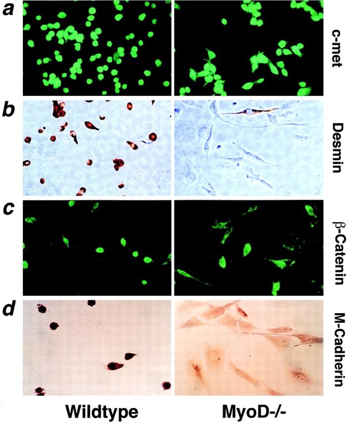
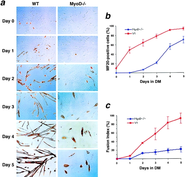
To gain insight into the regeneration deficit of MyoD-/- muscle, we investigated the growth and differentiation of cultured MyoD-/- myogenic cells. Primary MyoD-/- myogenic cells exhibited a stellate morphology distinct from the compact morphology of wild-type myoblasts, and expressed c-met, a receptor tyrosine kinase expressed in satellite cells. However, MyoD-/- myogenic cells did not express desmin, an intermediate filament protein typically expressed in cultured myoblasts in vitro and myogenic precursor cells in vivo. Northern analysis indicated that proliferating MyoD-/- myogenic cells expressed fourfold higher levels of Myf-5 and sixfold higher levels of PEA3, an ETS-domain transcription factor expressed in newly activated satellite cells. Under conditions that normally induce differentiation, MyoD-/- cells continued to proliferate and with delayed kinetics yielded reduced numbers of predominantly mononuclear myocytes. Northern analysis revealed delayed induction of myogenin, MRF4, and other differentiation-specific markers although p21 was upregulated normally. Expression of M-cadherin mRNA was severely decreased whereas expression of IGF-1 was markedly increased in MyoD-/- myogenic cells. Mixing of lacZ-labeled MyoD-/- cells and wild-type myoblasts revealed a strict autonomy in differentiation potential. Transfection of a MyoD-expression cassette restored cytomorphology and rescued the differentiation deficit. We interpret these data to suggest that MyoD-/- myogenic cells represent an intermediate stage between a quiescent satellite cell and a myogenic precursor cell.


Figures

Figure 1
Figure 1
Primary MyoD−/− myogenic cells exhibit a premyoblastic cellular phenotype. (a) Immunostaining revealed that both wild-type (Wildtype) and MyoD-deficient myogenic cells (MyoD−/−) expressed the c-met receptor tyrosine kinase, a marker for satellite cells and proliferating myoblasts. (b) Immunohistochemical analysis for desmin expression revealed a marked reduction in the proportion of cells in MyoD−/− cultures expressed desmin relative to wild-type cultures. (c) Normal level and localization of β-catenin in MyoD−/− cells as revealed by immunofluorescence. (d) Immunohistochemical detection of M-cadherin revealed low level expression of M-cadherin in MyoD−/− cells relative to primary wild-type myoblasts.
Figure 8
Figure 8
Rescue of the differentiation deficiency of MyoD−/− myogenic cells by forced expression of MyoD. (a) Western blot analysis revealed the expression of MyoD protein in wild-type cells (WT) and no expression in MyoD−/− cultures or MyoD−/− cells transfected with a selectable vector alone (PGK-Puro). However, MyoD-transfected pools of MyoD−/− cells (MyoD+) expressed readily detectable MyoD protein as did C2C12 myoblasts. (b) In growth medium, MyoD+ cells exhibited a refractile compact morphology typical of wild-type primary myoblasts and distinct from the stellate fibroblastlike morphology of MyoD−/− cells (growth). MyoD+ cultures exposed to differentiation medium for 3 d exhibited increased numbers of differentiated myocytes as detected with antiserum MF20 reactive with MHC, and restored formation of elongated bipolar multinucleated myotubes (day 3/MF20). (c) Determination of fusion index indicated a fivefold increase in the fusion potential of MyoD+ cells relative to untransfected MyoD−/− cells and similar to wild-type (WT) cultures. Fusion indices were calculated as described in Fig. 4.
Figure 4
Figure 4
Upregulation of Myf-5 and delayed expression of myogenin and MRF4 in MyoD−/− myogenic cells. Northern analysis revealed an absence of MyoD mRNA (a) in MyoD−/− cells (−/−), and a fourfold upregulation of Myf-5 mRNA (b) relative to wild-type myoblasts (WT). Upon differentiation, myogenin expression was upregulated and reduced (c). Similarly, MRF4 expression was delayed about 1 d in MyoD−/− cultures after transfer to differentiation medium (d). Day 0 denotes samples isolated from cells in growth medium. RNA samples were prepared 1, 2, 3, 4, and 5 d after the transfer of the cells to differentiation medium (DM). The fold activation in arbitrary units for each mRNA species is shown graphically beside each Northern blot. Fold activation was measured by densitometry and was normalized to 18S rRNA. (e) Western analysis of lysates prepared from wild-type or MyoD−/− cultures in growth medium with anti–Myf-5, anti-MyoD, and antimyogenin antibodies.
Figure 2
Figure 2
Reduced differentiation potential of MyoD−/− myogenic cells. (a) Differentiated myocytes were detected by immunostaining with antibody MF20 reactive with MHC. Incubation of wild-type (WT) myoblast cultures in differentiation medium resulted in a rapid increase in MHC synthesis and formation of elongated multinucleated myotubes. By contrast MyoD-deficient cells (MyoD−/−) differentiated with reduced kinetics and failed to form multinucleated elongated myotubes. Note the 100-fold reduced rate of spontaneous differentiation observed in MyoD−/− cultures under growth conditions (day 0). Days correspond to the time spent in differentiation medium before staining, whereas day 0 represents cultures in growth medium. (b) Percent MF20 positive cells was determined by enumeration of MHC expressing differentiated myocytes by immunostaining with antibody MF20. Note the delayed and reduced kinetics of differentiation in the absence of MyoD. (c) Calculation of fusion indices as percent cells containing two or more nuclei within a differentiated myocyte confirmed that MyoD−/− myoblasts were severely deficient in fusion capacity with the majority of differentiated myocytes containing a single nuclei. The error bars represent the standard error of the mean from three independently derived primary cultures.
Figure 3
Figure 3
Enhanced proliferative potential of primary MyoD−/− myogenic cells in growth and differentiation. (a) Analysis of [3H]thymidine incorporation during differentiation revealed that MyoD−/− myogenic cells continued to synthesize DNA after mitogen withdrawal. Incorporation was normalized to protein concentration. The error bars represent the standard error of the mean for three different isolates. (b) Immunodetection of BrdU incorporation in MyoD−/− cultures revealed continued DNA synthesis after mitogen withdrawal. Differentiation was assessed by immunostaining with antibody MF20. Taken together, these data indicate that MyoD−/− myogenic cells inefficiently withdraw from the cell cycle under differentiation promoting conditions.
Figure 5
Figure 5
Reduced expression of differentiation-specific markers in MyoD−/− myogenic cells. Northern analysis of α-skeletal (a) and α-cardiac actin (b) mRNA levels revealed reduced and delayed kinetics of induction upon differentiation of MyoD−/− cultures. Similarly, acetylcholine receptor δ subunit (AchR δ) (c) and M-cadherin mRNA levels were found to be reduced (d). A marked reduction in the expression of Musk (e) and adhalin (f) mRNAs was also observed. The numbered arrows for Musk correspond to the specific isoforms quantitated on the associated graphs. The fold activation in arbitrary units for each mRNA species is shown graphically beside each Northern blot. Fold activation was measured by densitometry and was normalized to 18S rRNA.
Figure 6
Figure 6
Northern analysis of growth-associated gene products. (a) Primary wild-type myoblasts expressed abundant β-catenin under growth conditions. These levels increased threefold after 2 d of differentiation and subsequently decreased. In MyoD−/− cultures, β-catenin levels were found to continuously increase to levels that were comparable to that of wild-type cells by day 5 of differentiation. (b) Wild-type myoblasts during growth expressed very low levels of PEA3 mRNA, and these levels increased about twofold by day 5 of differentiation. Growing MyoD−/− cells displayed sixfold higher levels of PEA3 mRNA, which declined steadily to wild-type levels by day 4 of differentiation. (c) No significant differences were observed in p21 mRNA levels between wild-type and MyoD−/− cells. (d) Wild-type myoblasts in growth conditions expressed low levels of IGF-I mRNA and these levels were rapidly extinguished after mitogen withdrawal. Mutant MyoD−/− myogenic cells expressed over threefold higher levels of the small IGF-I mRNA isoforms (1 and 2) and the 7-kb pre-IGF-I mRNA (isoform 3) was rapidly upregulated after 3 d of differentiation. The numbered arrows adjacent to IGF-I denote specific isoforms depicted on the corresponding graph. (e) Expression levels as determined by densitometry were normalized to 18S rRNA. Differentiation and graphical representation of the fold activation is as described in Fig. 5.
Figure 7
Figure 7
Strict cell autonomy in differentiated cocultures of lacZ-expressing MyoD−/− myogenic cells and wild-type myoblasts. Primary cells were plated in ratios of 1:0 (a), 1:4 (b), 4:1 (c), and 0:1 (d) of MyoD−/− to wild-type cells. After 5 d of differentiation, cells were fixed, and stained for β-galactosidase and with antibody MF20 reactive with MHC. Note the complete absence of lacZ-labeled nuclei in myotubes containing greater than two nuclei and the normal differentiation of wild-type myotubes.
Figure 9
Figure 9
Role of MyoD in regulating satellite cell function. RT-PCR analysis of single cells on cultured myofibers reveals quiescent satellite cells express c-met but no detectable MRFs. Activated satellite cells first express Myf-5 or MyoD before coexpressing both Myf-5 and MyoD, and progressing through their normal developmental program leading to terminal differentiation (Cornelison and Wold, 1997). In the absence of MyoD, satellite cells appear to exhibit a propensity for self-renewal rather than progression through the differentiation program (Megeney et al., 1996). Therefore, expression of Myf-5 alone may allow self-renewal of satellite cells either before returning to quiescence (yellow arrows) or upregulating MyoD and formation of proliferative myogenic precursor cells (mpc) (white arrows). The potential and replicative capacity of cells expressing MyoD alone is unknown; however, these cells likely irreversibly progress through the myogenic program. Taken together, we interpret these data to suggest that MyoD−/− myogenic cells represent an intermediate stage between myogenic stem cell and a myogenic precursor cell.

References

    1. Allen RE, Sheehan SM, Taylor RG, Kendall TL, Rice GM. Hepatocyte growth factor activates quiescent skeletal muscle satellite cells in vitro. J Cell Physiol. 1995;165:307–312. - PubMed
    1. Bader D, Masaki T, Fischman DA. Immunochemical analysis of myosin heavy chain during avian myogenesis in vivo and in vitro. J Cell Biol. 1982;95:763–770. - PMC - PubMed
    1. Barth AI, Nathke IS, Nelson WJ. Cadherins, catenins and APC protein: interplay between cytoskeletal complexes and signaling pathways. Curr Opin Cell Biol. 1997;9:683–690. - PubMed
    1. Birnboim HC. Rapid extraction of high molecular weight RNA from cultured cells and granulocytes for Northern analysis. Nucleic Acids Res. 1988;16:1487–1497. - PMC - PubMed
    1. Bischoff, R. 1994. The satellite cell and muscle regeneration. In Myogenesis. Vol. 2. A.G. Engel and C. Franszini-Armstrong, editors. McGraw-Hill Inc., New York. 97–118.

Publication types