. 1999 Feb 22;144(4):789-801.
doi: 10.1083/jcb.144.4.789.

LYVE-1, a new homologue of the CD44 glycoprotein, is a lymph-specific receptor for hyaluronan

Affiliations

LYVE-1, a new homologue of the CD44 glycoprotein, is a lymph-specific receptor for hyaluronan

S Banerji et al. J Cell Biol. .

Abstract

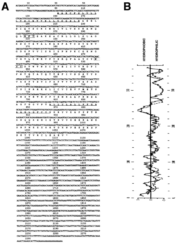
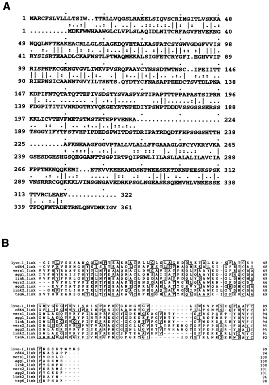
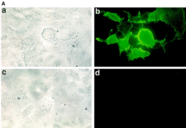
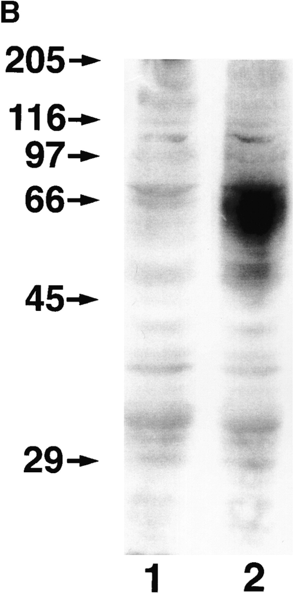
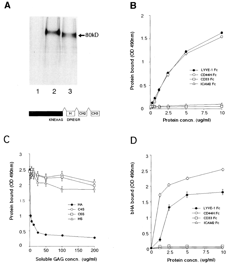
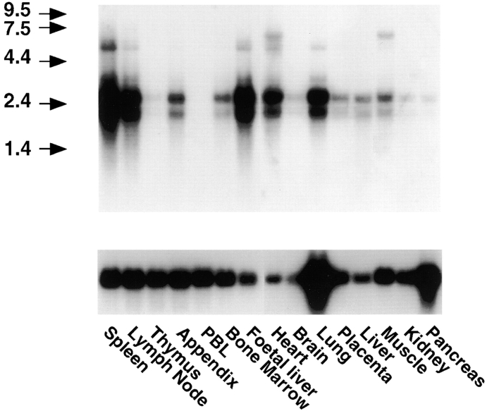
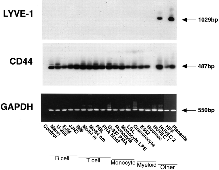
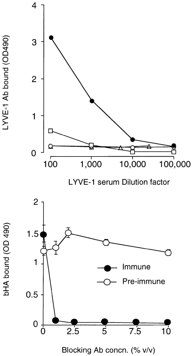
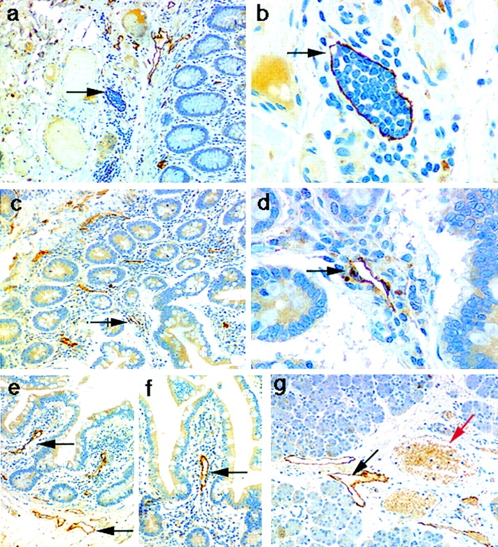
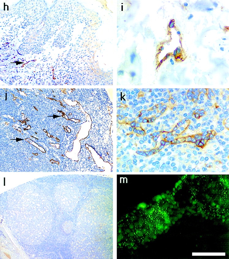
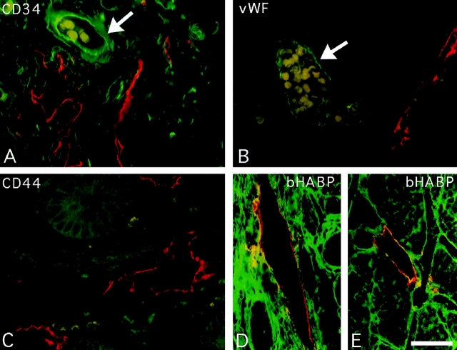
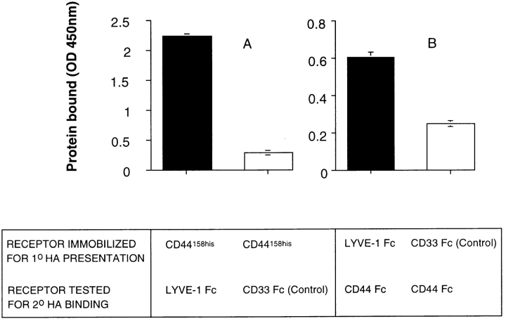
The extracellular matrix glycosaminoglycan hyaluronan (HA) is an abundant component of skin and mesenchymal tissues where it facilitates cell migration during wound healing, inflammation, and embryonic morphogenesis. Both during normal tissue homeostasis and particularly after tissue injury, HA is mobilized from these sites through lymphatic vessels to the lymph nodes where it is degraded before entering the circulation for rapid uptake by the liver. Currently, however, the identities of HA binding molecules which control this pathway are unknown. Here we describe the first such molecule, LYVE-1, which we have identified as a major receptor for HA on the lymph vessel wall. The deduced amino acid sequence of LYVE-1 predicts a 322-residue type I integral membrane polypeptide 41% similar to the CD44 HA receptor with a 212-residue extracellular domain containing a single Link module the prototypic HA binding domain of the Link protein superfamily. Like CD44, the LYVE-1 molecule binds both soluble and immobilized HA. However, unlike CD44, the LYVE-1 molecule colocalizes with HA on the luminal face of the lymph vessel wall and is completely absent from blood vessels. Hence, LYVE-1 is the first lymph-specific HA receptor to be characterized and is a uniquely powerful marker for lymph vessels themselves.


Figures

Figure 1
Figure 1
Nucleotide sequence and derived amino acid sequence of LYVE-1, a novel human HA receptor. A shows the complete nucleotide and derived amino acid sequence for the 2,313-bp LYVE-1 cDNA clone in pBluescript. A cleavable NH2-terminal leader predicted using the −3 +1 rule (56) and a probable transmembrane anchor are underlined. The positions of two putative N-glycosylation sites (N X S/T) are boxed. The sequence predicts a 322–amino acid residue polypeptide with a 26-residue leader peptide, a 21-residue hydrophobic membrane anchor, and a 63-residue cytoplasmic tail. B depicts Kyte-Doolittle (solid line) and Goldman (stippled line) hydropathy plots of the derived amino acid sequence, both of which confirm the position of the predicted hydrophobic transmembrane anchor. These sequence data are available from GenBank under accession number AF118108.
Figure 2
Figure 2
Comparison of the LYVE-1 receptor Link module with other Link superfamily members. In A, the derived amino acid sequence of LYVE-1 (top line) is shown aligned with that of the full-length human CD44 cDNA (bottom line) using the GCG program GAP. Positions where amino acid residues are identical are depicted with a line; semiconservative and conservative differences are depicted with one or two dots, respectively. The two sequences have an overall similarity of 41% and a similarity of 57% within the immediate Link homology unit (LYVE-1 residues 61–128). B shows a Prettyplot (GCG) comparison of sequence encompassing the “Link modules” (HA-binding domains) from LYVE-1 (corresponding to residues 40–138 in A), human CD44, the soluble human tumor necrosis factor–inducible TSG-6 molecule, and both tandem repeats (Links 1 and 2) of human Aggrecan, human Versican, and human Cartilage Link protein.
Figure 3
Figure 3
Cell surface expression of LYVE-1 receptor on transfected COS cells. In A, COS 1 cells were transiently transfected with either full-length LYVE-1 cDNA in the expression vector pRcCMV (a and b), or with a control empty pRcCMV vector (c and d) using DEAE dextran followed by surface immunofluorescent staining with rabbit polyclonal LYVE-1 antiserum (1:100 dilution) and FITC goat anti–rabbit IgG. In B, control and LYVE-1 transfected COS cells were electrophoresed on a 10% polyacrylamide SDS-PAGE gel, transferred to nitrocellulose, and Western blotted with LYVE-1 antiserum and peroxidase-conjugated goat anti–rabbit IgG (see Materials and Methods). Samples were control transfected COS (lane 1) and LYVE-1 transfected COS (lane 2). The positions and sizes in kilodaltons of the molecular mass calibration markers myosin (205 kD), β-galactosidase (116 kD), phosphorylase b (97 kD), BSA (66 kD), ovalbumin (45 kD), and carbonic anhydrase (29 kD) are indicated on the left.
Figure 3
Figure 3
Cell surface expression of LYVE-1 receptor on transfected COS cells. In A, COS 1 cells were transiently transfected with either full-length LYVE-1 cDNA in the expression vector pRcCMV (a and b), or with a control empty pRcCMV vector (c and d) using DEAE dextran followed by surface immunofluorescent staining with rabbit polyclonal LYVE-1 antiserum (1:100 dilution) and FITC goat anti–rabbit IgG. In B, control and LYVE-1 transfected COS cells were electrophoresed on a 10% polyacrylamide SDS-PAGE gel, transferred to nitrocellulose, and Western blotted with LYVE-1 antiserum and peroxidase-conjugated goat anti–rabbit IgG (see Materials and Methods). Samples were control transfected COS (lane 1) and LYVE-1 transfected COS (lane 2). The positions and sizes in kilodaltons of the molecular mass calibration markers myosin (205 kD), β-galactosidase (116 kD), phosphorylase b (97 kD), BSA (66 kD), ovalbumin (45 kD), and carbonic anhydrase (29 kD) are indicated on the left.
Figure 4
Figure 4
LYVE-1 binds both immobilized and soluble HA. LYVE-1, expressed as a soluble IgFc fusion protein in transiently transfected human COS fibroblasts, was compared with CD44 for binding to HA and other glycosaminoglycans. A shows LYVE-1 and CD44H Fc fusion proteins isolated from the supernatants of [35S] methionine/cysteine-labeled transfectants and electrophoresed on a 7.5% polyacrylamide SDS-PAGE gel. Samples were the protein A–Sepharose adsorbed proteins from control untransfected cells (lane 1), CD44H Fc transfected cells (lane 2), and LYVE-1 transfected cells (lane 3). The LYVE-1 fusion protein comprises residues 1–232 of the extracellular domain fused to the hinge (H), CH2, and CH3 domains of human IgG1. Details of the CD44H Fc protein, which includes residues 1–200 of the extracellular domain, have been published previously (1). For ligand binding assays, LYVE-1 Fc was compared with CD44H Fc and the negative control fusion proteins CD33 Fc and ICAM-2 Fc for adhesion to immobilized and soluble HA in 96-well microtiter plates (see Materials and Methods). B shows binding of the fusion proteins to immobilized HA, in the absence of competing glycosaminoglycans; C shows binding (LYVE-1 Fc only) in the presence of free chondroitin-4-SO4, chondroitin-6-SO4, or heparin; and D shows binding to soluble biotinylated HA. Detection of bound fusion protein and biotinylated HA was carried out using peroxidase-conjugated anti–human IgFc antibody and peroxidase-conjugated streptavidin, respectively. Values are the mean ± SEM of at least three replicates.
Figure 5
Figure 5
Northern blot hybridization analysis of LYVE-1 receptor mRNA. RNA blots containing 2 μg poly(A)+ RNA per lane from each of the tissues shown were hybridized to a 32P-labeled full-length LYVE-1 DNA probe (top), or glyceraldehyde-3-phosphate dehydrogenase probe (bottom), and washed at high stringency before autoradiography (see Materials and Methods). The migration positions of RNA calibration markers (kilodaltons) are shown to the left of the figure.
Figure 6
Figure 6
RT-PCR analysis of LYVE-1 mRNA expression in tissue culture cell lines. LYVE-1 and CD44 mRNA levels were compared within a panel of tissue culture cell lines representing different lineages. Samples of cDNA prepared from each of the cell lines shown were amplified with primers specific for either LYVE-1 (top), CD44 (middle), or glyceraldehyde-3-phosphate dehydrogenase (control, bottom) followed by electrophoresis on 1.25% agarose gels, staining with ethidium bromide (glyceraldehyde-3-phosphate dehydrogenase only), or Southern blotting and hybridization with the appropriate 32P-labeled detection oligonucleotides as described in Materials and Methods. Arrows depict the sizes of the PCR products in each case.
Figure 7
Figure 7
Specificity of a LYVE-1 receptor polyclonal serum. The specificity of an affinity-purified rabbit polyclonal antiserum generated against soluble LYVE-1 receptor and its capacity to block HA binding were assessed in microtiter plate binding assays. In the top panel, wells coated with either LYVE-1 Fc (filled circles), the CD44H ectodomain fragment CD44158his (triangles), or the control fusion protein ICAM-2 Fc (squares) were incubated with appropriately diluted LYVE-1 specific polyclonal antiserum, and binding was detected with peroxidase-conjugated anti–human Ig (see Materials and Methods). As a control, a second set of LYVE-1–coated wells was reacted with appropriately diluted preimmune serum (open circles). In the bottom panel, wells coated with LYVE-1 Fc were incubated with soluble biotinylated HA (5 μg/ml), in the presence of increasing concentrations of either LYVE-1 antiserum or control preimmune serum followed by detection of bound HA as described in Materials and Methods. Data in each case are the mean ± SEM of triplicate determinations.
Figure 8
Figure 8
Localization of the LYVE-1 HA receptor in human tissues. Paraffin-embedded human tissue sections were stained with rabbit polyclonal LYVE-1 antiserum (1:100 dilution), followed by peroxidase-conjugated goat anti–rabbit IgG as described in Materials and Methods. Tissues shown are colon (a and b), small intestine (c–f), salivary gland (g), skin (h and i), lymph node (j), spleen (k), and tonsil (l). m shows cultured HUVEC stained with LYVE-1 and FITC-conjugated anti–rabbit Ig (see Materials and Methods). Bars, j and l, 500 μm; a, c, and e–h, 200 μm; b, d, and k, 50 μm; and i and m, 20 μm. Black arrows depict lymphatic vessels, both empty (d–g) and containing lymphocytes (b), and red arrows depict blood vessels clearly identified by their content of weakly stained erythrocytes (g and i).
Figure 8
Figure 8
Localization of the LYVE-1 HA receptor in human tissues. Paraffin-embedded human tissue sections were stained with rabbit polyclonal LYVE-1 antiserum (1:100 dilution), followed by peroxidase-conjugated goat anti–rabbit IgG as described in Materials and Methods. Tissues shown are colon (a and b), small intestine (c–f), salivary gland (g), skin (h and i), lymph node (j), spleen (k), and tonsil (l). m shows cultured HUVEC stained with LYVE-1 and FITC-conjugated anti–rabbit Ig (see Materials and Methods). Bars, j and l, 500 μm; a, c, and e–h, 200 μm; b, d, and k, 50 μm; and i and m, 20 μm. Black arrows depict lymphatic vessels, both empty (d–g) and containing lymphocytes (b), and red arrows depict blood vessels clearly identified by their content of weakly stained erythrocytes (g and i).
Figure 9
Figure 9
Immunofluorescent staining of LYVE-1 in lymphatic vessels and colocalization with HA. Sections of human small intestine were double stained for LYVE-1, CD44, the vascular endothelial molecules CD34 and vWF, and for HA, by indirect immunofluorescence microscopy, using Texas red or fluorescein-conjugated secondary antibodies or bHABC as described in Materials and Methods. The antibody combinations were as follows: A, LYVE-1 (red) and CD34 (green); B, LYVE-1 (red) and vWF (green); C, LYVE-1 (red) and CD44 (green); and D and E, LYVE-1 (red) and bHABC (green). Arrows depict small blood vessels containing erythrocytes (pale yellow/ green), which are LYVE-1-ve. Lymph vessel endothelium positive for both LYVE-1 and HA in D and E is stained yellow/orange. Bar, 50 μm.
Figure 10
Figure 10
LYVE-1 molecules support HA-mediated binding to CD44. The capacity of LYVE-1 and CD44 to form ternary complexes with HA was tested using a modification of the HA-binding assay in Fig. 4 B. A shows binding of LYVE-1 Fc and CD33 Fc (control) to HA, presented by immobilized CD44158his protein. B shows binding of CD44 Fc to HA, presented by immobilized LYVE-1 Fc or CD33 Fc (control). Bound fusion proteins were detected with peroxidase-conjugated anti–human IgFc antibody, or with peroxidase-conjugated CD44 mAb A3D8, respectively, as described in Materials and Methods. No binding was observed in the absence of HA (not shown). Data are the mean ± SEM (n = 3).

References

    1. Aruffo A, Stamenkovic I, Melnick M, Underhill CB, Seed B. CD44 is the principal cell surface receptor for hyaluronate. Cell. 1990;61:1303–1313. - PubMed
    1. Bajorath J, Greenfield B, Munro SB, Day AJ, Aruffo A. Identification of CD44 residues important for hyaluronan binding and delineation of the binding site. J Biol Chem. 1998;273:338–343. - PubMed
    1. Banerji S, Day AJ, Kahmann JD, Jackson DG. Characterization of a functional hyaluronan-binding domain from the human CD44 molecule expressed in Escherichia coli. . Protein Exp Purification. 1998;14:371–381. - PubMed
    1. Bennett K, Jackson DG, Simon JC, Tanczos E, Peach R, Modrell B, Stamenkovic I, Plowman G, Aruffo A. CD44 isoforms containing exon v3 are responsible for the presentation of heparin binding growth factor. J Cell Biol. 1995;128:687–698. - PMC - PubMed
    1. Bennett K, Modrell B, Greenfield B, Bartolazzi A, Stamenkovic I, Peach R, Jackson D, Spring F, Aruffo A. Regulation of CD44 binding to hyaluronan by glycosylation of variably spliced exons. J Cell Biol. 1995;131:1623–1633. - PMC - PubMed

Publication types

MeSH terms

Associated data