. 1999 Mar 1;80(5):781-90.
doi: 10.1002/(sici)1097-0215(19990301)80:5<781::aid-ijc24>3.0.co;2-a.

Natural variation of the expression of HLA and endogenous antigen modulates CTL recognition in an in vitro melanoma model

Affiliations

Natural variation of the expression of HLA and endogenous antigen modulates CTL recognition in an in vitro melanoma model

J N Cormier et al. Int J Cancer. .

Abstract

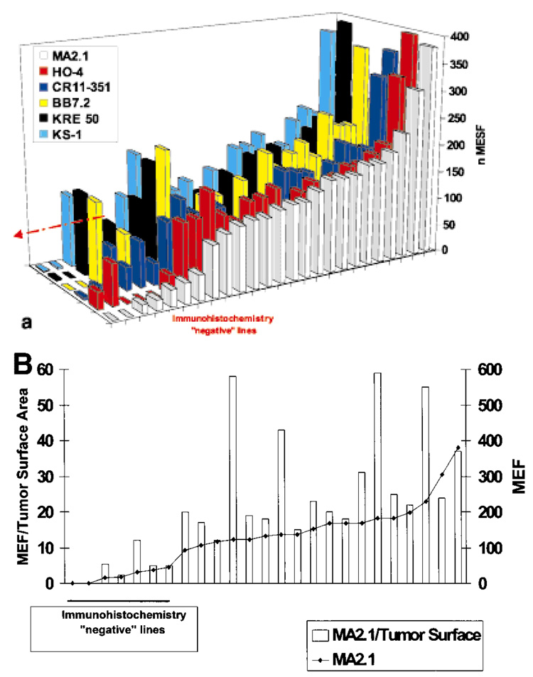
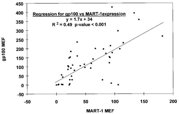
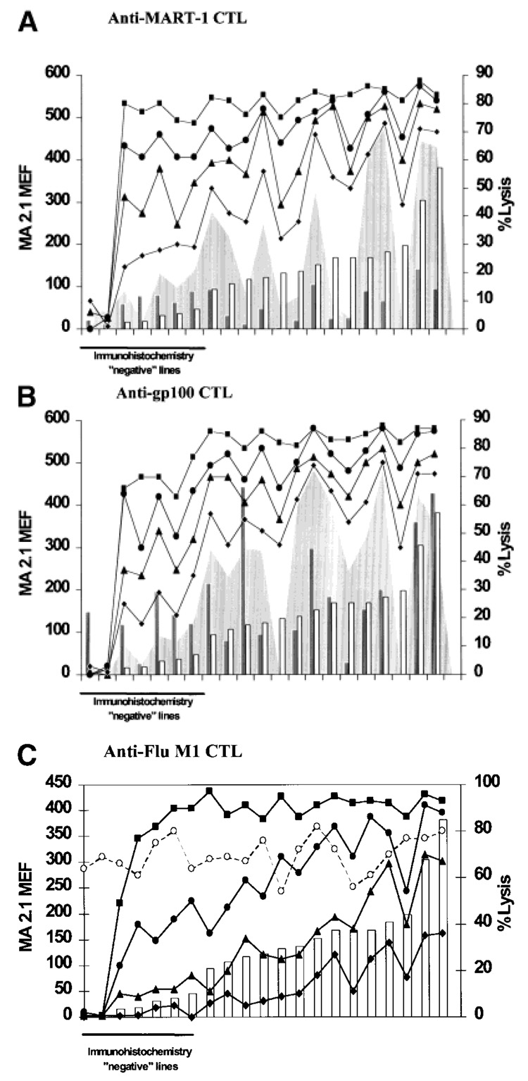
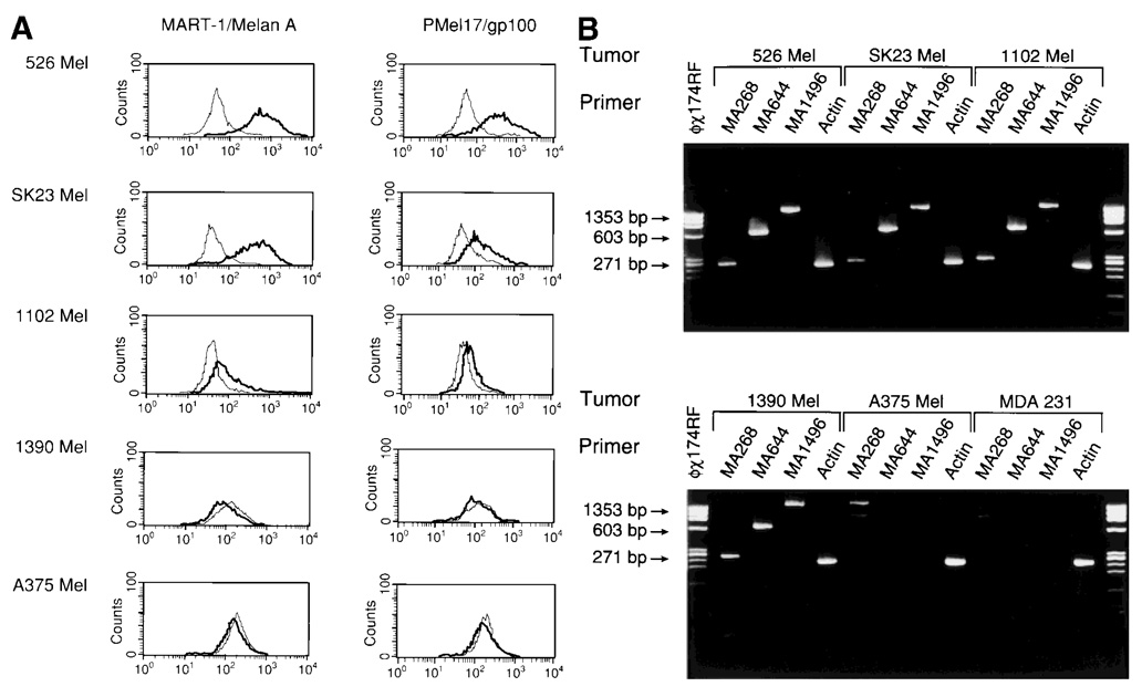
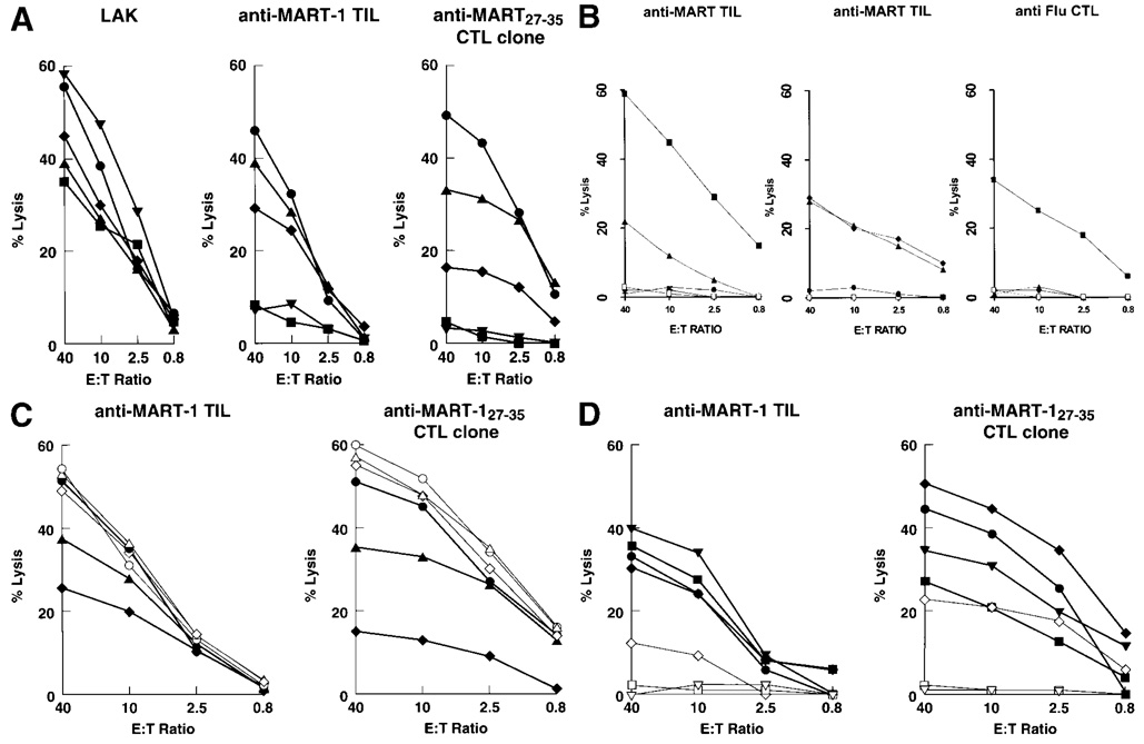
Increasing attention has been devoted to elucidating the mechanism of lost or decreased expression of MHC or melanoma-associated antigens (MAAs), which may lead to tumor escape from immune recognition. Loss of expression of HLA class I or MAA has, as an undisputed consequence, loss of recognition by HLA class I-restricted cytotoxic T cells (CTLs). However, the relevance of down-regulation remains in question in terms of frequency of occurrence. Moreover the functional significance of epitope down-regulation, defining the relationship between MHC/epitope density and CTL interactions, is a matter of controversy, particularly with regard to whether the noted variability of expression of MHC/epitope occurs within a range likely to affect target recognition by CTLs. In this study, bulk metastatic melanoma cell lines originated from 25 HLA-A*0201 patients were analyzed for expression of HLA-A2 and MAAs. HLA-A2 expression was heterogeneous and correlated with lysis by CTLs. Sensitivity to lysis was also independently affected by the amount of ligand available for binding at concentrations of 0.001 to 1 mM. Natural expression of MAA was variable, independent from the expression of HLA-A*0201, and a significant co-factor determining recognition of melanoma targets. Thus, the naturally occurring variation in the expression of MAA and/or HLA documented by our in vitro results modulates recognition of melanoma targets and may (i) partially explain CTL-target interactions in vitro and (ii) elucidate potential mechanisms for progressive escape of tumor cells from immune recognition in vivo.


Figures

FIGURE 1
FIGURE 1
(a) Variability of expression of HLA class I in melanoma cell lines. Heterogeneity in the staining by 6 MAbs [MA2.1 (white), HO-4 (red), CR11–351 (dark blue), BB7.2 (yellow), KRE 50 (black) and KS-1 (light blue)] recognizing distinct determinants of HLA-A2 antigens of 25 melanoma cell lines derived from HLA-A*0201 patients with metastatic melanoma. Data are expressed as normalized MEF (nMEF) as described “Material and Methods”. A significant proportion of cell lines displayed either loss of expression of HLA-A2 related to genomic loss (first 2 lanes) or greatly reduced level of expression (lanes 3–8). (b) Expression of HLA-A2 detected by MAb MA2.1 in the 25 melanoma cell lines shown in Figure 3a. Data are presented as MEF (◆) as well as MEF corrected for the calculated surface area (MEF/S, gray bars) for each cell line, providing a relative value of HLA-A2 molecule density. In 4 cell lines of very small cell size, the corrected values were quite different from the uncorrected MEF.
FIGURE 2
FIGURE 2
Correlation between expression of MART-1/MelanA and Pmel17/gp100 expression (MEF) in 46 melanoma cell lines derived from patients with metastatic melanoma (Surgery Branch, NCI). Data points represent the average of 2 experiments.
FIGURE 3
FIGURE 3
Effect of the level of HLA-A2 antigen expression on the recognition of cultured melanoma cells by HLA-A2-restricted CTLs. The cell lines described in Table I (with the exclusion of 624-MEL, 1143-MEL, 677-MEL and 1182-MEL) were derived from HLA-A*0201 melanoma patients and ranked according to different surface expression of HLA-A2 molecules (open bars). Expression of the relevant MAAs (gray bars) is also shown. These lines were tested for sensitivity to lysis by anti-MART-127–35-, anti-gp100209–217- anti-Flu M158–66-specific, HLA-A*0201-restricted CTLs. Cytotoxicity was tested in natural conditions (shaded areas) in the presence of different concentrations of the relevant peptide (■ = 1 µM, ● = 0.1 µM, ▲ = 0.01 µM, ◆ = 0.001 µM). The data summarize the percent of lysis obtained at an effector to target ratio of 10:1 (for MART-1- and gp100-specific CTLs) or 20:1 (for Flu-specific CTLs) and represent the mean of 3 separate experiments. Lysis by LAK cells (40:1 E:T ratio) is similar among different cell lines and is represented by the dashed line and the open circles in (c).
FIGURE 4
FIGURE 4
(a) Intracellular expression of MART-1/Melan A and Pmel17/gp100. MAA expression (bold lines) of 5 tumor lines (526-MEL, SK23-MEL, 1102-MEL, 1390-MEL and A375-MEL) as detected by intracellular FACS analysis (fine lines represent negative control, which is an isotype-matched IgG control plus secondary Goat Anti-mouse FITC (GAMF) MAb). (b) Expression of MART-1/MelanA by RT-PCR. mRNA expression of MART-1/Melan A in 5 melanoma lines (526-MEL, SK23-MEL, 1102-MEL, 1390-MEL and A375-MEL) and in the MDA 231 breast cancer cell line. Three different sets of primers (MA268, MA644 and MA1496) were used, encompassing different sequences of the MART-1/MelanA coding region. The m.w. marker ϕ ✕ 174 RF DNA/HaeIII was used for size confirmation, and β-actin-specific primer pairs were used in each PCR.
FIGURE 5
FIGURE 5
(a) Effect of the natural variation of MART-1/Melan A expression on the lysis of melanoma cells by anti-MART-1/Melan A CTLs. The 5 melanoma cell lines (A375, SK23, 1102-MEL, 526-MEL and 1390-MEL) express similar amounts of the HLA-A2 allele but varying amounts of MART-1/MelanA antigen [A375 (▼), MEF 0; SK23 (●), MEF 62; 1102-MEL (◆), MEF 11; 526-MEL (▲), MEF 93; 1390-MEL (■), MEF 0]. These cell lines were tested for lysis by LAK cells (left panel), anti-MART-1/MelanA TILs (center panel) and an anti-MART-127–35 CTL clone originated from a TIL culture (right panel). The data represent the average of 3 experiments (SEM <10% for all data points). Cumulative linear correlation analysis between lysis and MAA expression was significant for TILs and the anti-MART-1 CTL clone at E:T ratios of 40, 10, 2.5 and 0.8:1 (p < 0.01 for all data points). The early TIL culture used for recognition of melanoma targets (TIL 1235) is relatively oligoclonal, recognizes MART-127–35 and does not recognize most Pmel17/gp100-associated epitopes with the exception of gp100154 pulsed on T2 cells. Such specificity was lost in subsequent passages in culture (Kawakami et al., 1995). (b) Lysis of different targets by the early MART-1-specific TIL culture used for the experiments described in Figure 6a,c. In the left panel, the TIL culture was tested against T2 cells pulsed with MART-127–35 (■), gp100154 (▲), gp100209 (●), gp100280 (▼) and unpulsed T2 (□). In the central panel, lysis of MDA231 (breast cancer cell line) alone (□) or in the presence of unlabeled 1102-MEL (●) and lysis of MDA231 pulsed with 1 µM MART-127–35 in the presence (▲) or absence (◆) of unlabeled 1102-MEL. Susceptibility of 1102-MEL to non-specific killing by activated CTLs was tested in the right panel. 1102-MEL was tested for lysis by Flu-specific CTLs: alone (□), alone and pulsed with FluM158–66 peptide (■) and in the presence of unlabeled MDA231 pulsed (▲) or not pulsed (◆) with FluM158–66 peptide. Data are the average of 3 experiments (SEM <10% for all data points). (c) Effect of exogenous MART-1/MelanA peptide on the lysis of melanoma cells by anti-MART-1/Melan A CTLs. Functional saturation of HLA-A2-binding sites equalized the recognition of melanoma targets by anti-MART-1/MelanA CTLs. The 3 melanoma cell lines express similar amounts of HLA-A2 alleles but varying amounts of MART-1 (SK23, MEF 62; 1102-MEL, MEF 11; and 526-MEL, MEF 93). Lysis was compared in the presence of natural processing and presentation of MART-1/MelanAby the cell lines [SK23 (●), 1102-MEL (◆) and 526-MEL (▲)] or after exogenous loading (5 µg/ml) of the same targets with MART-127–35 (open symbols). The anti-MART-1/MelanA TILs (left panel) and anti-MART-127–35 CTL clone originated from a TIL culture (right panel) were used as effectors. Average of 2 experiments, SEM <10 % for all data points. (d) Effect of endogenously processed MART-1/MelanA peptide on the lysis of melanoma cells by anti-MART-1/Melan A CTLs. The 3 melanoma cell lines 1102-MEL, 1390-Mel and A375-MEL express similar amounts of HLA-A2 alleles but varying amounts of MART-1/Melan A. The anti-MART-1/MelanA TILs (left panel) and anti- MART-127–35 CTL clone originated from a TIL culture (right panel) were used as effectors. Lysis was tested against endogenous processing and presentation of MART-1/Melan A by the cell lines after infection with recombinant vaccinia virus encoding MART-1/Melan A (rVV-MART) [A375 (▼), 1390 (■) and 1102 (◆)] or with the irrelevant recombinant vaccinia virus encoding Pmel17/gp100 (rVV-gp100) (open symbols). As a control for maximal lysis in natural conditions (without viral infection), SK23-MEL (●) cells were added to the panel of targets.

Similar articles

Cited by

References

    1. Chen Y-T, Stockert E, Tsang S, Coplan KE, Old LJ. Immunophenotyping of melanomas for tyrosinase: implications for vaccine development. Proc. nat. Acad. Sci. USA. 1995;92:8125–8129. - PMC - PubMed
    1. Cormier JN, Hijazi YM, Abati A, Fetsch P, Bettinotti M, Steinberg SM, Rosenberg SA, Marincola FM. Heterogeneous expression of melanoma-associated antigens (MAA) and HLA-A2 in metastatic melanoma in vivo. Int. J. Cancer. 1998;75:517–524. - PubMed
    1. Cormier JN, Salgaller ML, Prevette T, Barracchini KC, Rivoltini L, Restifo NP, Rosenberg SA, Marincola FM. Enhancement of cellular immunity in melanoma patients immunized with a peptide from MART-1/MelanA. Cancer J. Sci. Am. 1997;3:37–44. - PMC - PubMed
    1. Ferrone S, Marincola FM. Loss of HLA class I antigens by melanoma cells: molecular mechanisms, functional significance and clinical relevance. Immunol. Today. 1995;16:487–494. - PubMed
    1. Garrido F, et al. Proceedings of the XII HLA International Workshop. France: EDK, Sèvres; 1997. HLA and cancer.

MeSH terms