. 1999 Feb 15;13(4):449-61.
doi: 10.1101/gad.13.4.449.

ELAV tumor antigen, Hel-N1, increases translation of neurofilament M mRNA and induces formation of neurites in human teratocarcinoma cells

Affiliations

ELAV tumor antigen, Hel-N1, increases translation of neurofilament M mRNA and induces formation of neurites in human teratocarcinoma cells

D Antic et al. Genes Dev. .

Abstract

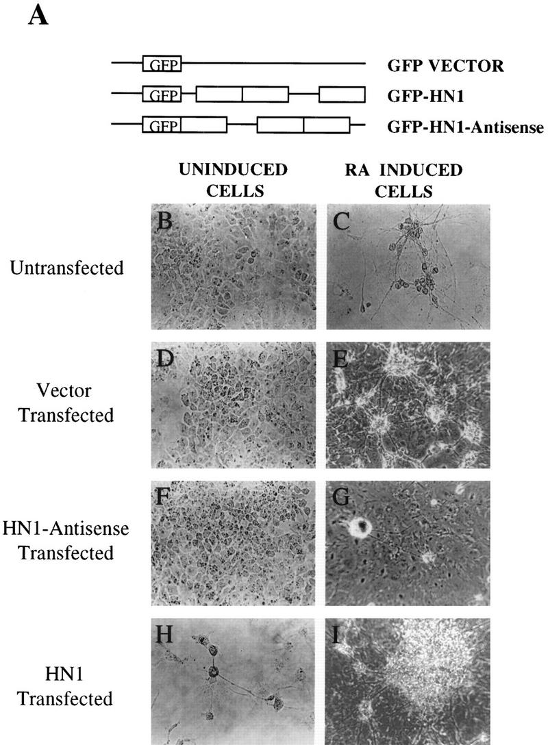
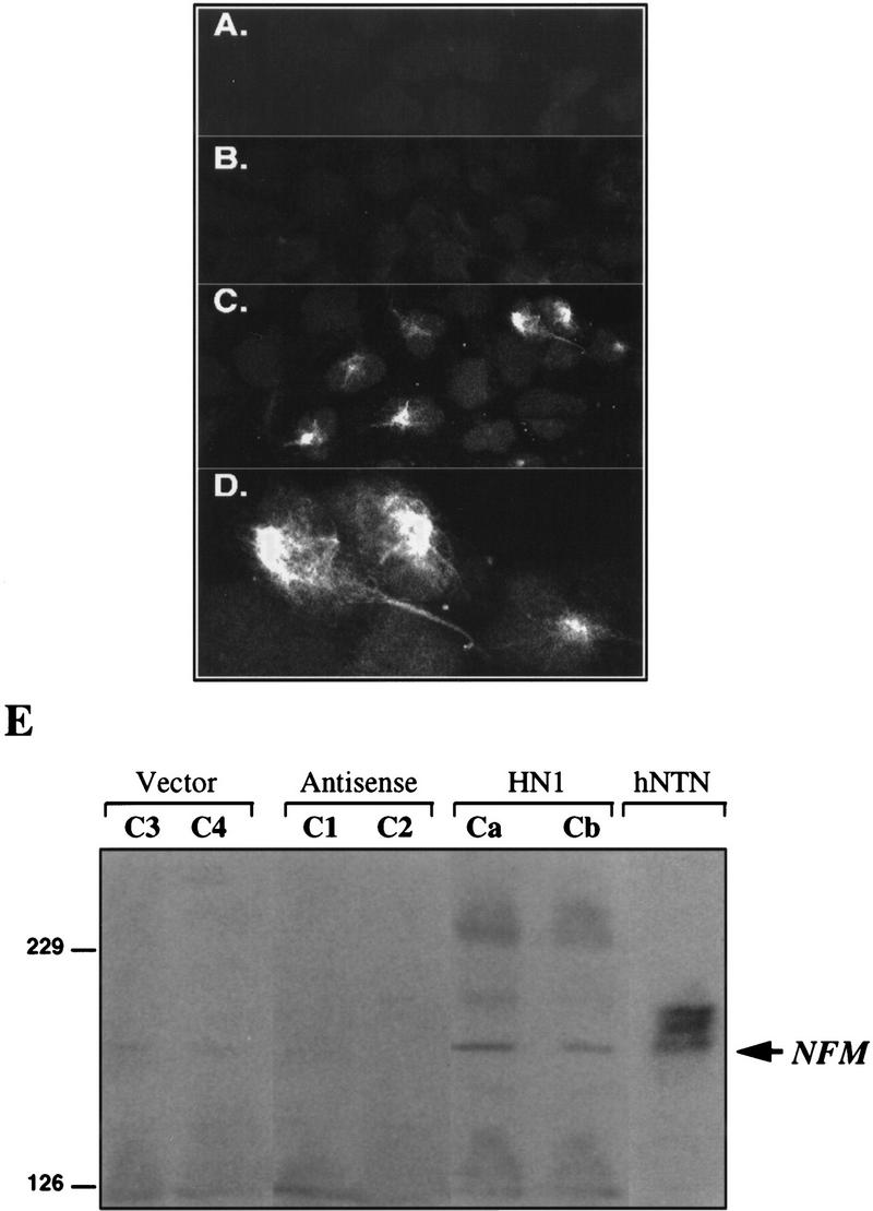
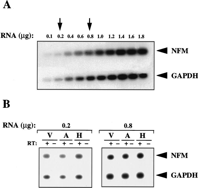
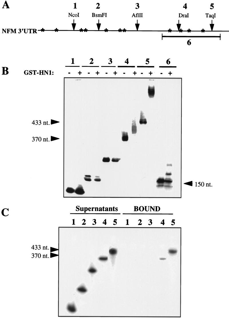
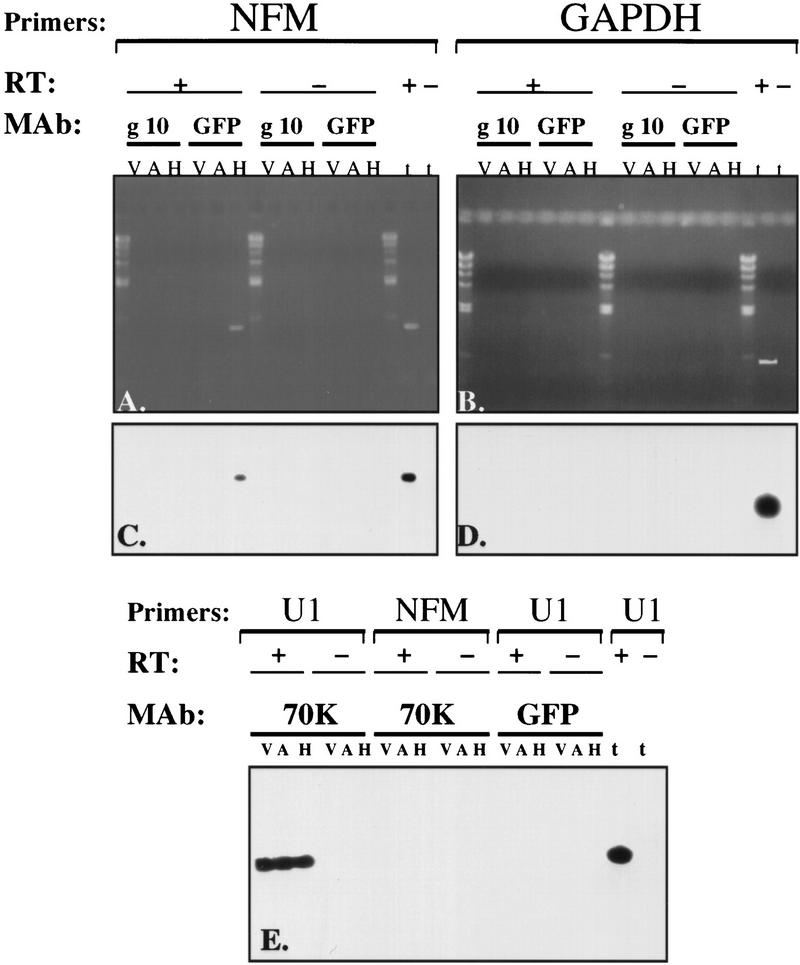
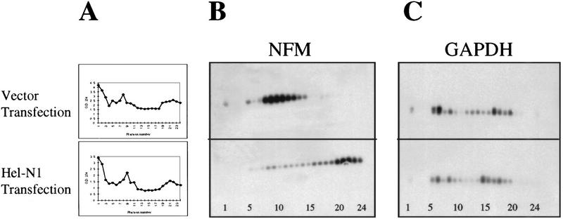
Human ELAV proteins are implicated in cell growth and differentiation via regulation of mRNA expression in the cytoplasm. In human embryonic teratocarcinoma (hNT2) cells transfected with the human neuronal ELAV-like protein, Hel-N1, neurites formed, yet cells were not terminally differentiated. Cells in which neurite formation was associated with Hel-N1 overexpression, also expressed increased levels of endogenous neurofilament M (NF-M) protein, which distributed along the neurites. However, steady-state levels of NF-M mRNA remained similar whether or not hNT2 cells were transfected with Hel-N1. These findings suggest that turnover of NF-M mRNA was not affected by Hel-N1 expression, despite the fact that Hel-N1 can bind to the 3' UTR of NF-M mRNA and was found directly associated with NF-M mRNA in transfected cells. Analysis of the association of NF-M mRNA with the translational apparatus in Hel-N1 transfectants showed nearly complete recruitment to heavy polysomes, indicating that Hel-N1 caused an increase in translational initiation. Our results suggest that the stability and/or translation of ARE-containing mRNAs can be regulated independently by the ELAV protein, Hel-N1, depending upon sequence elements in the 3' UTRs and upon the inherent turnover rates of the mRNAs that are bound to Hel-N1 in vivo.


Figures

Figure 1
Figure 1
ELAV proteins are up-regulated during neuronal differentiation of human embryonal teratocarcinoma (hNT) cells. (A) Uninduced hNT2 cells double stained with anti-Hel-N1 antibody (red) and anti-hnRNP C antibody (green); (B) hNT cells at day 5 of induction stained with anti-Hel-N1 antibody (red) and anti-NF-M antibody (green, and appears yellow because of overlap with red); (C) hNT cells at day 15 of induction stained with anti-Hel-N1 antibody (red) and anti-NF-M(P+++) (phosphorylated NF-M) antibody (green, and appears as yellow because of overlap with red). (D,E) hNTN cells at day 32 (4 weeks) of induction stained with (D) anti-Hel-N1 antibody (red) and anti-MAP2 antibody (green, and appears as yellow because of overlap with red) and (E) anti-Hel-N1 antibody (red) and anti-hnRNP C antibody (green, and yellow because of overlap with red). (D,E) Only mature neurons and not the supporting nonneuronal cells (stained green in E) are expressing ELAV proteins.
Figure 2
Figure 2
Morphology of stably transfected hNT2 cell lines. (A) Constructs used for transfections with ELAV protein, Hel-N1, were subcloned either into pHβAPr-1–neo or into pGFPC1 plasmids (see Materials and Methods). Untransfected (B,C), vector transfected (D,E), Hel-N1–antisense transfected (F,G), and Hel-N1-transfected (H,I) cells before (B,D,F,H) or after (C,E,G,I) the RA treatment. (C,H) Photographed with a 32× lens and are focused on the area containing isolated neurons (C) or neuron-like cells (H). Other panels were photographed with a 10× lens using a Zeiss inverted microscope and show clumps containing large numbers of neurons. Untransfected, RA-induced cells were morphologically indistinguishable at all magnifications from the vector-transfected, RA-induced cells shown in E.
Figure 3
Figure 3
Expression of NF-M protein in cells stably transfected with Hel-N1. Confocal microscopy images of vector-transfected cells (A), antisense-Hel-N1-transfected cells (B), and Hel-N1-transfected cells immunostained with anti-NF-M antibody(C,D). (D) Magnified top right corner of C. (E) Immunoblot stained with anti-NF-M antibody. Total cell extracts (150 μg) from two vector-transfected clones (C3 and C4), two antisense-Hel-N1-transfected clones (C1 and C2), two Hel-N1-transfected clones (Ca and Cb), and untransfected differentiated neurons (hNTN) were loaded onto 6% acrylamide gels and analyzed for NF-M expression by ECL immunoblotting.
Figure 4
Figure 4
Comparative levels of NF-M mRNA in stably transfected cell lines as determined by quantitative RT–PCR. (A) Southern blot—titration of RNA concentrations using total RNA from Hel-N1 transfected cells: dose-response for the NF-M (20 PCR cycles) and GAPDH mRNAs (15 PCR cycles). (B) Southern blots—quantitative analysis of NF-M mRNA in two pooled vector transfected clones (V), two pooled antisense-Hel-N1 transfected clones (A), or in two pooled Hel-N1 transfected clones (H) using 0.2 or 0.8 μg of total RNA. The RT step was performed either with (+) or without (−) reverse transcriptase. Values obtained for the NFM/GAPDH ratios were 0.5, 0.4, and 0.7 using 0.2 μg; and 0.7, 0.8, and 0.9 using 0.8 μg of V, A, and H RNA, respectively. These values were within experimental error based upon numerous repeats.
Figure 5
Figure 5
RNA binding by mobility shift or immunoprecipitation of NF-M 3′ UTR using recombinant GST–Hel–N1 fusion protein. (A) Partial restriction map and position of U-rich sequences (marked by *) in the 3′ UTR of NF-M mRNA. Numbers 1–6 indicate fragments of the 3′ UTR used for RNA-binding experiments. (B) Mobility shift of in vitro transcribed RNAs representing each fragment of the NF-M 3′ UTR (as in A). RNAs that were bound by recombinant GST–Hel-N1 fusion protein are marked by arrowheads and their size is labeled. RNAs were analyzed on nondenaturing polyacrylamide gels (C) Immunoprecipitation of in vitro-transcribed RNAs using recombinant GST–Hel-N1 fusion protein: RNAs representing fragments 1–5 were bound to GST–Hel-N1 protein and immunoprecipitated with anti-Hel-N1 antibody and protein A beads. Bound RNAs were extracted from the beads (bound) and remaining unbound RNAs were extracted from the supernatants. RNAs were analyzed on denaturing urea-polyacrylamide gels. Bound fragments 4 and 5 are marked by arrowheads.
Figure 6
Figure 6
Immunoprecipitation of NF-M mRNA GFP–Hel-N1 complexes from stably transfected cells demonstrates their direct association in vivo. Cell extracts of two pooled vector transfected clones (V), two pooled antisense Hel-N1 transfected clones (A), or two pooled Hel-N1-transfected clones (H) were used for immunoprecipitation with the following monoclonal antibodies (MAb): anti-gene 10 (g10), anti-GFP (GFP), or anti-U1–70K (70K). RNA was extracted from immunoprecipitated pellets and used for RT–PCR either with (+ RT) or without (−RT) reverse transcriptase. Total RNA lane (t) was used as a positive control. Primers specific for NF-M mRNA, GAPDH mRNA, or U1 RNA were employed both for the RT and for the PCR steps. (C,D) Southern blots of agarose gels shown in A and B probed with an oligonucleotide to NF-M (NFM probe) and GAPDH (GAPDH probe), respectively. (E) Southern blot probed simultaneously with an oligonucleotide specific for the U1 RNA (U1 probe) and with an oligonucleotide specific for the NF-M mRNA (NFM-probe).
Figure 7
Figure 7
NF-M mRNA is recruited to heavy polysomes in cells transiently transfected with Hel-N1. (A) Polysome profiles of cytoplasmic cell extracts from vector transfected or Hel-N1-transfected cells. (B) Distribution of NF-M mRNA and (C) GAPDH mRNA in polysome gradients that were loaded with vector or Hel-N1-transfected cytoplasmic cell extracts. Gradient fraction numbers (1–24) are shown at the bottom of the panels.

References

    1. Abe R, Yamamoto K, Sakamoto H. Target specificity of neuronal RNA-binding protein, Mel-N1: Direct binding to the 3′ untranslated region of its own mRNA. Nucleic Acids Res. 1996;24:2011–2016. - PMC - PubMed
    1. Andrews PW. Retinoic acid induces neuronal differentiation of a cloned human embryonal carcinoma cell line in vitro. Dev Biol. 1984;103:285–293. - PubMed
    1. Antic D, Keene JD. Embryonic lethal abnormal visual RNA-binding proteins involved in growth, differentiation, and posttranscriptional gene expression. Am J Hum Genet. 1997;61:273–278. - PMC - PubMed
    1. Antic D, Keene JD. Messenger ribonucleoprotein complexes containing human ELAV proteins: Interactions with cytoskeleton and translational apparatus. J Cell Sci. 1998;111:183–197. - PubMed
    1. Atasoy U, Watson J, Keene JD. Ubiquitously expressed ELAV RNA-binding protein, HuA, localizes to both the nucleus and cytoplasm and is upregulated during serum stimulation and T cell activation. J Cell Sci. 1998;111:3145–3156. - PubMed

Publication types

MeSH terms

LinkOut - more resources