. 1999 Feb 15;13(4):472-83.
doi: 10.1101/gad.13.4.472.

The Caenorhabditis elegans gene ham-2 links Hox patterning to migration of the HSN motor neuron

Affiliations

The Caenorhabditis elegans gene ham-2 links Hox patterning to migration of the HSN motor neuron

P D Baum et al. Genes Dev. .

Abstract

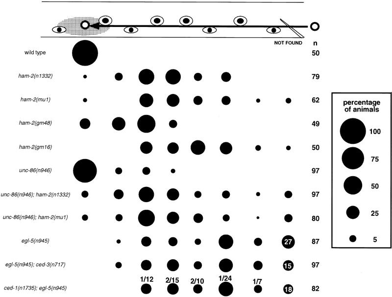
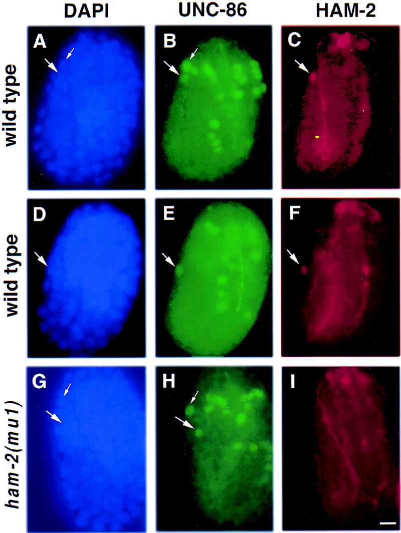
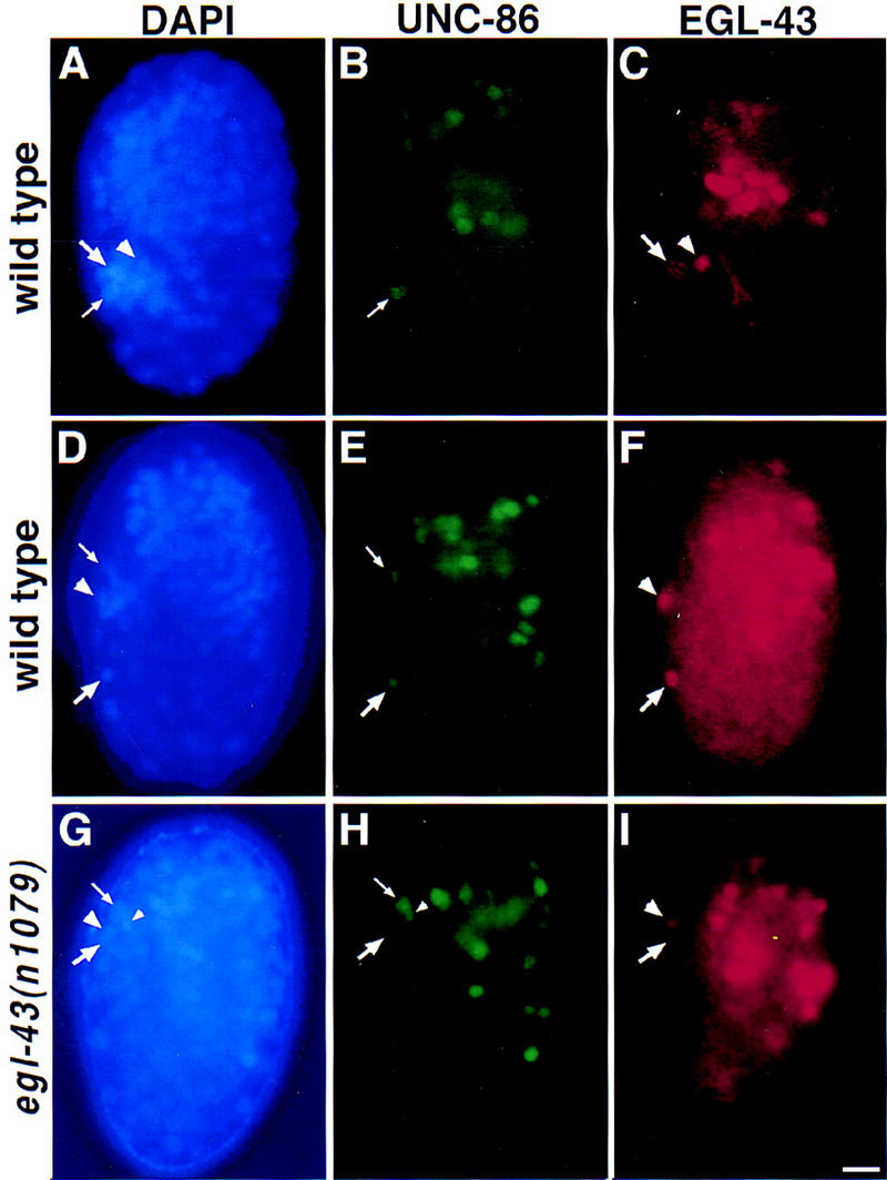
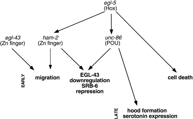
The Caenorhabditis elegans HSN motor neurons permit genetic analysis of neuronal development at single-cell resolution. The egl-5 Hox gene, which patterns the posterior of the embryo, is required for both early (embryonic) and late (larval) development of the HSN. Here we show that ham-2 encodes a zinc finger protein that acts downstream of egl-5 to direct HSN cell migration, an early differentiation event. We also demonstrate that the EGL-43 zinc finger protein, also required for HSN migration, is expressed in the HSN specifically during its migration. In an egl-5 mutant background, the HSN still expresses EGL-43, but expression is no longer down-regulated at the end of the cell's migration. Finally, we find a new role in early HSN differentiation for UNC-86, a POU homeodomain transcription factor shown previously to act downstream of egl-5 in the regulation of late HSN differentiation. In an unc-86; ham-2 double mutant the HSNs are defective in EGL-43 down-regulation, an egl-5-like phenotype that is absent in either single mutant. Thus, in the HSN, a Hox gene, egl-5, regulates cell fate by activating the transcription of genes encoding the transcription factors HAM-2 and UNC-86 that in turn individually control some differentiation events and combinatorially affect others.


Figures

Figure 1
Figure 1
HSN migration and cell-death defects of transcription factor mutants. The HSN normally migrates from the tail to the side of the gonad primordium in embryos. This figure shows the final positions of HSNs in L1 larvae from various strains, as scored by Nomarski optics. At the top, the positions of landmark hypodermal cells (eye-shaped symbols) and the gonad (gray oval) are depicted. The arrow indicates the HSN migration route. The area of each circle in the chart is proportional to the percentage of HSNs in that position along the anteroposterior axis of the worm (see key at right). In some strains, some HSNs could not be found in a percentage of the sides scored; these are represented at the right of the figure in the not found column. HSNs that could not be found along the migratory route either failed to migrate from their birthplace in the tail, where we cannot distinguish them from other neurons of the lumbar ganglia, or were missing. (n) The number of HSNs scored. We scored approximately equal numbers of left and right HSNs for each strain. The ced-3 mutation was used to reveal HSNs missing because of programmed cell deaths. The distribution of the HSNs in ced-1 and ced-3 mutants was the same as in wild type and is not shown. The percentage of HSNs that could not be found in egl-5, egl-5; ced-3, and ced-1; egl-5 mutants are indicated. The fractions above the circles for ced-1; egl-5 indicate the number of cell corpses/total number of cell corpses and surviving HSNs at each position. No cell corpses were seen along the HSN migratory route in ced-1 mutants. The unc-86 alleles e1416, n306, n843, and n844 had effects similar to unc-86(n946) on HSN cell-body position, both as single mutants and as double-mutant combinations with the ham-2(mu1) mutation (not shown). 12% of the HSNs in egl-43(n997) mutants migrated out of the tail between V5 (the fourth marker cell from the right) and P11/12 (the most right marker cell) (n = 50). 4% of the HSNs in egl-43(n997); egl-5(n945) migrated out of the tail between P9/10 (the third marker cell from the right) and P11/12 (n = 50). None of the HSNs in egl-43(n997) unc-4(e120); ham-2(n1332) migrated out of the tail (n = 40).
Figure 2
Figure 2
The Pun phenotype. Nomarski photomicrographs of L1 (first stage) larvae. The anterior (white arrowhead) and posterior (black arrowhead) ends of the pharynx are indicated. (A) wild type; (B) ham-2(gm16) mutant.
Figure 3
Figure 3
Two ham-2 transcripts encode zinc finger proteins. (A) Sequence and predicted product of two ham-2 cDNA products are shown. The longer cDNA is trans-spliced to SL1 or SL2 (italics). A shorter transcript is trans-spliced to SL2 at the beginning of the second exon (nucleotide 161). (&) The beginning of each exon. Cysteines and histidines predicted to form zinc fingers are in bold. An acidic region (amino acids 248–263) or a proline-rich region (amino acids 344–364) could potentially act as activation domains. In ham-2(gm16), nucleotide 89 is changed from c to t (underlined). This mutation changes amino acid 30 from S to F. The amino acid in this position of the finger motif is predicted to determine DNA-binding specificity (Berg and Shi 1996). In ham-2(gm48), nucleotide 115 is changed from c to t (underlined). This mutation changes amino acid 39, a histidine predicted to coordinate the zinc atom in the first zinc finger, to a tyrosine. (B) Transposon insertions in the first intron of ham-2. Only a portion of the 2260 nucleotide first intron is shown. Nucleotides are numbered starting with the first nucleotide of the intron. The ham-2(n1332) and ham-2(mu1) transposon insertion sites are indicated.
Figure 4
Figure 4
HAM-2 protein is expressed in the HSN nucleus during migration. Fluorescence photomicrographs of wild-type (A–F) and ham-2(mu1) mutant (G–I) embryos that have been stained with DAPI (blue), an UNC-86 antiserum (green) and a HAM-2 antiserum (red). Each panel presents a left lateral view of the embryo, with the developing tail to the left, the head to the right and the end of the tail and the top of the head both oriented upwards. The right sides are out of focus. (A–C) A wild-type embryo at ∼410 min after first cleavage, in which the HSN has not migrated out of the tail. (A) DAPI staining. The nuclei of the HSN (large arrow) and the ALN/PLM precursor (small arrow) are shown. (B) Anti-UNC-86 staining. Both the HSN (large arrow) and the ALN/PLM precursor (small arrow) express UNC-86. (C) Anti-HAM-2 staining. The HSN nucleus (large arrow) expresses HAM-2; several HAM-2 expressing cells in the head are out of focus. (D–F) A wild-type embryo at ∼430 min after first cleavage. (D) DAPI staining reveals the position of the HSN nucleus (arrow). The ALN/PLM precursor nucleus is out of the plane of focus. (E) Anti-UNC-86 staining. Shortly after commencing its anteriorly directed migration, the HSN begins expressing UNC-86 (arrow; Finney and Ruvkun 1990). (F) Anti-HAM-2 staining. During its migration, the HSN also expresses HAM-2 (arrow). (G–I) A ham-2(mu1) mutant embryo at ∼430 min after first cleavage. (G) DAPI staining reveals the positions of the HSN (large arrow) and the ALN/PLM precursor (small arrow) nuclei. (H) Anti-UNC-86 staining. The HSN (large arrow) and ALN/PLM precursor (small arrow) show normal expression of UNC-86. (I) Anti-HAM-2 staining. Although expression of HAM-2 in the head (mostly out of focus) and in the hypodermis (at an earlier stage not shown here) are normal, the HSN does not express HAM-2 in ham-2(mu1) embryos. The HSNs also lack HAM-2 expression in ham2(n1332) animals (not shown). Scale bar, 5 μm.
Figure 5
Figure 5
EGL-43 protein is expressed in the HSN nucleus during migration. Fluorescence photomicrographs of wild-type (A–F) and egl-43(n1079) mutant (G–I) embryos that have been stained with DAPI (blue), an UNC-86 antiserum (green), and an EGL-43 antiserum (red). Each panel presents a left lateral view of the embryo, with the developing tail to the left, the head to the right and the end of the tail and the top of the head both oriented upwards. The right sides are out of focus. (A–C) A wild-type embryo at ∼400 min after first cleavage. (A) DAPI staining was used to visualize cell nuclei. The HSN/PHB precursor (large arrow), PHA sensory neuron (arrowhead) and ALN/PLM precursor (small arrow) nuclei are located in the tail. (B) Anti-UNC-86 staining. At this time, only the ALN/PLM precursor nucleus (small arrow) expresses UNC-86 in the tail (Finney and Ruvkun 1990). (C) Anti-EGL-43 staining. At this time, only the HSN/PHB precursor (large arrow), which will divide to generate an HSN and PHB neuron, and the PHA sensory neuron (arrowhead) express EGL-43 in the tail. (D–F) Wild-type embryo at ∼430 min after first cleavage. (D) DAPI staining reveals the positions of the HSN nucleus (large arrow) which has migrated out of the tail, as well as a phasmid neuron nucleus (arrowhead) and an ALN/PLM precursor nucleus (small arrow) in the tail. (E) Anti-UNC-86 staining. Shortly after commencing its anteriorly directed migration, the HSN begins expressing UNC-86 (large arrow; Finney and Ruvkun 1990). (F) Anti-EGL-43 staining. During its migration, the HSN also expresses EGL-43-(large arrow). One of the EGL-43-expressing phasmid neuron nuclei (arrowhead) is also visible. (G–I) egl-43(n1079) mutant embryo at ∼430 min after first cleavage. (G) DAPI staining of this egl-43(n1079) mutant embryo shows the position of the HSN nucleus in the tail (large arrow). The phasmid neuron (large arrowhead), ALN/PLM precursor (small arrow) and the sister cell of the ALN/PLM precursor (small arrowhead) nuclei are indicated. (H) UNC-86 expression. The HSN neuron expressing UNC-86 (large arrow) has not migrated from its birthplace in the tail. The nuclei of the ALN/PLM precursor (small arrow) and its sister cell (small arrow) are indicated. (I) Anti-EGL-43 staining. One of the phasmid neurons (large arrowhead) but not the HSN neuron (large arrow) is expressing EGL-43. Scale bar, 5 μm.
Figure 6
Figure 6
Some HSN migration mutants fail to down-regulate EGL-43 and repress srb-6. (A–D) Fluorescence photomicrographs of wild-type (A,B) and egl-5(n945) mutant (C,D) L1 larvae that have been fixed and stained with UNC-86 and EGL-43 antisera. All four panels present left lateral views of the animals with anterior to the left. The right sides are out of focus. (A) In wild-type animals, the HSN neuron is located in the midbody and expresses UNC-86 (arrow). (B) EGL-43 expression in the same larva as in A. The PHA and PHB phasmid neuron nuclei (small arrow) express EGL-43 but the HSN neuron (arrow) does not. (C) In egl-5(n945) L1 larvae, posteriorly displaced HSNs (arrow) often fail to express UNC-86 (Baumeister et al. 1996). (D) EGL-43 expression in the same larva pictured in C. In contrast to wild-type, the posteriorly displaced HSN does express EGL-43 (arrow). The phasmid neurons also express EGL-43 (small arrow). Scale bar, 15 μm. (E) Table quantifying persistence of EGL-43 expression and presence of srb-6–gfp in the HSNs of transcription factor mutants.
Figure 7
Figure 7
A transcriptional network regulates HSN development. This figure shows transcription factors expressed in the HSN and required for HSN differentiation. The arrows designate functional relationships determined by genetic criteria; these relationships may be direct or indirect. The zinc finger protein encoded by the gene sem-4, which acts in late HSN differentiation (Basson and Horvitz 1996), is not depicted in this chart because its relationships to egl-5 and unc-86 have not been determined. Consistent with egl-43 and egl-5/ham-2 acting independently to regulate HSN migration, HSN migration in egl-43; egl-5 and egl-43; ham-2 double mutants is more severely defective than in any of the single mutants.

References

    1. Bang AG, Goulding MD. Regulation of vertebrate neural cell fate by transcription factors. Curr Opin in Neurobiol. 1996;6:25–32. - PubMed
    1. Basson M, Horvitz HR. The Caenorhabditis elegans gene sem-4 controls neuronal and mesodermal cell development and encodes a zinc finger protein. Genes & Dev. 1996;10:1953–1965. - PubMed
    1. Baumeister R, Liu Y, Ruvkun G. Lineage-specific regulators couple cell lineage asymmetry to the transcription of the Caenorhabditis elegans POU gene unc-86 during neurogenesis. Genes & Dev. 1996;10:1395–1410. - PubMed
    1. Berg JM, Shi Y. The galvanization of biology: A growing appreciation for the roles of zinc. Science. 1996;271:1081–1085. - PubMed
    1. Blumenthal T. Trans-splicing and polycistronic transcription in Caenorhabditis elegans. Trends Genet. 1995;11:132–136. - PubMed

Publication types

MeSH terms

Associated data

LinkOut - more resources