. 1999 Feb 15;13(4):484-94.
doi: 10.1101/gad.13.4.484.

Role of the Bicoid-related homeodomain factor Pitx1 in specifying hindlimb morphogenesis and pituitary development

Affiliations

Role of the Bicoid-related homeodomain factor Pitx1 in specifying hindlimb morphogenesis and pituitary development

D P Szeto et al. Genes Dev. .

Abstract

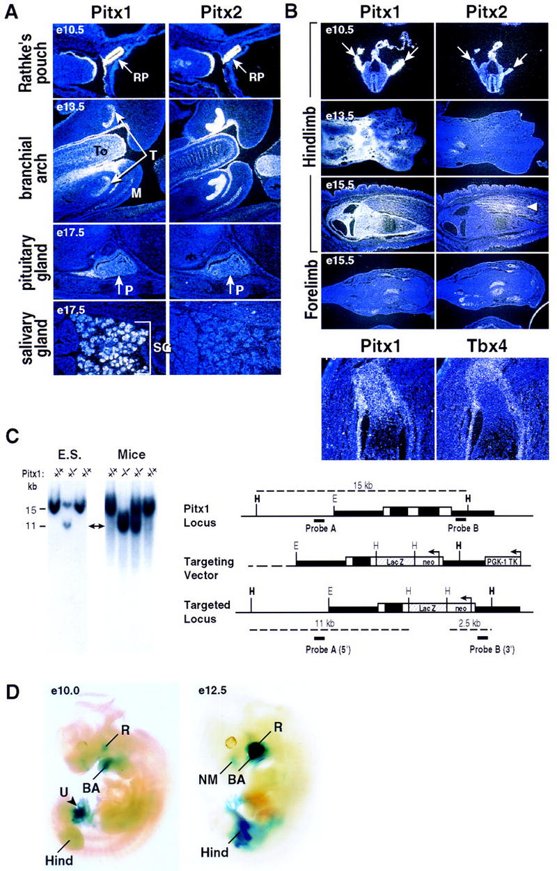
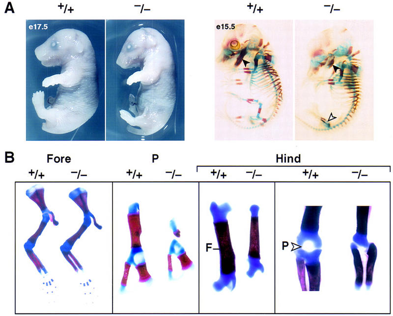
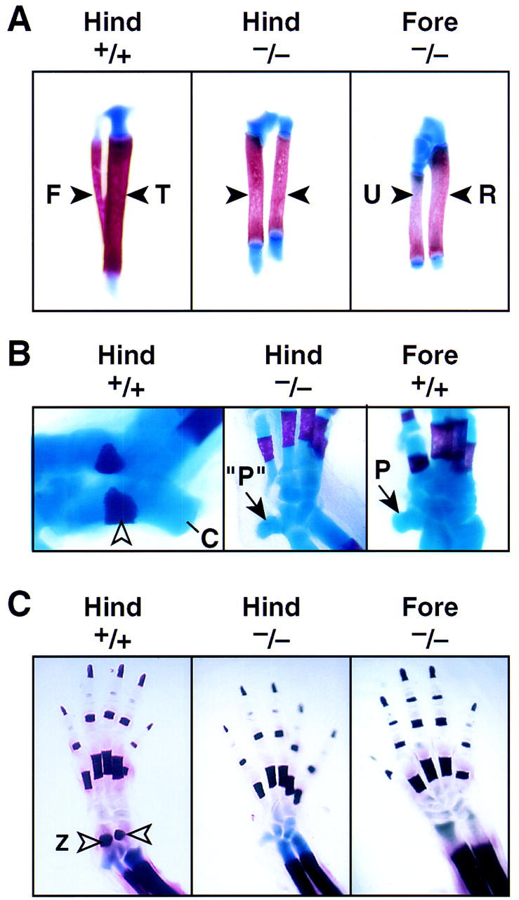
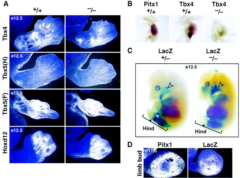
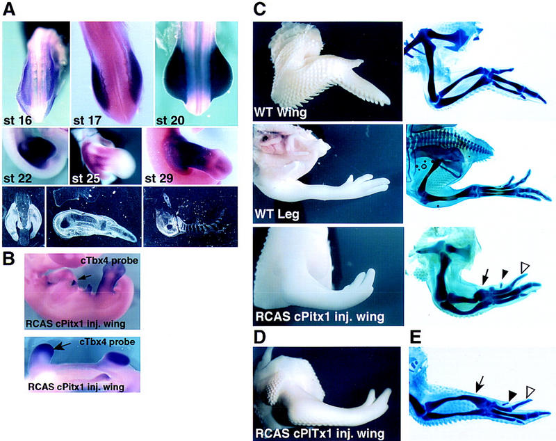
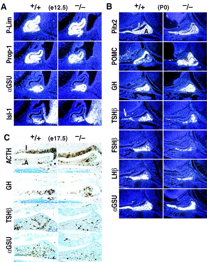
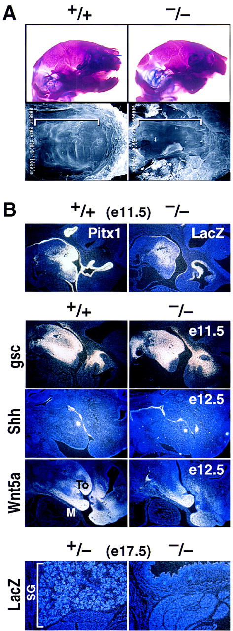
Pitx1 is a Bicoid-related homeodomain factor that exhibits preferential expression in the hindlimb, as well as expression in the developing anterior pituitary gland and first branchial arch. Here, we report that Pitx1 gene-deleted mice exhibit striking abnormalities in morphogenesis and growth of the hindlimb, resulting in a limb that exhibits structural changes in tibia and fibula as well as patterning alterations in patella and proximal tarsus, to more closely resemble the corresponding forelimb structures. Deletion of the Pitx1 locus results in decreased distal expression of the hindlimb-specific marker, the T-box factor, Tbx4. On the basis of similar expression patterns in chick, targeted misexpression of chick Pitx1 in the developing wing bud causes the resulting limb to assume altered digit number and morphogenesis, with Tbx4 induction. We hypothesize that Pitx1 serves to critically modulate morphogenesis, growth, and potential patterning of a specific hindlimb region, serving as a component of the morphological and growth distinctions in forelimb and hindlimb identity. Pitx1 gene-deleted mice also exhibit reciprocal abnormalities of two ventral and one dorsal anterior pituitary cell types, presumably on the basis of its synergistic functions with other transcription factors, and defects in the derivatives of the first branchial arch, including cleft palate, suggesting a proliferative defect in these organs analogous to that observed in the hindlimb.


Figures

Figure 1
Figure 1
Pitx1 and Pitx2 expression patterns and targeted disruption of the Pitx1 locus. (A) Expression of Pitx1 and Pitx2 analyzed by in situ hybridization. The distinct and overlapping expression patterns of Pitx1 and Pitx2 Rathke’s pouch (RP) (E10.5) and branchial arch structures (E13.5) (T) tooth; (To) tongue; (M) mandible; salivary gland (SG); (E17.5) and pituitary gland (P). (B) Expression of Pitx1, Pitx2, and Tbx4. Pitx1 is highly expressed in early limb bud, decreasing distally by E12.5; there is a differential expression of Pitx1, in the sheath and growth plate of the long bones, whereas Pitx2 is expressed in muscle (arrow). Note the similar, limited expression of Pitx1 and Pitx2 in forelimb. Pitx1 is robustly expressed in the developing hindlimb (arrows, top), throughout development with serial restriction from proximal, and then distal regions (second panel), finally in a pattern in perichondral regions, at the end of long bones, whereas Pitx2 is selectively expressed in muscle (arrow) as well as transiently in developing pelvis (not shown), Hind (hindlimb) and Fore (forelimb). Pitx1 expression overlaps with the Tbx4 expression in limb development, and is shown in the perichondral region and growth plate of the femur (P0, bottom right). (C) Targeted deletion of the Pitx1 genomic locus. Schematic representation of the Pitx1 locus (top), the targeting vector (middle), and the Pitx1 targeted allele (bottom). White and black boxes represent exons and introns, respectively, and restriction enzymes: HindIII (H); EcoRI (E). Analysis of transfected ES cells by genomic Southern blot analysis with the 5′-external probe A identifies a 11-kb HindIII fragment in the mutant allele and a 15-kb HindIII fragment in the wild-type allele. A 3′-external probe B was used to identify a 2.5-kb HindIII fragment in the mutant allele, while hybridizing to the 11-kb HindIII fragment in the wild-type mouse (not shown). Homozygous and heterozygous mice were identified by Southern blot analysis with probes A and B. (D) The normal Pitx1 expression patterns analyzed by whole-mount lacZ staining at E10–E12.5. (R) Rathke’s pouch; (NM) nasal mesenchyme; (BA) branchial arch; (U) umbilical cord; (Hind) hindlimb.
Figure 2
Figure 2
Morphological alterations of Pitx1+/+ and Pitx1−/− mice at different stages of development. (A) Pitx1+/+ and Pitx1−/− mice at E17.5; and E15.5 skeletal structures. Note the foreshortening of the mandible (solid arrowhead) and altered hindlimb (open arrowhead). (B) Details of skeletal structures of hindlimb and forelimb of Pitx1+/+ and Pitx1−/− mice at P0. The Forelimb (Fore) is unaltered, Pelvis (P) is abnormal with particular loss of the femur (F). The hindlimb (Hind) is altered in overall length and size. The patella (P) is absent (open arrowhead).
Figure 3
Figure 3
Analysis of the structural components of hindlimb development of Pitx1+/+ and Pitx1−/− at P0. (A) Skeletal structures of the tibia (T) and fibula (F) in P0 Pitx1+/+ and Pitx1−/− mice. There is alteration in size of the femur, whereas the growth and relative size of the tibia and fibula (solid arrowheads) now more closely resembles the morphological resemblance of the relationship between radius (R) and ulna (U) of the forelimb. (B) Tarsal development of the Pitx1+/+ and Pitx1−/− mice. Note the normal development of the Zucker nodes (open arrowhead) and calcaneous (C) in Pitx1+/+ mouse. The hindlimb (Hind) proximal tarsus of the Pitx1−/− mouse now contains an element that resembles the pisiform (P) seen in forelimb (Fore) carpus development of Pitx1+/+ littermate, with a very small calcaneous. (C) The development of the hindlimb distal components of Pitx1+/+ and Pitx1−/− mice at P0. Digit morphology is not reproducibly altered. Zucker nodes (open arrowhead) are absent in the hindlimb (Hind) of the Pitx1−/− mouse.
Figure 4
Figure 4
In situ hybridization and whole-mount analysis of limbs in Pitx1+/+ and Pitx1−/− mice. (A) In situ hybridization analysis of markers known to be expressed during limb development. Tbx4 expression is diminished distally, and no alteration in Tbx5 or Hoxd12 expression is noted in the hindlimb [Tbx5 (H)] or forelimb [Tbx5 (F)]. The expression of Hoxd12, PTHrP, PTHR, and Ihh are also similar in +/+ and −/− hindlimb at E12.5 and P0 (data not shown). (B) The expression patterns of Pitx1 and Tbx4 in the hindlimb by whole-mount in situ hybridization at E10.5. The Tbx4 expression overlaps with that of Pitx1 in the hindlimb of Pitx1+/+ mice and is reduced in the hindlimb of Pitx1−/− mice. (C) Whole-mount lacZ staining analysis. lacZ (marker of targeted gene) expression is present in the mandibular structures (open arrow) and is subtly but reproducibly restricted in an anterior/distal portion of the hindlimb (Hind) of Pitx1−/− mice (solid arrow in the bracket). (D) In situ hybridization analysis of Pitx1 and lacZ expression in hindlimb at E11.5, showing anterior/distal restriction of Pitx1/lacZ expression, consistent with findings in the whole-mount LacZ staining (C).
Figure 5
Figure 5
(A) Expression of cPitx1 in the developing chick hindlimb. Whole-mount and radioactive in situ hybridizations at different stages of hindlimb development. cPitx1 transcripts start to be detected in the presumptive leg bud cells. At later stages, transcripts are detected throughout the hindlimb bud cells until stage 24 where they start to disappear from the condensing cartilage (panels 4–8). The bottom right panel shows the transient expression of cPitx1 in the pelvis. (B) As seen by the ectopic patches (top) or broader expression (bottom) of Tbx4, retroviral infection of cPitx1 in the presumptive wing cells can, later on, induce the expression of the hindlimb-specific gene, Tbx4. (C) Misexpression of cPitx1 perturbs the outgrowth and patterning of the chick wing. Shown are whole-mount and skeletal preparations of control, and not injected, chick wing and leg. The chick wing has three digits of variable length, is covered by feathers, and displays a characteristic downward flexure at the wrist level. Instead, the chick leg is straight in its most distal part, not covered by feathers but by small scales, and has three digits of similar length and an additional small four digits located toward the back of the foot. Ectopic expression of cPitx1 in the wing transforms the curvature of the wing into an almost straight position, increases the size of digit 2 (open arrowhead) and induces the formation of a fourth digit (solid arrowhead). All of these changes in growth and patterning, together with the disappearance of feathers from the distal side of the wing, induce the infected wing to resemble a leg. Some of the infected embryos showed alterations in the size and morphology of the radius and ulna (solid arrow). (D,E) Results of a second experiment, to illustrate the range of phenotypic variations.
Figure 6
Figure 6
Effects of Pitx1 deletion on development of the anterior pituitary gland. (A) Effects of Pitx1 gene deletion on P-Lim, Prop-1, αGSU, and Isl-1 expression in the pituitary gland. No differences were observed in the expression of these factors in Pitx1−/− embryos as compared with that of the wild-type at E12.5. (B,C) Pitx1 gene deletion alters expression of ventral pituitary-specific cell types at P0 in mice analyzed by in situ hybridization (B) or by immunohistochemical analysis at E17.5 (C). The expression of TSHβ, FSHβ, and LHβ are markedly decreased, with some reduction of αGSU. Note particularly the loss of TSHβ in the rostral tip (arrow). Pitx2 and growth hormone (GH) gene expression are unchanged. POMC transcripts and ACTH immuno activity in anterior lobe are consistently increased. Because of saturation of the film with the POMC probe, the region is artificially black in a portion of the intermediate lobe (asterisk, B). Note that ACTH staining is similar on wild-type and −/− glands in the intermediate lobe (I), while clearly increased in the anterior lobe (A) (C).
Figure 7
Figure 7
Effects of Pitx1 gene deletion on mandible, palate, and submandibular gland development at E17.5 and P0. (A) Marked reduction in size of mandible analyzed by skeletal staining (top) and failure of palatal closure (see brackets) analyzed by scanning electron microscopy in the Pitx1−/− mouse (bottom). (B) Mandibular expression of Pitx1 and lacZ as a marker in Pitx1+/+ and Pitx1−/− mice, respectively at E11.5. Markers of mandibular development analyzed at E11.5 and E12.5 by in situ hybridization included gsc, Shh, and Wnt5a. Submandibular gland (SG) development in Pitx1+/− and Pitx1−/− mice at E17.5 marked by the lacZ probe. (To) tongue; (M) mandible.

References

    1. Acampora D, Mazan S, Lallemand Y, Avantaggiato V, Maury M, Simeone A, Brûlet P. Forebrain and midbrain regions are deleted in Otx2-/- mutants due to a defective anterior neuroectoderm specification during gastrulation. Development. 1995;121:3279–3290. - PubMed
    1. Acampora D, Mazan S, Avantaggiato V, Barone P, Tuorto F, Lallemand Y, Brûlet P, Simeone A. Epilepsy and brain abnormalities in mice lacking the Otx1 gene. Nat Genet. 1996;14:218–222. - PubMed
    1. Acampora D, Mazan S, Tuorto F, Avantaggiato V, Tremblay JJ, Lazzaro D, di Carlo A, Mariano A, Macchia PE, Corte G, Macchia V, Drouin J, Brulet P, Simeone A. Transient dwarfism and hypogonadism in mice lacking Otx-1 reveal prepubescent stage-specific control of pituitary levels of Gh, FSH and LH. Development. 1998;125:1229–1239. - PubMed
    1. Ang SL, Jin O, Rhinn M, Daigle N, Stevenson L, Rossant J. A targeted mouse Otx2 mutation leads to severe defects in gastrulation and formation of axial mesoderm and to deletion of rostral brain. Development. 1996;122:243–252. - PubMed
    1. Bermingham JR, Jr, Scherer SS, O’Connell S, Arroyo E, Kalla KA, Powell FL, Rosenfeld MG. Tst-1/Oct-6/SCIP regulates a unique step in peripheral myelination and is required for normal respiration. Genes & Dev. 1996;10:1751–1762. - PubMed

Publication types

MeSH terms