. 1999 Mar;181(5):1555-61.
doi: 10.1128/JB.181.5.1555-1561.1999.

Peptidoglycan-hydrolyzing activity of the FlgJ protein, essential for flagellar rod formation in Salmonella typhimurium

Affiliations

Peptidoglycan-hydrolyzing activity of the FlgJ protein, essential for flagellar rod formation in Salmonella typhimurium

T Nambu et al. J Bacteriol. 1999 Mar.

Abstract

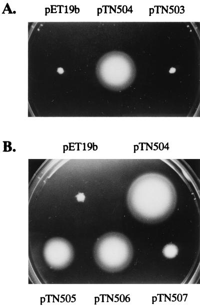
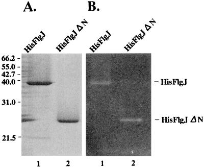
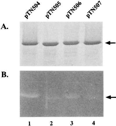
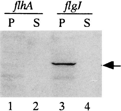
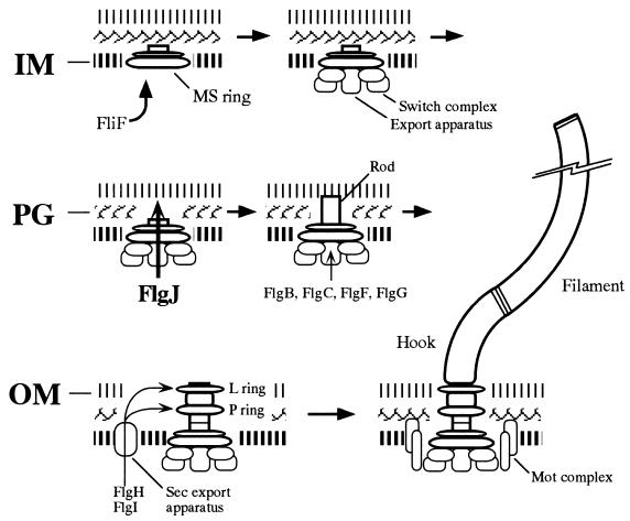
Because the rod structure of the flagellar basal body crosses the inner membrane, the periplasmic space, and the outer membrane, its formation must involve hydrolysis of the peptidoglycan layer. So far, more than 10 genes have been shown to be required for rod formation in Salmonella typhimurium. Some of them encode the component proteins of the rod structure, and most of the remaining genes are believed to encode proteins involved in the export process of the component proteins. Although FlgJ has also been known to be involved in rod formation, its exact role has not been understood. Recently, it was suggested that the C-terminal half of the FlgJ protein has homology to the active center of some muramidase enzymes from gram-positive bacteria. In this study, we showed that the purified FlgJ protein from S. typhimurium has a peptidoglycan-hydrolyzing activity and that this activity is localized in its C-terminal half. Through oligonucleotide-directed mutagenesis, we constructed flgJ mutants with amino acid substitutions in the putative active center of the muramidase. The resulting mutants produced FlgJ proteins with reduced enzymatic activity and showed poor motility. These results indicate that the muramidase activity of FlgJ is essential for flagellar formation. Immunoblotting analysis with the fractionated cell extracts revealed that FlgJ is exported to the periplasmic space, where the peptidoglycan layer is localized. On the basis of these results, we conclude that FlgJ is the flagellum-specific muramidase which hydrolyzes the peptidoglycan layer to assemble the rod structure in the periplasmic space.


Figures

FIG. 1
FIG. 1
Structures of the wild-type (A), His-tagged (B), and mutant (C) FlgJ proteins. Asterisks indicate the amino acids which are conserved in the putative active center of the enzymes of the muramidase family. Numbers above the bars indicate amino acid residues from the N terminus. Shaded and solid boxes indicate the His10 and FLAG tags, respectively, in the FlgJ proteins encoded by the recombinant plasmids. Hatched areas represent amino acids created by the frameshift mutations, and the numbers of amino acids added are shown in parentheses. Because all the Tn10-induced flgJ mutants (KK strains) carry Tn10 inserted in the identical site, only the structure of the FlgJ protein encoded by KK2017 is shown.
FIG. 2
FIG. 2
Motility recovery of the flgJ mutants by the His-tagged FlgJ-encoding plasmids. (A) Plasmids were introduced into SJW1488 and KK2017 by transformation. Single colonies of the resulting transformants were stabbed onto motility agar plates and incubated for 4 h at 37°C. In this figure, only the result with SJW1488 is shown. The same result was obtained with KK2017. (B) Experiments were carried out as for panel A, but only SJW1488 was used.
FIG. 3
FIG. 3
SDS-PAGE and zymogram analyses of the His-tagged FlgJ proteins. (A) The purified proteins were separated in an SDS–12% polyacrylamide gel. After electrophoresis, the gel was stained with 0.25% Coomassie brilliant blue. The molecular masses of the marker proteins are indicated in kilodaltons on the left. (B) The purified proteins were separated in an SDS–12% polyacrylamide gel containing M. lysodeikticus cells. After electrophoresis, the gel was treated as described in Materials and Methods for the zymogram analysis. Equal molar amounts of protein (54 pmol) were applied to each lane. The proteins used were His-FlgJ (lane 1) and His-FlgJΔN (lane 2).
FIG. 4
FIG. 4
SDS-PAGE (A) and zymogram (B) analyses of the FlgJ proteins with amino acid substitutions in the putative active center of muramidase. The procedures used were the same as those described in the legend to Fig. 3. The arrows indicate the positions of the His-tagged FlgJ proteins. The proteins used were His-FlgJ (lane 1), His-FlgJ(E223Q) (lane 2), His-FlgJ(D248N) (lane 3), and His-FlgJ(E223Q-D248N) (lane 4).
FIG. 5
FIG. 5
Export assay of FlgJ. Proteins from the periplasmic content (P) and culture supernatant (S) were prepared from the 1 mM IPTG-induced culture of the cells harboring pMM505. After separation by SDS-PAGE, the proteins were transferred to a nitrocellulose membrane and visualized immunologically with anti-FLAG antibody. The arrow indicates the position of the His-FLAG-tagged FlgJ protein. The strains used were SJW1364 (flhA) (lanes 1 and 2) and SJW1437 (flgJ) (lanes 3 and 4).
FIG. 6
FIG. 6
Model of the flagellar assembly pathway with special emphasis on the processes involved in the crossing of three envelope barriers by the rod structure. The rod is composed of at least four proteins, FlgB, FlgC, FlgF, and FlgG, which are exported into the periplasmic space via the flagellum-specific export pathway. Rod assembly proceeds by the formation of holes in the inner membrane (IM) by FliF (36), in the peptidoglycan (PG) by FlgJ (this study), and in the outer membrane (OM) by FlgH (16). Details are described in the text. Other assembly processes are drawn on the basis of the model described previously (1, 19).

References

    1. Aizawa S-I. Flagellar assembly in Salmonella typhimurium. Mol Microbiol. 1996;19:1–5. - PubMed
    1. Amann E, Ochs B, Abel K-J. Tightly regulated tac promoter vectors useful for the expression of unfused and fused proteins in Escherichia coli. Gene. 1988;69:301–314. - PubMed
    1. Baba T, Schneewind O. Targeting of muralytic enzymes to the cell division site of gram-positive bacteria: repeat domains direct autolysin to the equatorial surface ring of Staphylococcus aureus. EMBO J. 1998;17:4639–4646. - PMC - PubMed
    1. Buist G, Kok J, Leenhouts K J, Dabrowska M, Venema G, Haandrikman A J. Molecular cloning and nucleotide sequence of the gene encoding the major peptidoglycan hydrolase of Lactococcus lactis, a muramidase needed for cell separation. J Bacteriol. 1995;177:1554–1563. - PMC - PubMed
    1. DePamphilis M L, Adler J. Fine structure and isolation of the hook-basal body complex of flagella from Escherichia coli and Bacillus subtilis. J Bacteriol. 1971;105:384–395. - PMC - PubMed

Publication types

LinkOut - more resources