Comparative Study
. 1999 Mar;181(5):1652-63.
doi: 10.1128/JB.181.5.1652-1663.1999.

Characterization of the exopolygalacturonate lyase PelX of Erwinia chrysanthemi 3937

Affiliations
Comparative Study

Characterization of the exopolygalacturonate lyase PelX of Erwinia chrysanthemi 3937

V E Shevchik et al. J Bacteriol. 1999 Mar.

Abstract

Erwinia chrysanthemi 3937 secretes several pectinolytic enzymes, among which eight isoenzymes of pectate lyases with an endo-cleaving mode (PelA, PelB, PelC, PelD, PelE, PelI, PelL, and PelZ) have been identified. Two exo-cleaving enzymes, the exopolygalacturonate lyase, PelX, and an exo-poly-alpha-D-galacturonosidase, PehX, have been previously identified in other E. chrysanthemi strains. Using a genomic bank of a 3937 mutant with the major pel genes deleted, we cloned a pectinase gene identified as pelX, encoding the exopolygalacturonate lyase. The deduced amino acid sequence of the 3937 PelX is very similar to the PelX of another E. chrysanthemi strain, EC16, except in the 43 C-terminal amino acids. PelX also has homology to the endo-pectate lyase PelL of E. chrysanthemi but has a N-terminal extension of 324 residues. The transcription of pelX, analyzed by gene fusions, is dependent on several environmental conditions. It is induced by pectic catabolic products and affected by growth phase, oxygen limitation, nitrogen starvation, and catabolite repression. Regulation of pelX expression is dependent on the KdgR repressor, which controls almost all the steps of pectin catabolism, and on the global activator of sugar catabolism, cyclic AMP receptor protein. In contrast, PecS and PecT, two repressors of the transcription of most pectate lyase genes, are not involved in pelX expression. The pelX mutant displayed reduced pathogenicity on chicory leaves, but its virulence on potato tubers or Saintpaulia ionantha plants did not appear to be affected. The purified PelX protein has no maceration activity on plant tissues. Tetragalacturonate is the best substrate of PelX, but PelX also has good activity on longer oligomers. Therefore, the estimated number of binding subsites for PelX is 4, extending from subsites -2 to +2. PelX and PehX were shown to be localized in the periplasm of E. chrysanthemi 3937. PelX catalyzed the formation of unsaturated digalacturonates by attack from the reducing end of the substrate, while PehX released digalacturonates by attack from the nonreducing end of the substrate. Thus, the two types of exo-degrading enzymes appeared complementary in the degradation of pectic polymers, since they act on both extremities of the polymeric chain.


Figures

FIG. 1
FIG. 1
Physical map of the E. chrysanthemi 3937 pelX gene. The restriction map of the three PelX-encoding plasmids is indicated. The arrows below the MunI-PvuII fragment indicate the position and the transcription direction of the pelX gene and of adjacent genes. The flag shows the site of insertion of the uidA-Km cassette.
FIG. 2
FIG. 2
Comparison of the amino acid sequences of the PelX and PelL proteins. The PelX sequence of E. chrysanthemi EC16 is from reference , and PelL of strain 3937 was described by Lojkowska et al. (23). Colons indicates identical residues. The signal sequence of the proteins are underlined. D1 and D2 indicate the ends of the two deletions on 3937 PelX.
FIG. 3
FIG. 3
Alignment of the pelX promoters of E. chrysanthemi 3937 and EC16. The PelX sequence of E. chrysanthemi EC16 is from reference . Vertical lines indicate identical bases. The putative sequences corresponding to the translation start, Shine-Dalgarno (S.D.), −10 and −35 promoter, KdgR, and CRP-binding sites are underlined.
FIG. 4
FIG. 4
Growth of E. chrysanthemi pelX mutants on polygalacturonate. The sole carbon source was polygalacturonate (4 g · liter−1) in M63 medium. Growth was monitored by measuring the optical density of the culture at 600 nm (OD600). A350 is the parental strain of the two pelX mutants A2524 and A2699.
FIG. 5
FIG. 5
Location of the pelX gene on the genetic map of E. chrysanthemi 3937. Localization was performed by chromosomal mobilization with plasmid pULB110. The numbers indicate the percentage of cotransfer between two markers. Arrowheads point to the unselected marker.
FIG. 6
FIG. 6
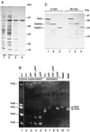
Identification of the PelX protein. (A) SDS-PAGE separation was followed by staining with Coomassie blue. Lanes: Whole-cell extract of BL21(DE3)/pTaX before (lane 1) and after (lane 2) IPTG induction; periplasmic fraction of BL21(DE3)/pTaX after IPTG induction (lane 3); pure PelX protein (lane 4). Molecular mass markers are indicated. (B) Electrofocusing was performed either at 2,500 V-h (lane 1) or at 1,500 V-h (lanes 2 to 11) and followed by specific revelation of pectate lyase activity in the presence of either CaCl2 for 10 min (lane 1) or MnCl2 for 40 min (lanes 2 to 11). Culture supernatants (lanes 1 to 5) and periplasmic fractions (lanes 6 to 9) of the E. chrysanthemi strains are shown: A837 (parental strain) (lanes 1, 2, and 6), A3497 (Δpeh) (lanes 3 and 7), A3498 (Δpeh pelX::Cm) (lanes 4 and 8), and A2737 (pelX::Km) (lanes 5 and 9). Lanes 10 and 11 contain purified PehX and PelX, respectively. The positions of the five major pectate lyases (PelA to PelE), PelX, and PehX are indicated. w.t., wild type. (C) Stability of the PelX deletion derivatives. E. coli K38/pGP1.2 cells carrying pTaX (lane 1), pTaXD2 (lane 2), or pTaXD1 (lane 3) were labelled with [35S]cysteine-[35S]methionine. An excess of “cold” cysteine-methionine was added 10 min later, and the cells were collected immediately (0 min) or incubated for additional 30 min at 37°C (30 min).
FIG. 7
FIG. 7
Enzymatic properties of PelX. (A) The influence of Ca2+ was tested by using various concentrations of CaCl2 in 50 mM Tris-HCl (pH 8.5)–0.5 g of polygalacturonate per liter. The cation requirement was confirmed by addition of 0.5 and 1 mM EDTA. (B) The effect of pH was tested with 0.5 g of polygalacturonate per liter as substrate, in the presence of 0.1 mM CaCl2 in 50 mM ACES-NaOH buffer (pH 5.5 to 7.5 [open symbols]) or in 50 mM Tris-HCl buffer (pH 7 to 9 [solid symbols]). (C) The thermostability of PelX was monitored at 45°C after various incubation times in 50 mM Tris-HCl (pH 8.5) without (control) or with the addition of CaCl2 (0.5 mM), EDTA (0.5 mM), or polygalacturonate (0.5 g · liter−1). The residual activity is given as a percentage of the initial activity. Activity toward pectins presenting various degrees of methylation (D) was determined in 50 mM Tris-HCl (pH 8.5)–0.1 mM CaCl2–0.5 g of substrate liter−1.
FIG. 8
FIG. 8
Separation of the PelX and PehX reaction products by thin-layer chromatography. The reaction mixtures contained 0.1 M Tris-HCl (pH 8), 0.1 mM CaCl2, 5 U of enzymatic activity per ml, and 1.5 mg of the following substrates per ml: digalacturonic acid (G2), trigalacturonic acid (G3), a mixture of unsaturated di- and trigalacturonic acids (uG2, uG3), or polygalacturonic acid (PGA). Incubations were performed at 30°C for 3 h after addition of PelX or PehX or without enzyme (−). A 5-μl sample from each reaction mixture was applied to chromatogram sheets. The positions of the individual compounds are indicated by arrows. It should be noted that the staining method used is not sensitive enough to detect the unsaturated monomer which appears by degradation of uG3 with PelX.
FIG. 9
FIG. 9
Analysis of PelX and PehX reaction products on hexagalacturonate (A) and reduced hexagalacturonate (B) by high-pressure liquid chromatography. For PelX, the reaction mixtures (0.5 ml) contained 0.5 mM substrate in 20 mM Tris-HCl (pH 8)–1 mM CaCl2. A 10- or 200-ng sample of PelX was used in a 30-min reaction for G6 (A) and rG6 (B), respectively. For PehX, the reaction mixtures (0.5 ml) contained 0.5 mM substrate in 20 mM Tris-HCl pH 7. 25 ng or 500 ng of PehX were used in a 5 min reaction for G6 (A) and rG6 (B), respectively. Reactions were carried out at 30°C. The blanks contained the substrate prior to enzyme addition. The reaction products were identified by using standard mixtures of uG2 and uG3 (curve 1), oligogalacturonates G1 to G6 (curve 3), and reduced oligogalacturonates rG1 to rG6 (curve 2).
FIG. 10
FIG. 10
Sites of attack of the E. chrysanthemi exo-enzymes PelX and PehX. The arrows indicate the site of cleavage of the polymer, polygalacturonate, for each type of enzyme produced by E. chrysanthemi, PehX, PelX, and the endo-Pels (PelA, PelB, PelC, PelD, PelE, PelI, PelL, and PelZ). The sole product resulting from the action of the exo-polygalacturonase, PehX, or the exopolygalacturonate lyase, PelX, is indicated below the corresponding arrow.

Similar articles

Cited by

References

    1. Alfano J R, Ham J H, Collmer A. Use of Tn5tac1 to clone a pel gene encoding a highly alkaline, asparagine-rich pectate lyase isozyme from an Erwinia chrysanthemi EC16 mutant with deletions affecting the major pectate lyase isozymes. J Bacteriol. 1995;177:4553–4556. - PMC - PubMed
    1. Bardonnet N, Blanco C. uidA antibiotic resistance cassettes for insertion mutagenesis, gene fusion and genetic constructions. FEMS Microbiol Lett. 1992;93:243–248. - PubMed
    1. Barras F, Van Gijsegem F, Chatterjee A K. Extracellular enzymes and pathogenesis of soft-rot Erwinia. Annu Rev Phytopathol. 1994;32:201–234.
    1. Beaulieu C, Boccara M, Van Gijsegem F. Pathogenic behaviour of pectinase-defective Erwinia chrysanthemi mutants on different plants. Mol Plant-Microbe Interact. 1993;6:197–202.
    1. Beer, S. Personal communication.

Publication types

MeSH terms

Associated data

LinkOut - more resources