. 1999 Mar;65(3):1061-70.
doi: 10.1128/AEM.65.3.1061-1070.1999.

Induction of defense responses in cucumber plants (Cucumis sativus L. ) By the biocontrol agent trichoderma harzianum

Affiliations

Induction of defense responses in cucumber plants (Cucumis sativus L. ) By the biocontrol agent trichoderma harzianum

I Yedidia I et al. Appl Environ Microbiol. 1999 Mar.

Abstract

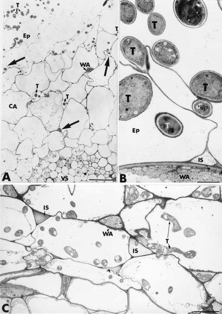
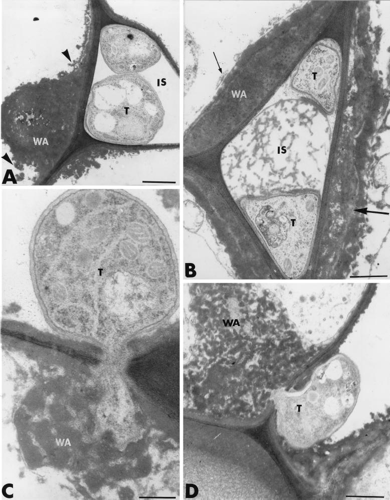
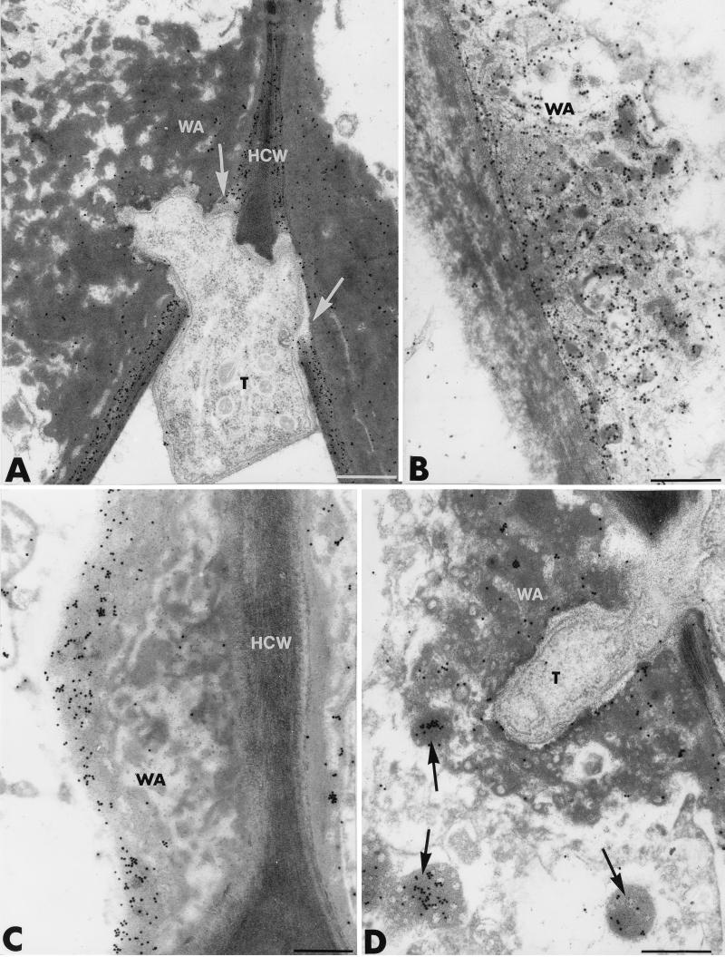
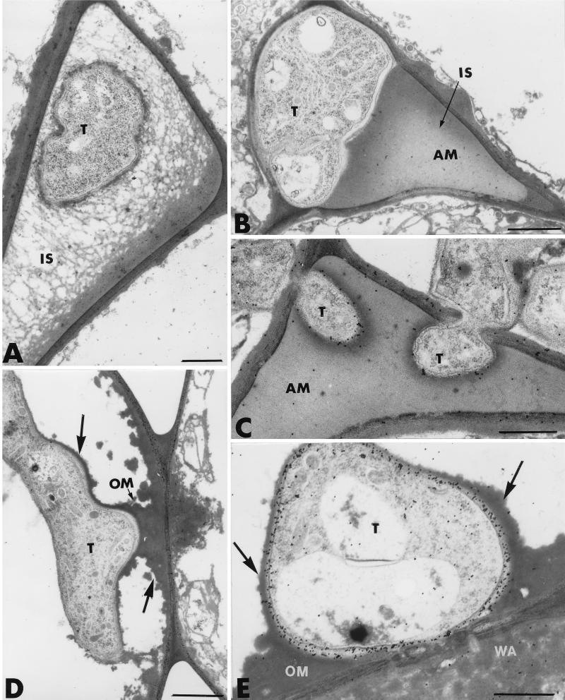
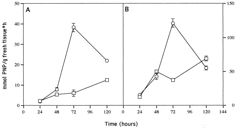
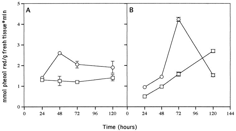
The potential of the biocontrol agent Trichoderma harzianum T-203 to trigger plant defense responses was investigated by inoculating roots of cucumber seedlings with Trichoderma in an aseptic, hydroponic system. Trichoderma-treated plants were more developed than nontreated plants throughout the experiment. Electron microscopy of ultrathin sections from Trichoderma-treated roots revealed penetration of Trichoderma into the roots, restricted mainly to the epidermis and outer cortex. Strengthening of the epidermal and cortical cell walls was observed, as was the deposition of newly formed barriers. These typical host reactions were found beyond the sites of potential fungal penetration. Wall appositions contained large amounts of callose and infiltrations of cellulose. The wall-bound chitin in Trichoderma hyphae was preserved, even when the hyphae had undergone substantial disorganization. Biochemical analyses revealed that inoculation with Trichoderma initiated increased peroxidase and chitinase activities within 48 and 72 h, respectively. These results were observed for both the roots and the leaves of treated seedlings, providing evidence that T. harzianum may induce systemic resistance mechanisms in cucumber plants.


Figures

FIG. 1
FIG. 1
Transmission electron micrographs of T. harzianum-inoculated cucumber root tissues. A large number of fungal hyphae (T) develop at the root surface. Trichoderma hyphae penetrate the root epidermis (Ep) and progress toward the cortical area (CA), mainly by intercellular growth (arrows in A). Wall appositions (WA) are seen in noninvaded host cells beneath the colonized areas. Fungal colonization of the epidermis and cortex is not associated with host cell alterations or cell wall digestion (C). IS, intercellular spaces; VS, vascular stele. Bars: A, 10 μm; B, 1.5 μm; C, 5 μm.
FIG. 2
FIG. 2
Transmission electron micrographs of T. harzianum-inoculated cucumber root tissues. (A and B) Heterogeneous wall appositions (WA) are formed in noninfected host cells adjacent to invaded cells. The material accumulating at the junction of adjacent host cells was either stratified and bordered by a layer of aggregated material (large arrow in B) or granular and of very high electron density (small arrow in B). Frequently, the appositions were delimited by a band of osmiophilic material that released small droplets into the cell lumen (arrowheads in A). (C and D) Unsuccessful attempts by the fungus to penetrate WA are observed. Trichoderma hyphae (T) penetrating such appositions show marked signs of alteration characterized by morphological changes and even necrosis of the penetration peg. IS, intercellular spaces. Bars: A and D, 1 μm; B, 0.5 μm; C, 0.25 μm.
FIG. 3
FIG. 3
Transmission electron micrographs of T. harzianum-inoculated cucumber root tissues. (A) Labeling with the β-1,4-exoglucanase–gold complex to localize cellulose. The host cell walls (HCW) are heavily labeled. Labeling also occurs over the wall appositions (WA) but is irregularly distributed. Trichoderma hyphae (T) display the ability to locally disrupt the cellulose-enriched HCW (arrows). Bar, 0.25 μm. (B to D) Labeling with the tobacco β-1,3-glucanase to localize callose. Young WA, mainly characterized by their loose arrangement and their low density (B), are more intensely labeled than the more mature ones, labeling of which occurs preferentially over the outermost layer (C). Electron-opaque vesicles, either enclosed in the WA (arrow pointing up in D) or apparently free in the cytoplasm (arrows pointing down in D), are labeled by a substantial number of gold particles. Bars, 0.25 μm.
FIG. 4
FIG. 4
Transmission electron micrographs of T. harzianum-inoculated cucumber root tissues. Most intercellular spaces (IS) in the epidermis and outer root cortex are occluded with a dense material (AM) that shows various degrees of compactness and electron opacity. Fungal cells trapped in this material are morphologically altered. Upon incubation with the tobacco β-1,3-glucanase, this material appears unlabeled, except next to the fungal cells, where a few gold particles can be seen (C). An osmiophilic material (OM) coating the host cell walls surrounds the invading hyphae (arrows in D). Upon labeling with WGA–ovomucoid-gold complex for the localization of chitin, regular deposition of gold particles occurs over the coated fungal cell walls; arrows show the OM coating (E). T, Trichoderma hyphae; WA, wall appositions. Bars: A, D, and E, 0.5 μm; B and C, 1 μm.
FIG. 5
FIG. 5
Chitinase activity in roots (A) and leaves (B) of 7-day-old cucumber seedlings inoculated with Trichoderma (○) at 24 to 120 h and uninoculated controls (□). Bars represent 1 standard deviation.
FIG. 6
FIG. 6
Peroxidase activity in roots (A) and leaves (B) of 7-day-old cucumber seedlings inoculated with Trichoderma (○) at 24 to 120 h and uninoculated controls (□). Bars represent 1 standard deviation.

References

    1. Baker R. Improved Trichoderma spp. for promoting crop productivity. Trends Biotech. 1989;7:34–38.
    1. Benhamou N. Immunocytochemistry of plant defense mechanisms induced upon microbial attack. Microsc Res Tech. 1995;31:63–78. - PubMed
    1. Benhamou N. Preparation and application of lectin-gold complexes. In: Hayat M A, editor. Colloidal gold, principles, methods, and applications. Vol. 1. New York, N.Y: Academic Press, Inc.; 1989. pp. 95–143.
    1. Benhamou N. Ultrastructural detection of β-1,3-glucans in tobacco root tissues infected by Phytophthora parasitica var. nicotianae using a gold-complexed tobacco β-1,3-glucanase. Physiol Mol Plant Pathol. 1992;41:351–370.
    1. Benhamou N, Chamberland H, Ouellette G B, Pauzé F J. Ultrastructural localization of β-1,4-d-glucans in two pathogenic fungi and in their host tissues by means of an exoglucanase-gold complex. Can J Microbiol. 1987;33:405–417.

LinkOut - more resources