

In situ identification of cyanobacteria with horseradish peroxidase-labeled, rRNA-targeted oligonucleotide probes
- PMID: 10049892
- PMCID: PMC91173
- DOI: 10.1128/AEM.65.3.1259-1267.1999
In situ identification of cyanobacteria with horseradish peroxidase-labeled, rRNA-targeted oligonucleotide probes
Abstract
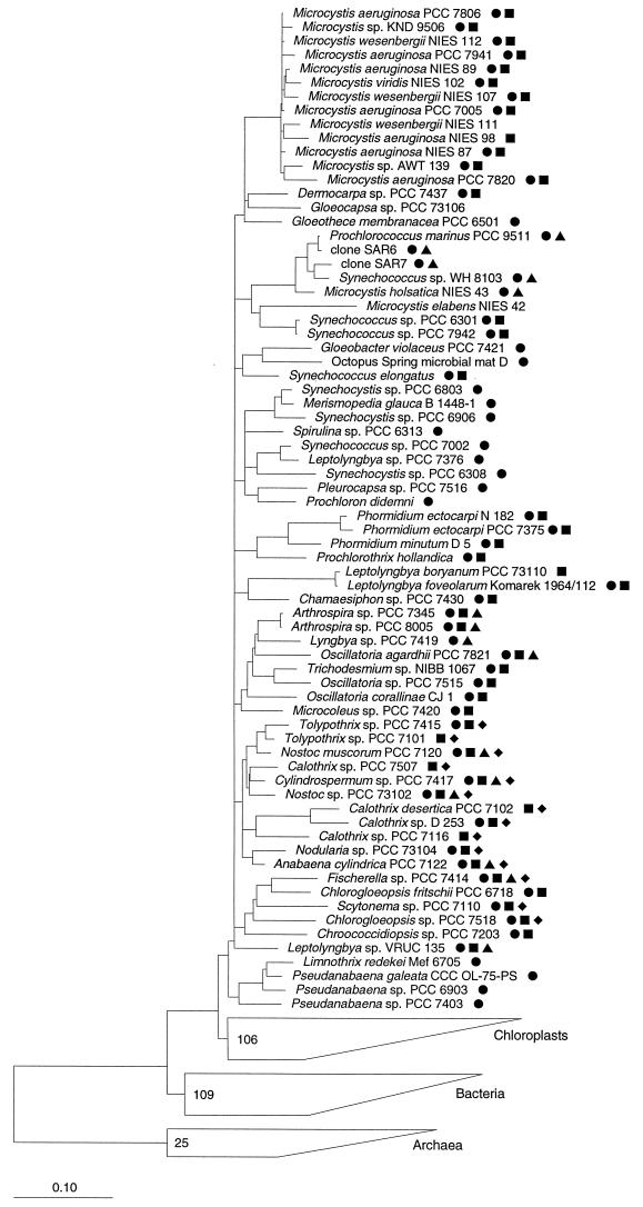
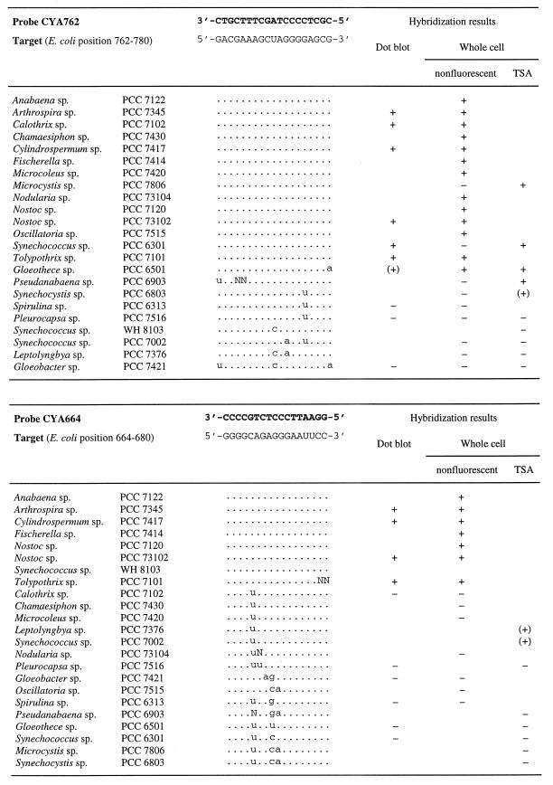
Individual cyanobacterial cells are normally identified in environmental samples only on the basis of their pigmentation and morphology. However, these criteria are often insufficient for the differentiation of species. Here, a whole-cell hybridization technique is presented that uses horseradish peroxidase (HRP)-labeled, rRNA-targeted oligonucleotides for in situ identification of cyanobacteria. This indirect method, in which the probe-conferred enzyme has to be visualized in an additional step, was necessary since fluorescently monolabeled oligonucleotides were insufficient to overstain the autofluorescence of the target cells. Initially, a nonfluorescent detection assay was developed and successfully applied to cyanobacterial mats. Later, it was demonstrated that tyramide signal amplification (TSA) resulted in fluorescent signals far above the level of autofluorescence. Furthermore, TSA-based detection of HRP was more sensitive than that based on nonfluorescent substrates. Critical points of the assay, such as cell fixation and permeabilization, specificity, and sensitivity, were systematically investigated by using four oligonucleotides newly designed to target groups of cyanobacteria.
Figures


Similar articles
-
Picobenthic cyanobacterial populations revealed by 16S rRNA-targeted in situ hybridization.Environ Microbiol. 2002 Jul;4(7):375-82. doi: 10.1046/j.1462-2920.2002.00307.x. Environ Microbiol. 2002. PMID: 12123473
-
Improved sensitivity of whole-cell hybridization by the combination of horseradish peroxidase-labeled oligonucleotides and tyramide signal amplification.Appl Environ Microbiol. 1997 Aug;63(8):3268-73. doi: 10.1128/aem.63.8.3268-3273.1997. Appl Environ Microbiol. 1997. PMID: 9251215 Free PMC article.
-
Closely related Prochlorococcus genotypes show remarkably different depth distributions in two oceanic regions as revealed by in situ hybridization using 16S rRNA-targeted oligonucleotides.Microbiology (Reading). 2001 Jul;147(Pt 7):1731-1744. doi: 10.1099/00221287-147-7-1731. Microbiology (Reading). 2001. PMID: 11429451
-
Single-cell identification in microbial communities by improved fluorescence in situ hybridization techniques.Nat Rev Microbiol. 2008 May;6(5):339-48. doi: 10.1038/nrmicro1888. Nat Rev Microbiol. 2008. PMID: 18414500 Review.
-
A survey of the relative abundance of specific groups of cellulose degrading bacteria in anaerobic environments using fluorescence in situ hybridization.J Appl Microbiol. 2007 Oct;103(4):1332-43. doi: 10.1111/j.1365-2672.2007.03362.x. J Appl Microbiol. 2007. PMID: 17897237 Review.
Cited by
-
Coexistence of bacterial sulfide oxidizers, sulfate reducers, and spirochetes in a gutless worm (Oligochaeta) from the Peru margin.Appl Environ Microbiol. 2005 Mar;71(3):1553-61. doi: 10.1128/AEM.71.3.1553-1561.2005. Appl Environ Microbiol. 2005. PMID: 15746360 Free PMC article.
-
Time-Resolved Visualization of Cyanotoxin Synthesis via Labeling by the Click Reaction in the Bloom-Forming Cyanobacteria Microcystis aeruginosa and Planktothrix agardhii.Toxins (Basel). 2025 Jun 3;17(6):278. doi: 10.3390/toxins17060278. Toxins (Basel). 2025. PMID: 40559856 Free PMC article.
-
Snow surface microbiome on the High Antarctic Plateau (DOME C).PLoS One. 2014 Aug 7;9(8):e104505. doi: 10.1371/journal.pone.0104505. eCollection 2014. PLoS One. 2014. PMID: 25101779 Free PMC article.
-
Fluorescence in situ hybridization and catalyzed reporter deposition for the identification of marine bacteria.Appl Environ Microbiol. 2002 Jun;68(6):3094-101. doi: 10.1128/AEM.68.6.3094-3101.2002. Appl Environ Microbiol. 2002. PMID: 12039771 Free PMC article.
-
Whole cell hybridisation for monitoring harmful marine microalgae.Environ Sci Pollut Res Int. 2013 Oct;20(10):6816-23. doi: 10.1007/s11356-012-1416-9. Epub 2013 Jul 9. Environ Sci Pollut Res Int. 2013. PMID: 23835584
References
-
- Anagnostidis K, Komárek J. Modern approach to the classification system of cyanophytes. 1. Introduction. Arch Hydrobiol Suppl. 1985;71:291–302.
Publication types
MeSH terms
Substances
LinkOut - more resources
Full Text Sources
Other Literature Sources
Research Materials
