. 1999 Mar;65(3):1268-79.
doi: 10.1128/AEM.65.3.1268-1279.1999.

Identification of Epichloë endophytes in planta by a microsatellite-based PCR fingerprinting assay with automated analysis

Affiliations

Identification of Epichloë endophytes in planta by a microsatellite-based PCR fingerprinting assay with automated analysis

C D Moon et al. Appl Environ Microbiol. 1999 Mar.

Abstract

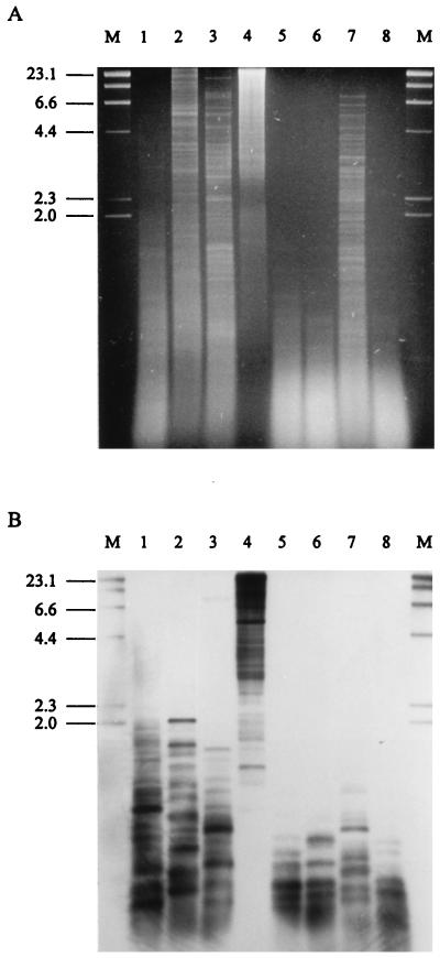
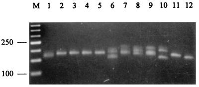
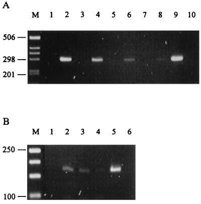
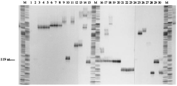
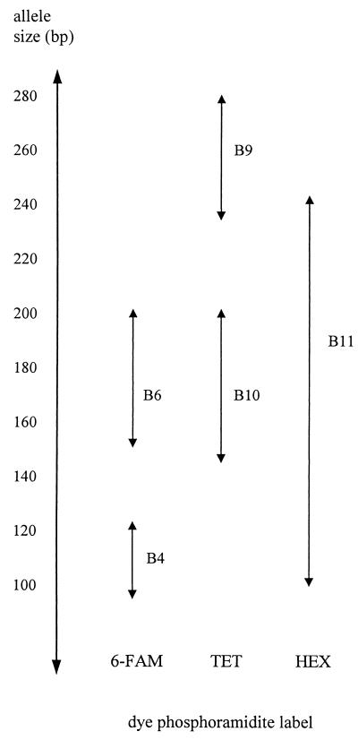
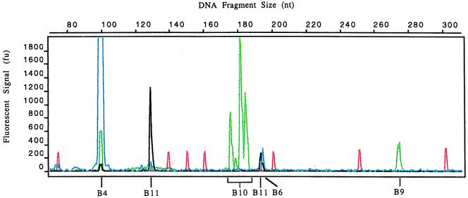
Epichloë endophytes are a group of filamentous fungi that include both sexual (Epichloë) and asexual (Neotyphodium) species. As a group they are genetically diverse and form both antagonistic and mutualistic associations with temperate grasses. We report here on the development of a microsatellite-based PCR system for fingerprinting this group of fungi with template isolated from either culture or infected plant material. M13mp19 partial genomic libraries were constructed for size-fractionated genomic DNA from two endophyte strains. These libraries were screened with a mixture of DIG-labeled dinucleotide and trinucleotide repeat probes. Positive clones were sequenced, and nine unique microsatellite loci were identified. An additional microsatellite was serendipitously identified in the 3' untranscribed region of the 3-hydroxy-3-methylglutaryl coenzyme A (HMG CoA) reductase gene from N. lolii Lp19. Primers were designed for each locus and a panel of endophytes, from different taxonomic groupings, was screened to determine the degree of polymorphism. On the basis of these results a multiplex assay was developed for strain identification with fluorescently labeled primers for five of these loci. Using this system the size of the products amplified can be precisely determined by automated analysis, and an allele profile for each strain can be readily generated. The assay was shown to resolve endophyte groupings to the level of known isozyme phenotype groupings. In a blind test the assay was used successfully to identify a set of endophytes in planta. A reference database of allele sizes has been established for the panel of endophytes examined, and this will be expanded as new strains are analyzed.


Figures

FIG. 1
FIG. 1
Hybridization of microsatellite probes to the Lp1 genome. (A) Ethidium bromide-stained agarose (1.25%) gel of Lp1 genomic DNA (1 μg) digested with AluI (lane 1); ThaI (lane 2); HaeIII (lane 3); BamHI (lane 4); AluI, ThaI, and BamHI (lane 5); AluI, HaeIII, and BamHI (lane 6); ThaI, HaeIII, and BamHI (lane 7); and AluI, ThaI, HaeIII, and BamHI (lane 8). (B) Autoradiograph of Southern blot of gel from panel A, which has been probed at low stringency with DIG-labeled (CAA)10, (GAA)10, and (ATC)10 oligonucleotides, followed by chemiluminescent detection. The numbers on the left of the figures indicate the sizes in kilobases of the λ-HindIII size markers in lane M.
FIG. 2
FIG. 2
Amplification of Epichloë endophyte microsatellite loci. Ethidium bromide-stained agarose (3% NuSieve) gel of PCR products amplified with primers B10.1 and B10.2 by using genomic DNA (40 pg μl−1) from Lp1 (lane 1), Lp5 (lane 2), Lp13 (lane 3), Lp19 (lane 4), Lp7 (lane 5), Tf27 (lane 6), Tf28 (lane 7), Tf13 (lane 8), Tf18 (lane 9), Fp2 (lane 10), E8 (lane 11), and Fr1 (lane 12). The numbers on the left of the figures indicate the sizes in base pairs of the 50-bp ladder in lane M.
FIG. 3
FIG. 3
Amplification of Epichloë microsatellite loci from endophyte-infected plant material. Ethidium bromide-stained agarose (3% NuSieve) gels of PCR products amplified from genomic DNA isolated from grass tissue by using primers to microsatellite loci B1 (A) and B10 (B). (A) PCR products generated with DNA prepared from AR1-infected L. perenne × L. multiflorum hybrid ryegrass (lanes 2, 4, 6, and 8) and endophyte-free hybrid ryegrass (lanes 1, 3, 5, and 7) by the CTAB method (18) at total DNA concentrations of 2 ng μl−1 (lanes 1 and 2), 400 pg μl−1 (lanes 3 and 4), 200 pg μl−1 (lanes 5 and 6), and 40 pg μl−1 (lanes 7 and 8); at an AR1 fungal genomic DNA concentration of 40 pg μl−1 (lane 9); and with no DNA (lane 10). Lane M, 1-kb ladder. (B) PCR products generated with DNA prepared from AR1-infected L. perenne × L. multiflorum hybrid ryegrass by the FastDNA Kit H minipreparation method at total DNA concentrations of 2 ng μl−1 (lane 1), 400 pg μl−1 (lane 2), 80 pg μl−1 (lane 3), and 16 pg μl−1 (lane 4); at an AR1 fungal genomic DNA concentration of 40 pg μl−1 (lane 5); and with no DNA (lane 6). Lane M, 50-bp ladder. The numbers on the left of the figures indicate the sizes in base pairs of the DNA size markers.
FIG. 4
FIG. 4
Autoradiograph of radiolabeled microsatellite products amplified from Epichloë genomic DNA. Fungal genomic DNA (40 pg μl−1) was amplified by PCR in the presence of [α-33P]dCTP with primers to the B11 microsatellite locus. These products were separated on a 6% denaturing polyacrylamide gel. Lanes: M, 1-bp ladder (pBSMB sequence) combining A and G reactions, and C with T; 1, Lp1; 2, Lp2; 3, Lp5; 4, Lp6; 5, Lp13; 6, Lp19; 7, Lp20; 8, Lp21; 9, Lp7; 10, Lp14; 11, Lp9; 12, AR1; 13, Tf5; 14, Tf27; 15, Tf28; 16, Tf13; 17, Tf15; 18, Tf20; 19, Tf16; 20, Tf18; 21, Fp1; 22, Fp2; 23, Fp4; 24, E8; 25, Fr1; 26, Frc5; 27, Frc7; 28, Frr1; 29, Fg1; 30, Fl1.
FIG. 5
FIG. 5
Strategy for the dye labeling of primers to microsatellite loci. The strategy shows how the products for microsatellite loci B4, B6, B9, B10, and B11 can be amplified by multiplex PCR and analyzed in a single lane of the automatic sequencer. Double-headed arrows indicate the observed size range, as determined by separation on polyacrylamide gels, of microsatellite PCR products generated for the Epichloë endophytes analyzed. Primers used for each locus were Bx.1 and Bx.2, Exception for B9, which uses B9.1 and B9.4.
FIG. 6
FIG. 6
Multiplex amplification of Epichloë endophyte microsatellite loci. Ethidium bromide-stained agarose (3% NuSieve) gel of the products generated from either a single or a multiplex PCR for endophytes Tf13 (lanes 1 to 6) and Fr1 (lanes 7 to 12). Lanes: M, 50-bp ladder; 1, multiplex; 2, B4; 3, B6; 4, B9; 5, B10; 6, B11; 7, multiplex; 8, B4; 9, B6; 10, B9; 11, B10; and 12, B11.
FIG. 7
FIG. 7
Electropherogram of an endophyte microsatellite fingerprint. Genomic DNA from Tf28 was amplified in the presence of fluorescent primers, and the products were separated by PAGE. GeneScan 2.1 software was used to generate the electropherogram, and the sizes of the products were determined by reference to GS-500 TAMRA internal size standards. Peaks in red, blue, green, and black correspond to the size standards: 6-FAM (B4 and B6), TET (B9 and B10), and HEX (B11)-labeled products, respectively.

References

    1. Arachevaleta M, Bacon C W, Hoveland C S, Radcliffe D E. Effect of the tall fescue endophyte on plant response to environmental stress. Agron J. 1989;81:83–90.
    1. Bacon C W, Lyons P C, Porter J K, Robbins J D. Ergot toxicity from endophyte-infected grasses: a review. Agron J. 1986;78:106–116.
    1. Bacon C W, Porter J K, Robbins J D, Luttrell E S. Epichloë typhina from toxic tall fescue grasses. Appl Environ Microbiol. 1977;34:576–581. - PMC - PubMed
    1. Boehringer Mannheim GmbH. The DIG system user’s guide for filter hybridization. Mannheim, Germany: Boehringer Mannheim GmbH; 1995.
    1. Bolton E T, McCarthy B J. A general method for the isolation of RNA complementary to DNA. Proc Natl Acad Sci USA. 1962;48:1390–1397. - PMC - PubMed

Publication types

Associated data