. 1999 Mar 1;189(5):767-78.
doi: 10.1084/jem.189.5.767.

Identification of MAGE-3 epitopes presented by HLA-DR molecules to CD4(+) T lymphocytes

Affiliations

Identification of MAGE-3 epitopes presented by HLA-DR molecules to CD4(+) T lymphocytes

P Chaux et al. J Exp Med. .

Abstract

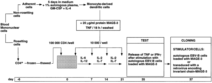
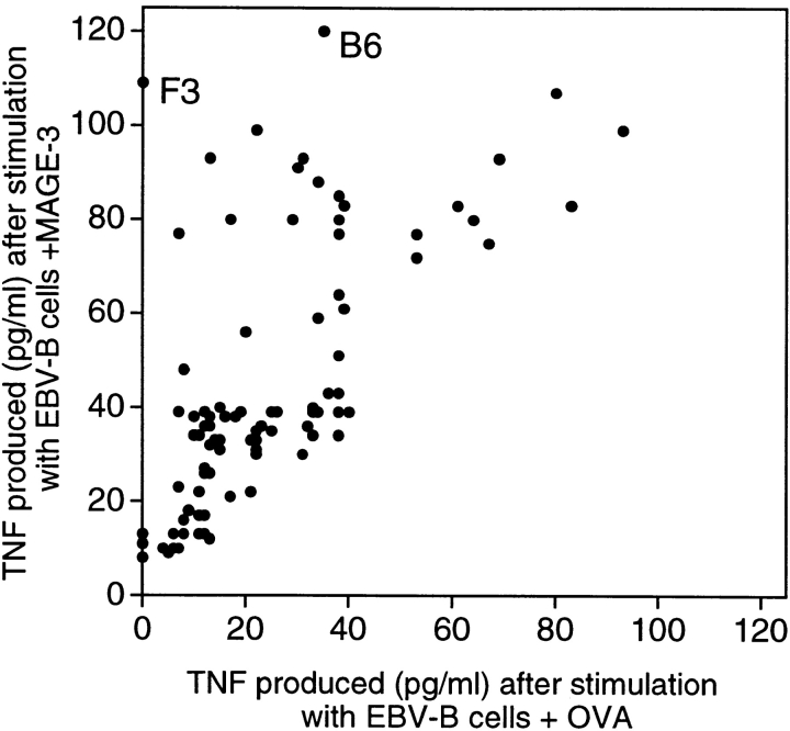
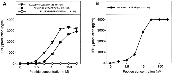
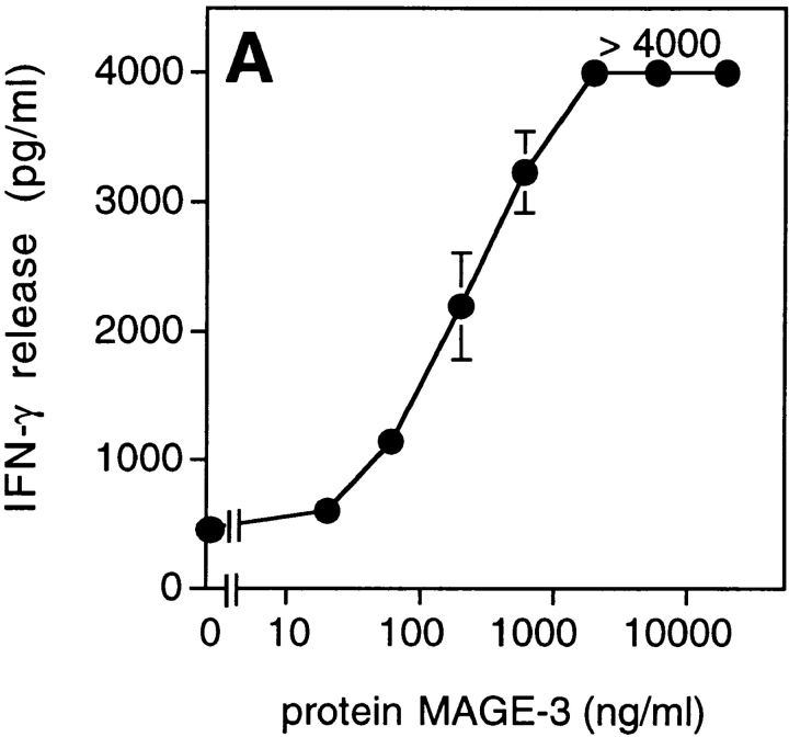
MAGE-type genes are expressed by many tumors of different histological types and not by normal cells, except for male germline cells, which do not express major histocompatibility complex (MHC) molecules. Therefore, the antigens encoded by MAGE-type genes are strictly tumor specific and common to many tumors. We describe here the identification of the first MAGE-encoded epitopes presented by histocompatibility leukocyte antigen (HLA) class II molecules to CD4(+) T lymphocytes. Monocyte-derived dendritic cells were loaded with a MAGE-3 recombinant protein and used to stimulate autologous CD4(+) T cells. We isolated CD4(+) T cell clones that recognized two different MAGE-3 epitopes, MAGE-3114-127 and MAGE-3121-134, both presented by the HLA-DR13 molecule, which is expressed in 20% of Caucasians. The second epitope is also encoded by MAGE-1, -2, and -6. Our procedure should be applicable to other proteins for the identification of new tumor-specific antigens presented by HLA class II molecules. The knowledge of such antigens will be useful for evaluation of the immune response of cancer patients immunized with proteins or with recombinant viruses carrying entire genes coding for tumor antigens. The use of antigenic peptides presented by class II in addition to peptides presented by class I may also improve the efficacy of therapeutic antitumor vaccination.


Figures

Figure 1
Figure 1
Overview of the procedure used to obtain anti–MAGE-3 CD4+ T cell clones.
Figure 2
Figure 2
Production of TNF by CD4+ T cells stimulated with autologous EBV-B cells incubated with protein MAGE-3. EBV-B cells from hemochromatosis patient LB 1555 were incubated with 20 μg/ml of OVA or MAGE-3 protein for 20 h. 5,000 cells were distributed in microwells to which an aliquot of each of the 96 CD4 microcultures was added (∼3,000 effector cells). TNF production was estimated after overnight culture, by the toxicity of the supernatants for the TNF-sensitive WEHI 164-13 cells. The dots represent the average result obtained with two aliquots of each CD4 microculture.
Figure 3
Figure 3
MAGE-3 peptides recognized by clone 37. (A) Stimulation of clone 37 by two overlapping MAGE-3 peptides. Autologous EBV-B cells were incubated for 2 h with different concentrations of the peptides. Clone 37 (2,500 cells) was then cocultured with 5,000 peptide-pulsed cells for 20 h. IFN-γ production in the supernatant was measured by ELISA. (B) Stimulation by the shortest peptide recognized by clone 37. Conditions were as in A.
Figure 4
Figure 4
Presentation of the MAGE-3 antigen by dendritic cells incubated with purified protein or cell lysates. (A) HLA-DR13 dendritic cells were cultured for 24 h with different concentrations of MAGE-3 protein. The cells were washed, then incubated with 2,500 cells per well of clone 37. IFN-γ production was measured after 20 h by ELISA. The results shown represent the average of triplicate cultures. (B) 293-EBNA cells (5 × 10⁵ cells per well) were transfected with different doses of pCEP4-MAGE-3 mixed with Lipofectamine®. 24 h after transfection, the transfected cells were lysed by freeze–thawing. HLA-DR13 dendritic cells (105 cells per well) were cultured with lysates at the equivalent of 5 293-EBNA cells per dendritic cell for 24 h. The experiment was pursued as in A.
Figure 4
Figure 4
Presentation of the MAGE-3 antigen by dendritic cells incubated with purified protein or cell lysates. (A) HLA-DR13 dendritic cells were cultured for 24 h with different concentrations of MAGE-3 protein. The cells were washed, then incubated with 2,500 cells per well of clone 37. IFN-γ production was measured after 20 h by ELISA. The results shown represent the average of triplicate cultures. (B) 293-EBNA cells (5 × 10⁵ cells per well) were transfected with different doses of pCEP4-MAGE-3 mixed with Lipofectamine®. 24 h after transfection, the transfected cells were lysed by freeze–thawing. HLA-DR13 dendritic cells (105 cells per well) were cultured with lysates at the equivalent of 5 293-EBNA cells per dendritic cell for 24 h. The experiment was pursued as in A.
Figure 5
Figure 5
Recognition by clone 37 of cells expressing an Ii MAGE-3 construct. (A) MZ2-MEL.43 melanoma cells express both HLA-DR13 and MAGE-3. Cells were pulsed for 2 h with 5 μg/ml of peptide MAGE-3115–130 and washed, or transduced with a retroviral construct encoding Ii.MAGE-3. (B) MZ2-EBV cells express HLA-DR13. As a positive control for stimulating cells, we incubated them for 20 h with 20 μg/ml of protein MAGE-3. MZ2-EBV cells were transduced with a retroviral construct encoding MAGE-3 alone or Ii.MAGE-3. Tumor cells (104) and EBV-B cells (5 × 10³) were distributed in microwells, and 2,500 cells of clone 37 were added. IFN-γ production was measured after 20 h of coculture by ELISA. The results shown represent the average of triplicate cultures.
Figure 6
Figure 6
Clone 2 recognizes a different MAGE-3 epitope than clone 37. (A) Production of IFN-γ by clone 2 stimulated with autologous EBV-B cells incubated with protein MAGE-3, or transduced with a retroviral vector encoding the Ii.MAGE-3 fusion protein. Autologous EBV-B cells were incubated for 20 h with 20 μg/ml of the recombinant MAGE-3 protein. Clone 2 (2,500 cells) was incubated with 5,000 autologous EBV-B cells loaded with MAGE-3, or transduced with Ii.MAGE-3. IFN-γ production was measured after 20 h by ELISA. The results shown represent the average of triplicate cultures. (B) HLA-DR13 EBV-B cells were incubated for 2 h with different concentrations of overlapping MAGE-3 peptides. The peptides were purified by HPLC, to compare accurately the efficacy of the different peptides used at the same concentrations. Clones (2,500 cells) were incubated with 5,000 peptide-pulsed cells for 20 h. IFN-γ production was measured by ELISA. The results shown represent the average of triplicate cultures.
Figure 7
Figure 7
Stimulation of CD4 clone 2 by the MAGE-3121–134 peptide. HLA-DR13 EBV-B cells were pulsed for 2 h with different concentrations of the peptide. Clones (2,500 cells) were incubated with 5,000 peptide-pulsed cells for 20 h. IFN-γ production was measured by ELISA.

Comment in

References

    1. Anichini A, Fossati G, Parmiani G. Clonal analysis of the cytolytic T-cell response to human tumors. Immunol Today. 1987;8:385–389. - PubMed
    1. Hérin M, Lemoine C, Weynants P, Vessière F, Van Pel A, Knuth A, Devos R, Boon T. Production of stable cytolytic T-cell clones directed against autologous human melanoma. Int J Cancer. 1987;39:390–396. - PubMed
    1. De Plaen E, Arden K, Traversari C, Gaforio JJ, Szikora J-P, De Smet C, Brasseur F, van der Bruggen P, Lethé B, Lurquin C, et al. Structure, chromosomal localization and expression of twelve genes of the MAGE family. Immunogenetics. 1994;40:360–369. - PubMed
    1. Van den Eynde B, van der Bruggen P. T cell defined tumor antigens. Curr Opin Immunol. 1997;9:684–693. - PubMed
    1. Takahashi K, Shichijo S, Noguchi M, Hirohata M, Itoh K. Identification of MAGE-1 and MAGE-4 proteins in spermatogonia and primary spermatocytes of testis. Cancer Res. 1995;55:3478–3482. - PubMed

Publication types