. 1999 Mar 1;515 ( Pt 2)(Pt 2):419-29.
doi: 10.1111/j.1469-7793.1999.419ac.x.

Presynaptic action of adenosine on a 4-aminopyridine-sensitive current in the rat carotid body

Affiliations

Presynaptic action of adenosine on a 4-aminopyridine-sensitive current in the rat carotid body

C Vandier et al. J Physiol. .

Abstract

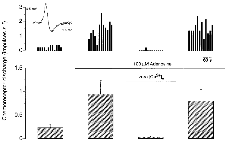
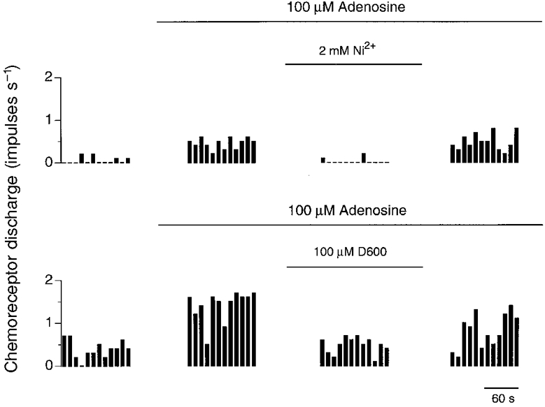
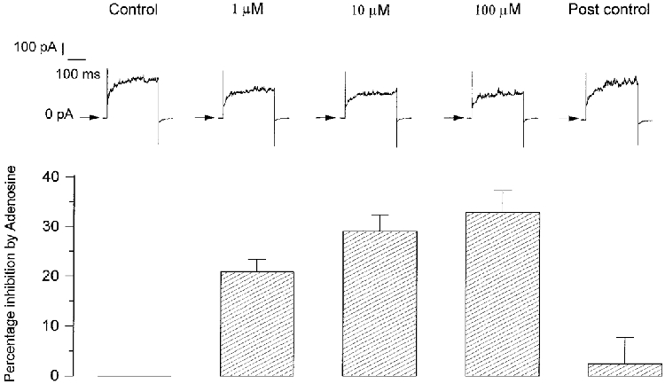
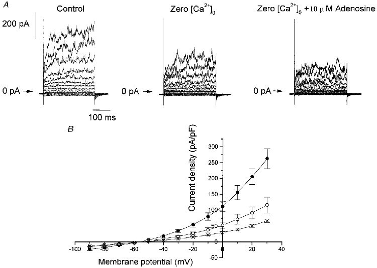
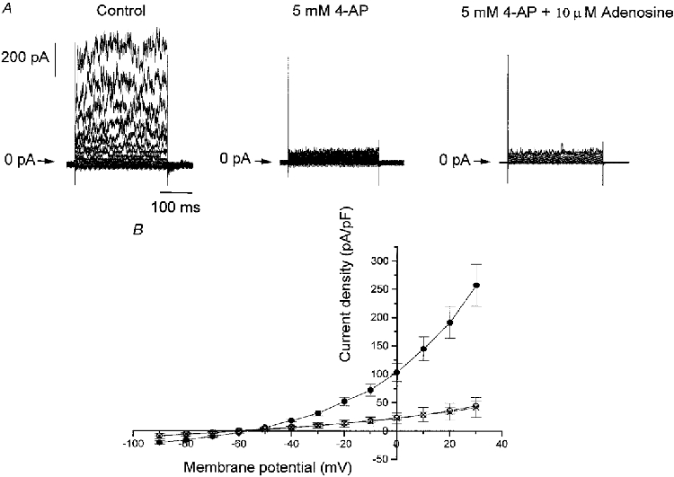
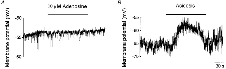
1. Plasma adenosine concentration increases during hypoxia to a level that excites carotid body chemoreceptors by an undetermined mechanism. We have examined this further by determining the electrophysiological responses to exogenous adenosine of sinus nerve chemoafferents in vitro and of whole-cell currents in isolated type I cells. 2. Steady-state, single-fibre chemoafferent discharge was increased approximately 5-fold above basal levels by 100 microM adenosine. This adenosine-stimulated discharge was reversibly and increasingly reduced by methoxyverapamil (D600, 100 microM), by application of nickel chloride (Ni2+, 2 mM) and by removal of extracellular Ca2+. These effects strongly suggest a presynaptic, excitatory action of adenosine on type I cells of the carotid body. 3. Adenosine decreased whole-cell outward currents at membrane potentials above -40 mV in isolated type I cells recorded during superfusion with bicarbonate-buffered saline solution at 34-36 C. This effect was reversible and concentration dependent with a maximal effect at 10 microM. 4. The degree of current inhibition induced by 10 microM adenosine was voltage independent (45.39 +/- 2. 55 % (mean +/- s.e.m.) between -40 and +30 mV) and largely ( approximately 75 %), but not entirely, Ca2+ independent. 4-Aminopyridine (4-AP, 5 mM) decreased the amplitude of the control outward current by 80.60 +/- 3.67 % and abolished the effect of adenosine. 5. Adenosine was without effect upon currents near the resting membrane potential of approximately -55 mV and did not induce depolarization in current-clamp experiments. 6. We conclude that adenosine acts to inhibit a 4-AP-sensitive current in isolated type I cells of the rat carotid body and suggest that this mechanism contributes to the chemoexcitatory effect of adenosine in the whole carotid body.


Figures

Figure 1
Figure 1. The stimulatory effect of adenosine is Ca2+ dependent
Single-fibre chemoreceptor activity recorded during steady-state control (left panel) and during exposure to 100 μM adenosine in the presence and absence of extracellular Ca2+ (middle panels) and after re-addition of Ca2+ (right panel). An example from one preparation is shown at the top, binned every 10 s and expressed in impulses s−1. The inset at top left shows three superimposed action potentials. The mean +s.e.m. of five preparations is shown below. Superfusate PO2 > 400 mmHg and PCO2≈35 mmHg throughout.
Figure 2
Figure 2. Effect of Ni2+ and D600 upon adenosine stimulated chemoreceptor discharge
Single-fibre chemoreceptor activity recorded from two preparations during steady-state control (left panel) and during exposure to 100 μM adenosine in the presence and absence of either 2 mM Ni2+ or 100 μM D600 (middle panels) and after removal of antagonist (right panel). Discharge was binned every 10 s.
Figure 3
Figure 3. Dose-dependent effect of adenosine on outward current
Top, example of outward currents elicited in one type I cell during 300 ms pulses to +30 mV from a holding potential of -70 mV in control conditions, at three different concentrations of adenosine and during post control conditions subsequent to the removal of adenosine. Currents during adenosine infusion were recorded 1 min after the calculated new solution equilibria would have been reached and 4 min after post control wash of adenosine. Bottom, means +s.e.m. (n = 6) of the percentage inhibition of outward currents at +30 mV induced by the three concentrations of adenosine.
Figure 4
Figure 4. Adenosine decreases outward currents above -40 mV
A, example of outward currents obtained during 300 ms pulses to membrane potentials between -90 and +30 mV from a holding potential of -70 mV in control conditions and with 10 μM adenosine in the bath. B, mean ±s.e.m. (n = 7) current density-voltage relation obtained in control conditions (•) and in the presence of 10 μM adenosine (○). The inset shows an expanded scale of the same current density-voltage relation between -75 and -45 mV which highlights the negligible effect of adenosine at membrane potentials where current density is equal to 0 pA pF−1. C, means ±s.e.m. (n = 7) of the adenosine-sensitive component of the total current density-voltage relation determined as the difference between the current density amplitudes in control conditions and in the presence of 10 μM adenosine.
Figure 5
Figure 5. Adenosine decreases outward currents in the absence of external Ca2+
A, example of outward currents obtained during 300 ms pulses to membrane potentials between -90 and +30 mV from a holding potential of -70 mV in control conditions, in the absence of external Ca2+ (Zero [Ca2+]o) and in the presence of 10 μM adenosine. B, means ±s.e.m. of the current density-voltage relation obtained in control conditions (•, n = 7), in the absence of external Ca2+ (○, n = 7) and in the presence of 10 μM adenosine (×, n = 4).
Figure 6
Figure 6. 4-AP decreases outward currents and abolishes the effect of adenosine
A, an example of outward currents obtained during 300 ms pulses to membrane potentials between -90 and +30 mV from a holding potential of -70 mV in control conditions, and in the presence of 5 mM 4-AP both in the absence and presence of 10 μM adenosine. B, mean ±s.e.m. current density-voltage relations obtained in control conditions (•, n = 10), in the presence of 5 mM 4-AP (○, n = 10) and in the last condition but with 10 μM adenosine (×, n = 3).
Figure 7
Figure 7. Adenosine has no effect at the resting membrane potential
A, an example of the negligible effect of 10 μM adenosine in one cell on the resting membrane potential during current clamp. B, an example of the depolarizing effect of acidosis (pHo 7.30) in one cell on the resting membrane potential during current clamp.

Similar articles

Cited by

References

    1. Bartrup J, Stone T. Dihydropyridines alter adenosine sensitivity in rat hippocampal slice. British Journal of Pharmacology. 1990;101:97–102. - PMC - PubMed
    1. Buckler KJ. A novel oxygen-sensitive potassium current in rat carotid body type I cells. The Journal of Physiology. 1997;498:649–662. - PMC - PubMed
    1. Buckler KJ, Vaughan-Jones RD. Effects of hypoxia on membrane potential and intracellular calcium in rat neonatal carotid body type I cells. The Journal of Physiology. 1994a;476:423–428. - PMC - PubMed
    1. Buckler KJ, Vaughan-Jones RD. Effects of hypercapnia on membrane potential and intracellular calcium in rat neonatal carotid body type I cells. The Journal of Physiology. 1994b;478:157–171. - PMC - PubMed
    1. Chen J, Dinger B, Fidone SJ. cAMP production in rabbit carotid body: Role of adenosine. Journal of Applied Physiology. 1997;82:1771–1775. - PubMed

Publication types

LinkOut - more resources