. 1999 Mar 1;515 ( Pt 2)(Pt 2):533-42.
doi: 10.1111/j.1469-7793.1999.533ac.x.

Expression of alpha2-adrenergic receptors in rat primary afferent neurones after peripheral nerve injury or inflammation

Affiliations

Expression of alpha2-adrenergic receptors in rat primary afferent neurones after peripheral nerve injury or inflammation

L A Birder et al. J Physiol. .

Abstract

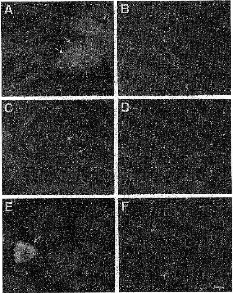
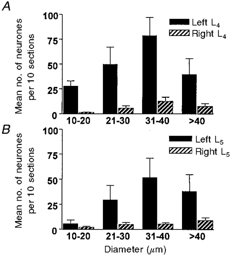
1. Immunocytochemistry with polyclonal antibodies directed against specific fragments of intracellular loops of alpha2A- and alpha2C-adrenergic receptors (alpha2A-AR, alpha2C-AR) was used to explore the possibility that expression of these receptors in dorsal root ganglion (DRG) neurones of rat alters as a result of peripheral nerve injury or localized inflammation. 2. Small numbers of neurones with positive alpha2A-AR immunoreactivity (alpha2A-AR-IR) were detected in DRG from normal animals or contralateral to nerve lesions. In contrast, after complete or partial sciatic nerve transection the numbers of ipsilateral L4 and L5 DRG somata expressing alpha2A-AR-IR sharply increased (>5-fold). There was no discernible change in the number of DRG neurones exhibiting alpha2A-AR-IR innervating a region in association with localized chemically induced inflammation. 3. After nerve injury, double labelling with Fluoro-Gold, a marker of retrograde transport from transected fibres, or by immunoreactivity for c-jun protein, an indicator of injury and regeneration, suggested that many of the neurones expressing alpha2A-AR-IR were uninjured by the sciatic lesions. 4. In general the largest proportionate increase in numbers of neurones labelled by alpha2A-AR-IR after nerve lesions appeared in the medium-large diameter range (31-40 microm), a group principally composed of cell bodies of low threshold mechanoreceptors. The number of small diameter DRG neurones labelled by alpha2A-AR-IR, a category likely to include somata of nociceptors, also increased but proportionately less. 5. Relatively few DRG neurones exhibited alpha2C-AR-IR; this population did not appear to change after either nerve lesions or inflammation. 6. These observations are considered in relation to effects of nerve injury on excitation of primary afferent neurones by sympathetic activity or adrenergic agents, sympathetically related neuropathy and reports of sprouting of sympathetic fibres in DRG.


Figures

Figure 7
Figure 7. Diameter distribution of neurones displaying c-jun-IR after nerve injury
Note that the ordinate is the proportion (percentage) of the total counted in L4 ganglia (±s.d.). A, 788 counted positive neurones from 2 animals following complete ipsilateral sciatic transection. B, 658 counted positive neurones from 2 animals after partial ipsilateral sciatic transection.
Figure 1
Figure 1. Epifluorescence image of sections from L4 dorsal root ganglia depicting α2-AR-IR
A and C, sections depicting α2A-AR-IR after partial sciatic nerve transection (A) and complete sciatic nerve transection (C). B and D, adjacent sections to A and C showing block of reaction product by addition of α2A-AR fusion protein. Arrows indicate positive reaction product. E, epifluorescence image of a section from L4 dorsal root ganglion depicting α2C-AR-IR (complete sciatic transection 14 days previously). Identical images were found in DRG of normal animals. F, block of reaction in a section adjacent to E by addition of α2C-AR fusion protein. Arrow indicates neurone with positive reaction product. Calibration bar, 10 μm for each panel.
Figure 2
Figure 2. Number of L4 DRG neurones expressing α2A
AR-IR in 8 unoperated animals (Control) or after injection into the ipsilateral hindpaw of the inflammatory agents Freund's complete adjuvant (n = 4), carrageenan (Carrag., n = 4) and formalin (n = 4). Data are means per 10 sections ±s.d.
Figure 3
Figure 3. Number of L4 DRG neurones expressing α2C-AR-IR in unoperated animals (n = 8) or after partial (n = 7) or complete transection (n = 11) of the sciatic nerve
Data are shown as means per 10 sections ±s.d. No change of α2C-AR expression was noted after injection of identical inflammatory agents as in Fig. 2.
Figure 4
Figure 4. Diameter distribution of DRG neurones expressing α2A-AR-IR after partial transection of the sciatic nerve 7-14 days previously
Data are means per 10 sections ±s.d. (n = 7). Filled bars, ipsilateral to lesion; hatched bars, contralateral to lesion. A, L4 DRG; B, L5 DRG. See Methods for additional details.
Figure 5
Figure 5. Diameter distribution of DRG neurones expressing α2A-AR-IR after complete transection of the sciatic nerve 7-14 days previously
Data are means per 10 sections ±s.d. (n = 11 animals). Filled bars, ipsilateral to the lesion; hatched bars, contralateral. A, L4 DRG; B, L5 DRG.
Figure 6
Figure 6. Diameter distribution of DRG neurones expressing α2A-AR-IR in relation to markers for transection or injury of peripheral fibres
A, mean number (±s.d.) of α2A-AR-IR neurones in 10 sections of the ipsilateral L4 DRG from 5 animals after complete sciatic nerve transection 7-14 days previously. Filled bars, α2A-AR-IR only; hatched bars, α2A-AR-IR and retrogradely transported Fluoro-Gold (FG). B, mean number (±s.d.) of α2A-AR-IR neurones in 10 sections of the ipsilateral L4 DRG from 6 animals after partial transection of the ipsilateral sciatic nerve. Filled bars, α2A-AR-IR only; hatched bars, α2A-AR-IR and c-jun-IR. See Methods and Results for additional details.

References

    1. Abdulla FA, Smith PA. Ectopic α2 adrenoceptors couple to N-type Ca2+ channels in axotomized rat sensory neurons. Journal of Neuroscience. 1997;17:1633–1641. - PMC - PubMed
    1. Akoev GN. Catecholamine, acetylcholine and excitability of mechanoreceptors. Progress in Neurobiology. 1981;15:269–294. 10.1016/0301-0082(80)90007-6. - DOI - PubMed
    1. Baranowski AP, Anand U, McMahon SB. Retrograde labelling of dorsal root ganglion cells in the rat: a quantitative and morphological comparison of Fluoro-Gold with horseradish peroxidase labelling. Neuroscience Letters. 1992;141:53–56. - PubMed
    1. Barasi S, Lynn B. Effects of sympathetic stimulation on mechanoreceptive and nociceptive afferent units from the rabbit pinna. Brain Research. 1986;378:21–27. - PubMed
    1. Birder LA, Perl ER. Upregulation of the α2A adrenergic receptor subtype after peripheral nerve injury. Society for Neuroscience Abstracts. 1996;22:1803.

Publication types

Substances

LinkOut - more resources