. 1999 Jan;126(1):349-57.
doi: 10.1038/sj.bjp.0702305.

Growth-inhibitory effect of cyclic GMP- and cyclic AMP-dependent vasodilators on rat vascular smooth muscle cells: effect on cell cycle and cyclin expression

Affiliations

Growth-inhibitory effect of cyclic GMP- and cyclic AMP-dependent vasodilators on rat vascular smooth muscle cells: effect on cell cycle and cyclin expression

N Kronemann et al. Br J Pharmacol. 1999 Jan.

Abstract

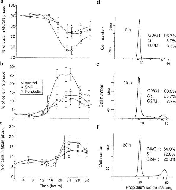
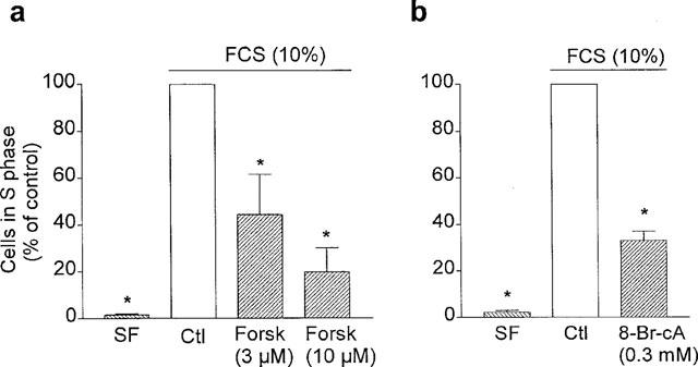
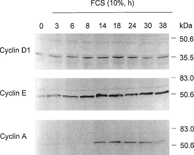
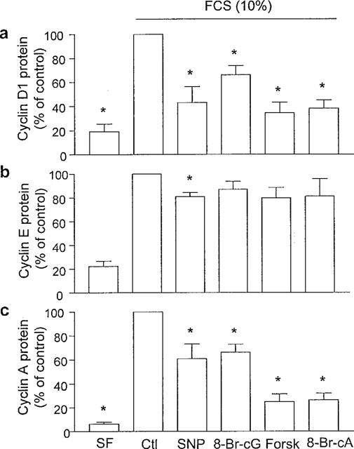
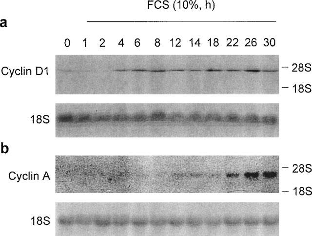
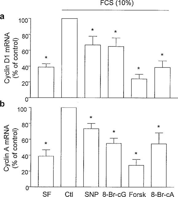
1. The possibility that the antiproliferative effect of cyclic GMP- and cyclic AMP-dependent vasodilators involves an impaired progression of vascular smooth muscle cells (VSMC) through the cell cycle and expression of cyclins, which in association with the cyclin-dependent kinases control the transition between the distinct phases of the cell cycle, was examined. 2. FCS (10%) stimulated the transition of quiescent VSMC from the G0/G1 to the S phase (maximum within 18-24 h and then to the G2/M phase (maximum within 22-28 h). Sodium nitroprusside and 8-Br-cyclic GMP, as well as forskolin and 8-Br-cyclic AMP markedly reduced the percentage of cells in the S phase after FCS stimulation. 3. FCS stimulated the low basal protein expression of cyclin D1 (maximum within 8-24 h) and E (maximum within 8-38 h) and of cyclin A (maximum within 14-30 h). The stimulatory effect of FCS on cyclin D1 and A expression was inhibited, but that of cyclin E was only minimally affected by the vasodilators. 4. FCS increased the low basal level of cyclin D1 mRNA after a lag phase of 2 h and that of cyclin A after 12 h. The vasodilators significantly reduced the FCS-stimulated expression of cyclin D1 and A mRNA. 5. These findings indicate that cyclic GMP- and cyclic AMP-dependent vasodilators inhibit the proliferation of VSMC by preventing the progression of the cell cycle from the G0/G1 into the S phase, an effect which can be attributed to the impaired expression of cyclin D1 and A.


Figures

Figure 1
Figure 1
Effect of (a) SNP and (b) forskolin (Forsk) on the FCS (5%)-stimulated proliferation of cultured rat aortic smooth muscle cells. Cell numbers were determined 24 h after initial plating (Day 0) and 3 days after exposure of VSMC to FCS (Day 3) in the absence or presence of either SNP or forskolin. Data shown as mean±s.e.mean of seven experiments for SNP and five experiments for forskolin. (*P<0.05 vs Day 0, #P<0.05 vs Day 3).
Figure 2
Figure 2
Flow cytometric analysis of cell cycle progression of cultured rat aortic smooth muscle cells. VSMC were arrested in the G0/G1 phase by FCS starvation for 48 h and then were stimulated to enter the cell cycle by the addition of FCS (10%). The effect of SNP (600 μM) and forskolin (3 μM) on the FCS-stimulated progression of VSMC through the cell cycle is also shown. In (a), (b) and (c) data shown as mean±s.e.mean of five experiments for SNP and seven experiments for forskolin. In (d), (e) and (f) representative DNA histograms of VSMC at times 0, 18 and 28 h after FCS stimulation are depicted.
Figure 3
Figure 3
Effect of (a) SNP and (b) 8-Br-cyclic GMP (8-Br-cG) on the FCS-stimulated S phase entry of cultured rat aortic smooth muscle cells. Following growth arrest VSMC were stimulated to enter the cell cycle by the addition of FCS (10%) for 18 h. VSMC were exposed to a cyclic GMP-dependent vasodilator 5 min prior to FCS. Data shown as mean±s.e.mean of six experiments for SNP and three experiments for 8-Br-cyclic GMP. (*P<0.05 vs Ctl). Serum-free: SF; Control: Ctl.
Figure 4
Figure 4
Effect of (a) forskolin (Forsk) and (b) 8-Br-cyclic AMP (8-Br-cA) on the FCS-stimulated S phase entry of cultured rat aortic smooth muscle cells. Following growth arrest VSMC were stimulated to enter the cell cycle by the addition of FCS (10%) for 18 h. VSMC were exposed to a cyclic AMP-dependent vasodilator 5 min prior to FCS. Data shown as mean±s.e.mean of three experiments for forskolin and five experiments for 8-Br-cyclic AMP. (*P<0.05 vs Ctl). Serum-free: SF; Control: Ctl.
Figure 5
Figure 5
Representative Western blots showing the time course of cyclin D1, E, and A protein expression induced by FCS (10%) in cultured rat aortic smooth muscle cells. Equal amounts of protein from nuclear extracts were subjected to SDS–PAGE, transferred to nitrocellulose membranes, and immunoblotted with polyclonal antibodies against G1 phase cyclin D1 (top), and cyclin E (center) and S phase cyclin A (bottom). Similar results were obtained in two additional experiments.
Figure 6
Figure 6
Cumulative data from Western blots showing the effect of cyclic GMP- and cyclic AMP-dependent vasodilators on the FCS-induced (a) cyclin D1, (b) cyclin E and (c) cyclin A protein expression in cultured rat aortic smooth muscle cells. VSMC were exposed to FCS (10%) for 22 h. The vasodilators were added 5 min prior to FCS. Results shown as mean±s.e.mean of at least four experiments. (*P<0.05 vs Ctl). Serum-free: SF; control: Ctl; SNP: 300 μM; 8-Br-cyclic GMP: 8-Br-cG, 3 mM; forskolin: Forsk, 10 μM; 8-Br-cyclic AMP: 8-Br-cA, 0.3 mM.
Figure 7
Figure 7
Representative Northern blots showing the time course of cyclin D1 and cyclin A mRNA expression induced by FCS (10%) in cultured rat aortic smooth muscle cells. (a) cyclin D1 cDNA probe (top), 18S ribosomal RNA (bottom), (b) cyclin A cDNA probe (top), 18S ribosomal RNA (bottom). Similar results were obtained in two additional experiments for cyclin D1 and in an additional experiment for cyclin A.
Figure 8
Figure 8
Cumulative data from Northern blots showing the effect of cyclic GMP- and cyclic AMP-dependent vasodilators on the FCS-induced (a) cyclin D1 and (b) cyclin A mRNA expression in cultured rat aortic smooth muscle cells. VSMC were exposed to FCS (10%) for 16 h. The vasodilators were added 5 min prior to FCS. Results shown as mean±s.e.mean of at least four experiments. (*P<0.05 vs Ctl). Serum-free: SF; control: Ctl; SNP: 300 μM; 8-Br-cyclic GMP: 8-Br-cG, 3 mM; forskolin: Forsk, 10 μM; 8-Br-cyclic AMP: 8-Br-cA, 0.3 mM.

Similar articles

Cited by

References

    1. ABELL T.J., RICHARDS A.M., IKRAM H., ESPINIER E.A., YANDLE T. Atrial natriuretic factor inhibits proliferation of vascular smooth muscle cells stimulated by platelet-derived growth factor. Biochem. Biophys. Res. Commun. 1989;160:1392–1396. - PubMed
    1. ASSENDER J.W., SOUTHGATE K.M., HALLETT M.B., NEWBY A.C. Inhibition of proliferation, but not of Ca2+ mobilization, by cyclic AMP and GMP in rabbit aortic smooth muscle cells. Biochem. J. 1992;288:527–532. - PMC - PubMed
    1. BARLAT I., HENGLEIN B., PLET A., LAMB N., FERNANDEZ A., MCKENZIE F., POUYSSÉGUR J., VIÉ A., BLANCHARD J.M. TGF-β1 and cyclic AMP attenuate cyclin A gene transcription via a cyclic AMP responsive element through independent pathways. Oncogene. 1995;11:1309–1318. - PubMed
    1. BEAVO J.A., REIFSYNDER D.H. Primary sequence of cyclic nucleotide phosphodiesterase isozymes and the design of selective inhibitors. Trends Pharmacol. Sci. 1990;11:150–155. - PubMed
    1. BUSSE R., FLEMING I. Endothelial dysfunction in atherosclerosis. J. Vasc. Res. 1996;331:181–194. - PubMed

Publication types

MeSH terms