. 1999 Mar;113(3):441-56.
doi: 10.1085/jgp.113.3.441.

ATP dependence of the ICl,swell channel varies with rate of cell swelling. Evidence for two modes of channel activation

Affiliations

ATP dependence of the ICl,swell channel varies with rate of cell swelling. Evidence for two modes of channel activation

T Bond et al. J Gen Physiol. 1999 Mar.

Abstract

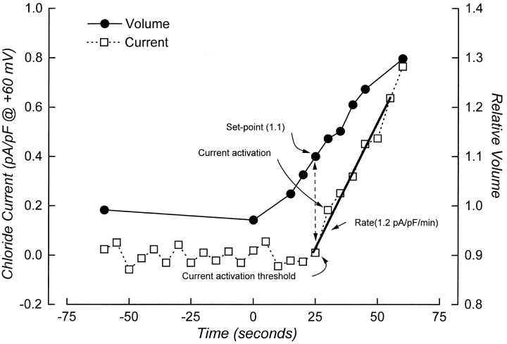
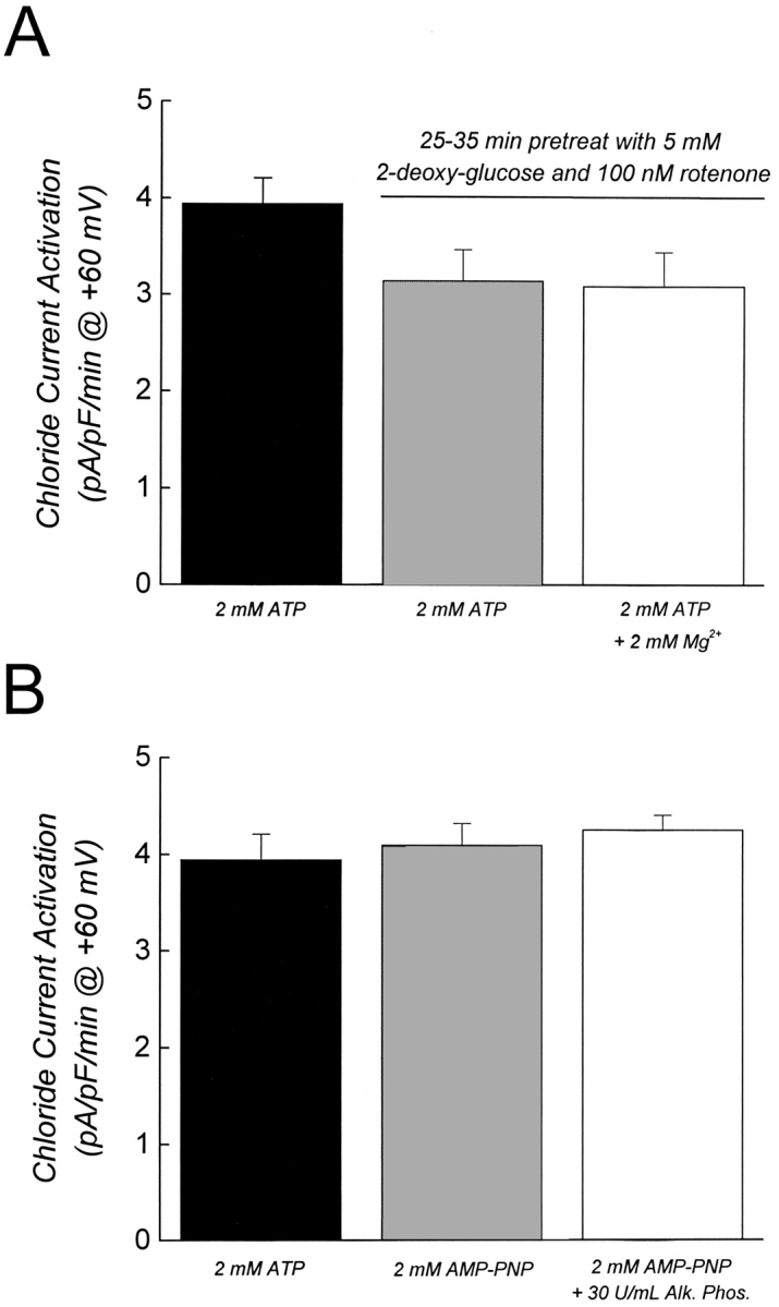
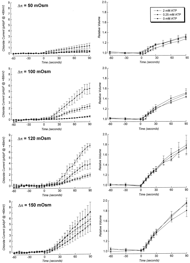
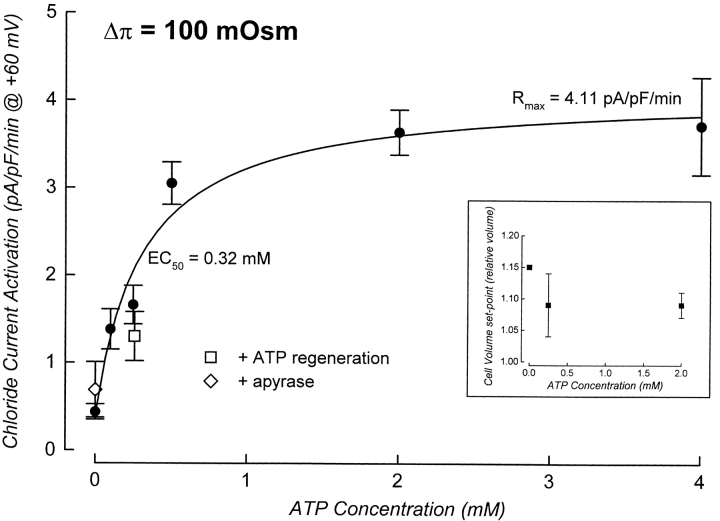
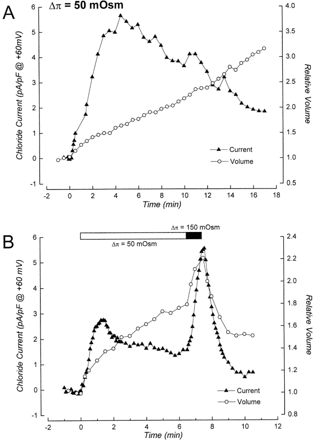
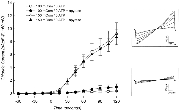
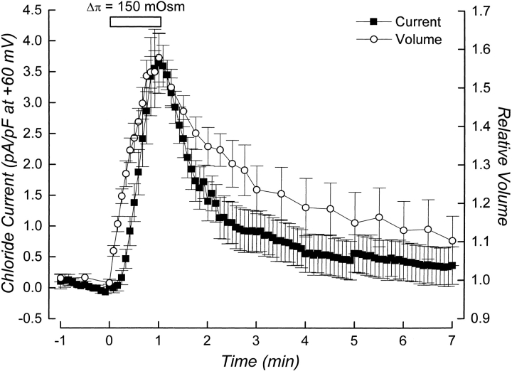
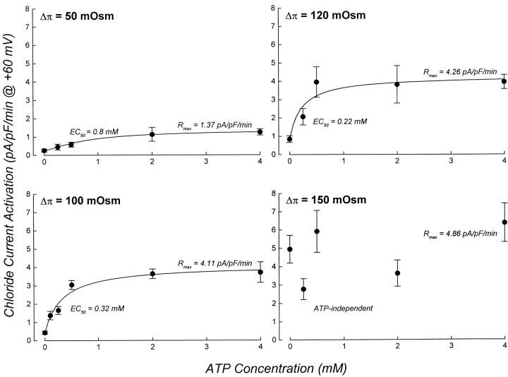
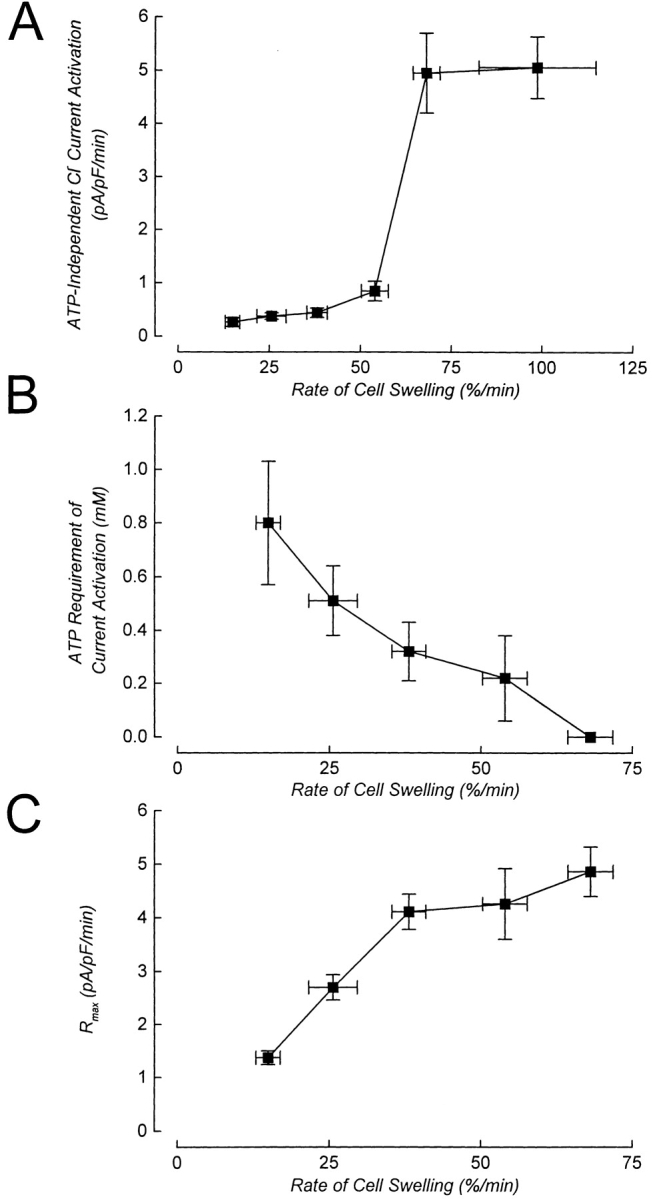
Swelling-induced activation of the outwardly rectifying anion current, ICl, swell, is modulated by intracellular ATP. The mechanisms by which ATP controls channel activation, however, are unknown. Whole cell patch clamp was employed to begin addressing this issue. Endogenous ATP production was inhibited by dialyzing N1E115 neuroblastoma cells for 4-5 min with solutions containing (microM): 40 oligomycin, 5 iodoacetate, and 20 rotenone. The effect of ATP on current activation was observed in the absence of intracellular Mg2+, in cells exposed to extracellular metabolic inhibitors for 25-35 min followed by intracellular dialysis with oligomycin, iodoacetate, and rotenone, after substitution of ATP with the nonhydrolyzable analogue AMP-PNP, and in the presence of AMP-PNP and alkaline phosphatase to dephosphorylate intracellular proteins. These results demonstrate that the ATP dependence of the channel requires ATP binding rather than hydrolysis and/or phosphorylation reactions. When cells were swollen at 15-55%/min in the absence of intracellular ATP, current activation was slow (0.3-0.8 pA/pF per min). ATP concentration increased the rate of current activation up to maximal values of 4-6 pA/pF per min, but had no effect on the sensitivity of the channel to cell swelling. Rate of current activation was a saturable, hyperbolic function of ATP concentration. The EC50 for ATP varied inversely with the rate of cell swelling. Activation of current was rapid (4-6 pA/pF per min) in the absence of ATP when cells were swollen at rates >/=65%/min. Intracellular ATP concentration had no effect on current activation induced by high rates of swelling. Current activation was transient when endogenous ATP was dialyzed out of the cytoplasm of cells swollen at 15%/min. Rundown of the current was reversed by increasing the rate of swelling to 65%/min. These results indicate that the channel and/or associated regulatory proteins are capable of sensing the rate of cell volume increase. We suggest that channel activation occurs via ATP-dependent and -independent mechanisms. Increasing the rate of cell swelling appears to increase the proportion of channels activating via the ATP-independent pathway. These findings have important physiological implications for understanding ICl, swell regulation, the mechanisms by which cells sense volume changes, and volume homeostasis under conditions where cell metabolism is compromised.


Figures

Figure 1
Figure 1
Example of simultaneous current and volume measurements performed on a single, patch-clamped cell. The figure illustrates how the cell volume set-point of the channel and the rate of current activation were quantified. Current activation is defined as the point at which there is a significant and continuous increase in current amplitude above the baseline current. Rate of current activation was quantified by performing linear regression analysis on whole cell currents measured for 30–60 s after activation was detected. The threshold for current activation is the intercept between the line-defining rate of current activation and the current before activation begins. The relative volume of the cell at the current activation threshold is defined as the cell volume set-point of the channel. Cell was swollen by exposure to a 75-mOsm reduction in bath at time zero.
Figure 2
Figure 2
Activation of ICl, swell does not require phosphorylation and/or ATP hydrolysis. (A) Effect of prolonged metabolic inhibition on rate of ICl, swell activation. Cells were maintained in the standard bath solution or were pretreated with 5 mM 2-deoxyglucose and 100 nM rotenone for 25–35 min. Metabolic inhibitors were maintained in the bath throughout the experiment. The three groups of cells were dialyzed for 4–5 min with patch pipette solutions containing metabolic inhibitors before swelling was induced. Except where indicated (open bar), all experiments were done in the absence of intracellular Mg2+ using EDTA buffered solutions. In the Mg2+-containing pipette solution, 1 mM EDTA was replaced with 1 mM EGTA. Values are means ± SEM (n = 4–28). Results were not statistically different (P > 0.1). (B) Effect of intracellular dialysis with alkaline phosphatase. Cells were dialyzed with the patch pipette solution for 4–5 min before swelling was induced. Values are means ± SEM (n = 5–28). Results were not statistically different (P > 0.8). In both A and B, swelling was induced by a 100-mOsm reduction of bath. Rates of cell swelling were similar under all experimental conditions (data not shown).
Figure 3
Figure 3
Effects of intracellular ATP concentration on rate of current activation and rate of cell swelling. Cells were swollen at different rates by a 50–150-mOsm reduction in bath at 0 min. Osmotic shock used is shown in bold in the upper left corner of the current plots. Data are plotted on the same scale to facilitate comparison. Current and volume measurements were made simultaneously in patch clamped cells (e.g., see Fig. 1). Values are means ± SEM (n = 3–9).
Figure 4
Figure 4
Increases in intracellular ATP concentration increase the rate of ICl, swell activation when cells are swollen by a 100-mOsm reduction in bath. Rate of ICl, swell activation is a saturable function of intracellular ATP concentration. The curve was fit to the data by nonlinear regression analysis (Microcal Origin 4.1; Microcal Software) using a the logistic dose–response equation, formula image where y is the rate of current activation, x is the ATP concentration, A 1 is the rate of current activation with 0 mM ATP, A 2 is the maximal rate of current activation (R max), and x 0 is the EC50. b is the so-called “slope factor” and is related to the extent of sigmoidicity (De Lean et al., 1978). Given that the relationship between ATP concentration and current activation was clearly a hyperbolic rather than a sigmoidal function, b was constrained to a value of 1 in the fitting routine. Current activation was also measured in the presence of 0.25 mM ATP and an ATP regeneration system (10 mM creatine phosphate and 80 U/ml of creatine kinase), and under ATP-free conditions in the presence of 2 U/ml apyrase and 0.5 mM CaCl2 (an essential cofactor for apyrase). Values are means ± SEM (n = 3–23). (Inset) Cell volume set-point of the channel is unaffected by intracellular ATP concentration. Values are single measurements or means ± SEM (n = 1–7).
Figure 9
Figure 9
Properties of swelling-induced anion current during washout of intracellular ATP and under ATP-free conditions. (A) Current rundown occurs during washout of intracellular ATP when the rate of cell swelling is slow. Cell was patch clamped with an ATP-free pipette solution containing metabolic inhibitors. Swelling was induced by exposure to a 50-mOsm reduction in bath 60 s after obtaining the whole cell configuration. Current rundown occurs despite continued cell swelling. Rundown is presumably due to loss of intracellular ATP (Lewis et al., 1993; Jackson et al., 1994; Oike et al., 1994). Data shown in graph are from a single cell. Similar results were obtained in experiments on four other cells (data not shown). (B) Current rundown is reversed by increasing the rate of cell swelling. Cell was patch clamped with an ATP-free pipette solution containing metabolic inhibitors. Swelling was induced by exposure to a 50-mOsm hypotonic shock (open bar) 90 s after obtaining the whole cell configuration. The rate of cell swelling was 12%/ min. Rundown began ∼95 s after current activation was observed. Bath osmolality was reduced by 150 mOsm (closed bar) when relative cell volume was 1.8, which increased the rate of cell swelling to 63%/min and induced a rapid activation of current. Data shown in graph are from a single cell. Similar results were obtained in experiments on five other cells (data not shown).
Figure 5
Figure 5
Activation of ICl, swell can occur in the absence of intracellular ATP. Cells were patch clamped and dialyzed for 4–5 min with ATP-free pipette solutions containing metabolic inhibitors. Swelling was induced at time 0 by reducing bath osmolality by either 100 or 150 mOsm. The presence of apyrase (2 U/ml plus 0.5 mM CaCl2) in the patch pipette had no effect on the rate of current activation. Values are means ± SEM (n = 4–17). (Insets) Examples of voltage ramps (−80 to +80 mV at 80 mV/s) recorded during current activation in cells swollen by exposure to a 150- (top) or 100- (bottom) mOsm reduction in bath.
Figure 6
Figure 6
Activation of ATP-independent current is reversed by cell shrinkage. Cells were patch clamped with an ATP-free pipette solution containing metabolic inhibitors. After dialysis for 4–5 min, cells were swollen by exposure to a 150-mOsm reduction in bath (open bar). Swelling was reversed by returning cells to control (300 mOsm) bath. Values are means ± SEM (n = 6).
Figure 7
Figure 7
ATP dependence of ICl, swell activation varies with rate of cell swelling. Cells were swollen at different rates (see Table I) by exposure to 50–150-mOsm reductions in bath. Osmotic shock used is shown in the top left corner of each graph. Data for the 100-mOsm reduction in bath are reproduced from Fig. 4. To facilitate comparison, data are plotted on the same scale. For the 50–120-mOsm reductions in bath, curves were fit to the data as described in Fig. 4 legend. Rates of current activation measured with various concentrations of ATP for cells swollen by a 150-mOsm reduction in bath were not significantly different (P > 0.2). R max value shown was determined by averaging rates of current activation measured in the presence of 0–4 mM ATP. Values are means ± SEM (n = 3–23).
Figure 8
Figure 8
Summary of the effect of rate of cell swelling on the ATP dependence of ICl, swell activation. (A) Rate of current activation in metabolically poisoned cells dialyzed with an ATP-free pipette solution and swollen at different rates. Values are means ± SEM (n = 4–17). (B) ATP requirement of current activation decreases with increasing rates of cell swelling. At rates of swelling between 15 and 55%/min (i.e., 50–120-mOsm reductions in bath), current activation is a saturable function of intracellular ATP concentration (see Figs. 4 and 7). For swelling within this range, the ATP requirement is defined as the EC50 value obtained from dose– response relationships shown in Fig. 7. Current activation at rates of swelling ≥65%/min (i.e., 150-mOsm reduction in bath) is ATP independent (Fig. 7), and the ATP requirement is plotted as 0 mM. (C) Maximal rate of current activation (R max) is a direct function of rate of cell swelling. EC50 and R max values were determined by nonlinear regression analysis as described in the Fig. 4 legend. Vertical error bars for the points in B and C are standard errors, which were generated as part of the curve fitting routine. Rate of cell swelling values are means ± SEM (obtained from Table I).

Similar articles

Cited by

References

    1. Boese SH, Kinne RHK, Wehner F. Single-channel properties of swelling-activated anion conductance in rat inner medullary collecting duct cells. Am J Physiol. 1996;271:F1224–F1233. - PubMed
    1. Bryan J, Aguilar-Bryan L. The ABCs of ATP-sensitive potassium channels: more pieces of the puzzle. Curr Opin Cell Biol. 1997;9:553–559. - PubMed
    1. Cannon CL, Basavappa S, Strange K. Intracellular ionic strength regulates the volume sensitivity of a swelling-activated anion channel. Am J Physiol. 1998;275:C416–C422. - PubMed
    1. Carpenter E, Peers C. Swelling- and cAMP-activated Cl−currents in isolated rat carotid body type I cells. J Physiol (Lond) 1997;503:497–511. - PMC - PubMed
    1. Chicurel ME, Chen CS, Ingber DE. Cellular control lies in the balance of forces. Curr Opin Cell Biol. 1998;10:232–239. - PubMed

Publication types