. 1999 Mar 2;96(5):1898-903.
doi: 10.1073/pnas.96.5.1898.

Small antibody-like proteins with prescribed ligand specificities derived from the lipocalin fold

Affiliations

Small antibody-like proteins with prescribed ligand specificities derived from the lipocalin fold

G Beste et al. Proc Natl Acad Sci U S A. .

Abstract

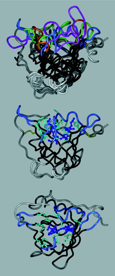
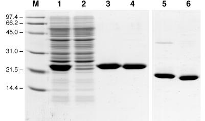
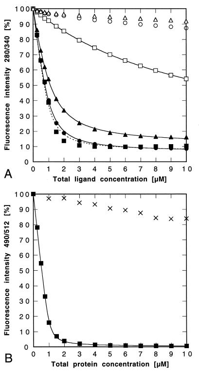
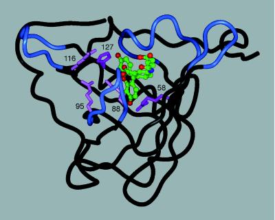
We demonstrate that the ligand pocket of a lipocalin from Pieris brassicae, the bilin-binding protein (BBP), can be reshaped by combinatorial protein design such that it recognizes fluorescein, an established immunological hapten. For this purpose 16 residues at the center of the binding site, which is formed by four loops on top of an eight-stranded beta-barrel, were subjected to random mutagenesis. Fluorescein-binding BBP variants were then selected from the mutant library by bacterial phage display. Three variants were identified that complex fluorescein with high affinity, exhibiting dissociation constants as low as 35.2 nM. Notably, one of these variants effects almost complete quenching of the ligand fluorescence, similarly as an anti-fluorescein antibody. Detailed ligand-binding studies and site-directed mutagenesis experiments indicated (i) that the molecular recognition of fluorescein is specific and (ii) that charged residues at the center of the pocket are responsible for tight complex formation. Sequence comparison of the BBP variants directed against fluorescein with the wild-type protein and with further variants that were selected against several other ligands revealed that all of the randomized amino acid positions are variable. Hence, a lipocalin can be used for generating molecular pockets with a diversity of shapes. We term this class of engineered proteins "anticalins." Their one-domain scaffold makes them a promising alternative to antibodies to create a stable receptor protein for a ligand of choice.


Figures

Figure 1
Figure 1
Tertiary structure of the bilin-binding protein (5), either alone (Middle), with the randomized amino acids depicted, or superimposed (Top) with three other lipocalins: retinol-binding protein, RBP (6); mouse major urinary protein, MUP (7); and epididymal retinoic acid-binding protein, EPA (8). The polypeptide backbone is shown in a ribbon representation (InsightII molecular modeling software), with the β-barrel in black, the four loops surrounding the ligand pocket colored (BBP, blue; RBP, magenta; MUP, green; EPA, amber), and the remainder of the structure in gray. BBP (Middle) is shown with its natural ligand, biliverdin IXγ, in a ball and stick representation (dark blue). The 16 amino acid positions at the center of the binding site that were subjected to random mutagenesis are depicted with their original side chains (light blue). The two disulfide bonds are indicated as well (yellow). (Bottom) The same molecule is shown in an orientation that permits viewing into the binding pocket.
Figure 2
Figure 2
A 0.1% SDS/15% polyacrylamide gel illustrating the purification of the bacterially produced FluA as a Strep-tag II fusion protein. Lane M, molecular size standard (masses labeled in kDa); lane 1, periplasmic protein extract of E. coli JM83 harboring pBBP21-FluA after overnight expression at 22°C; lane 2, flow-through of the streptavidin affinity column; lane 3, FluA eluted from the column; lane 4, BBP prepared in the same way after 3-h induction; lanes 5 and 6, as lanes 3 and 4 but without reduction of the disulfide bonds.
Figure 3
Figure 3
Ligand binding studies with the purified lipocalin variant FluA. (A) Fluorescence titration (λEx = 280 nm; λEm = 340 nm) of a 1 μM protein solution with fluorescein (■), 4-aminofluorescein (●), 4-glutarylamidofluorescein (▴), pyrogallol red (□), phenolphthalein (○), and rhodamine B (▵). The data were corrected and fitted as described in Materials and Methods. The resulting Kd values are given in Table 2. In the case of fluorescein the curve (dotted line) could not be fitted as perfectly as with the other ligands. (B) Fluorescence titration (λEx = 490 nm; λEm = 512 nm) of a 1 μM fluorescein solution with FluA (■) or with BBP (×). The data for FluA were fitted without correction, yielding Kd = 35.2 ± 3.2 nM.
Figure 4
Figure 4
Tentative three-dimensional model of FluA complexed with 4-glutarylamidofluorescein (for coloring cf. Fig. 1). The backbone conformation corresponds to the crystal structure of BBP, whereas the five basic residues were introduced with their preferred side-chain rotamers (25) by using InsightII modeling software. The fluorescein structure was taken from the coordinates of its complex with the antibody 4-4-20 (24) and equipped with the aliphatic substituent in an extended standard conformation. The ligand (C, green; O, red; N, blue) was manually placed such that its fluorescein moiety can form hydrogen bonds with the two critical His and Arg residues (magenta), similarly as in the crystal structure of the antibody complex. Another three Arg residues (pink) can participate in electrostatic interactions with the negatively charged hapten.

References

    1. Skerra A. Curr Opin Immunol. 1993;5:256–262. - PubMed
    1. Winter G, Milstein C. Nature (London) 1991;349:293–299. - PubMed
    1. Hoogenboom H R. Trends Biotechnol. 1997;15:62–70. - PubMed
    1. Flower D R. Biochem J. 1996;318:1–14. - PMC - PubMed
    1. Huber R, Schneider M, Mayr I, Müller R, Deutzmann R, Suter F, Zuber H, Falk H, Kayser H. J Mol Biol. 1987;198:499–513. - PubMed

Publication types

MeSH terms

LinkOut - more resources