. 1999 Mar 2;96(5):1947-52.
doi: 10.1073/pnas.96.5.1947.

The GTPase activating factor for transducin in rod photoreceptors is the complex between RGS9 and type 5 G protein beta subunit

Affiliations

The GTPase activating factor for transducin in rod photoreceptors is the complex between RGS9 and type 5 G protein beta subunit

E R Makino et al. Proc Natl Acad Sci U S A. .

Abstract

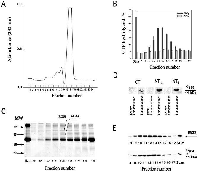
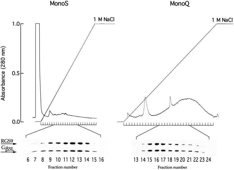
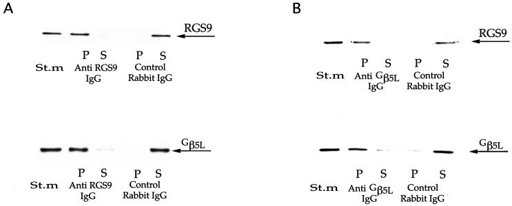
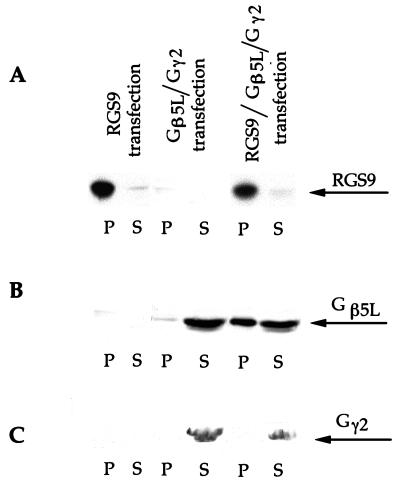
Proteins of the regulators of G protein signaling (RGS) family modulate the duration of intracellular signaling by stimulating the GTPase activity of G protein alpha subunits. It has been established that the ninth member of the RGS family (RGS9) participates in accelerating the GTPase activity of the photoreceptor-specific G protein, transducin. This process is essential for timely inactivation of the phototransduction cascade during the recovery from a photoresponse. Here we report that functionally active RGS9 from vertebrate photoreceptors exists as a tight complex with the long splice variant of the G protein beta subunit (Gbeta5L). RGS9 and Gbeta5L also form a complex when coexpressed in cell culture. Our data are consistent with the recent observation that several RGS proteins, including RGS9, contain G protein gamma-subunit like domain that can mediate their association with Gbeta5 (Snow, B. E., Krumins, A. M., Brothers, G. M., Lee, S. F., Wall, M. A., Chung, S., Mangion, J., Arya, S., Gilman, A. G. & Siderovski, D. P. (1998) Proc. Natl. Acad. Sci. USA 95, 13307-13312). We report an example of such a complex whose cellular localization and function are clearly defined.


Figures

Figure 1
Figure 1
Comigration of RGS9 and Gβ5L during gel filtration. Washed ROS membranes (containing 3 mg rhodopsin) were solubilized in 400 μl of buffer (20 mM Hepes adjusted to pH 7.4 by KOH, 100 mM NaCl, 2 mM MgCl2, 1 mM DTT, and 2% lauryl sucrose) and loaded on the Superose 6 column attached to the fast protein liquid chromatography (FPLC) system (Pharmacia). The column was equilibrated by the same buffer containing 0.5% lauryl sucrose, 5% glycerol, and 2 mg/ml soybean l-α-phosphatidylcholine (Sigma product P-5638). The elution rate was 0.4 ml/min; the fraction size was 0.4 ml. (A) Protein elution profile monitored at 280 nm. The major source of the UV absorbance is rhodopsin. (B) GAP activity of RGS9 in chromatography fractions. Single-turnover transducin GTPase measurements were performed in duplicate with and without PDEγ (see Materials and Methods). The y-axis value represents the percentage of GTP hydrolysis over the 5-s period; error bars indicate the range of determined values. The St.m bars represent the activity in the starting material after it was diluted to achieve the same lauryl sucrose, glycerol, and phosphatidylcholine concentration as in the fractions. (C) Coomassie staining of proteins in fractions surrounding the peak of RGS9 activity. (D) Western blot immunostaining of the 44-kDa protein band by three immune and pre-immune serums raised in sheep against the Gβ5L C-terminal peptide (CT), Gβ5L N-terminal peptide (NTL), and N-terminal peptide of the Gβ5 short splice variant (NTS). (E) Comigration of RGS9 and Gβ5L in the chromatography fractions. Western blots were probed with rabbit anti-RGS9c and anti-NTS antibodies.
Figure 2
Figure 2
Comigration of RGS9 and Gβ5L during cation-exchange and anion-exchange chromatography. Washed ROS membranes (containing 3 mg rhodopsin) were solubilized in 500 μl of 2% lauryl sucrose either in 20 mM Hepes-KOH (pH 6.0) with 2 mM MgCl2 (MonoS) or in 50 mM Tris⋅HCl (pH 7.8) with 2 mM MgCl2 (MonoQ). The columns were equilibrated by the corresponding buffers containing 0.5% lauryl sucrose. A 0–1 M gradient of NaCl was used to elute the bound proteins. The flow rate was 1 ml/min; the fraction size was 1 ml. Western blots of the chromatography fractions were probed by a mixture of rabbit anti-RGS9c and anti-NTS antibodies against Gβ5 at dilutions yielding similar intensities of immunostaining. No GAP activity in corresponding fractions was present in these cases, consistent with previous reports that this activity is extremely unstable in detergent solutions (32).
Figure 3
Figure 3
Reciprocal coimmunoprecipitation of RGS9 and Gβ5L by rabbit anti-RGS9c (A) and anti-Gβ5L NTL (B) antibodies. Washed ROS membranes, containing either 180 (A) or 8 (B) μg rhodopsin, were solubilized in 0.5% lauryl sucrose and subjected to the immunoprecipitation. The difference in the amounts of membranes used in A and B reflects ≈20-fold difference in precipitating capacities of the antibodies used in the assay. Western blots from the samples originated from the starting material (St.m), unbound proteins in the supernatant (S) and proteins bound to the pelleted beads (P) were probed with purified sheep anti-RGS9c and anti-NTS antibodies.
Figure 4
Figure 4
Coimmunoprecipitation of RGS9 and Gβ5L expressed in the HEK293 cells. HEK293 cells were transfected with pcDNA3-RGS9 plasmid, pcDNA3-Gβ5L and pEV1-Gγ2 plasmids, or all three plasmids. Membranes from each transfected line were solubilized in 0.5% lauryl sucrose and immunoprecipitated with rabbit anti-RGS9c antibodies. Three identical Western blots were performed with the aliquots of unbound proteins in the supernatant (S) and proteins bound to the pelleted beads (P). Each blot was probed with one of three antibodies: purified sheep anti-RGS9c (A), purified sheep anti-Gβ5L NTS antibodies (B), and commercial rabbit anti-Gγ2 antibodies (C).

References

    1. Neer E J. Cell. 1995;80:249–257. - PubMed
    1. Hildebrandt J D. Biochem Pharmacol. 1997;54:325–339. - PubMed
    1. Koelle M R. Curr Opin Cell Biol. 1997;9:143–147. - PubMed
    1. Dohlman H G, Thorner J. J Biol Chem. 1997;272:3871–3874. - PubMed
    1. Berman D M, Gilman A G. J Biol Chem. 1998;273:1269–1272. - PubMed

Publication types

MeSH terms