. 1999 Mar 2;96(5):2110-5.
doi: 10.1073/pnas.96.5.2110.

Regulation of G1 progression by the PTEN tumor suppressor protein is linked to inhibition of the phosphatidylinositol 3-kinase/Akt pathway

Affiliations

Regulation of G1 progression by the PTEN tumor suppressor protein is linked to inhibition of the phosphatidylinositol 3-kinase/Akt pathway

S Ramaswamy et al. Proc Natl Acad Sci U S A. .

Abstract

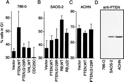
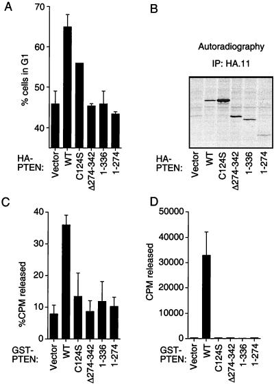
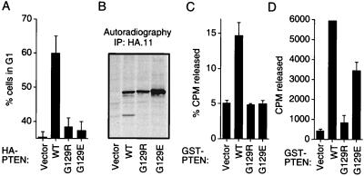
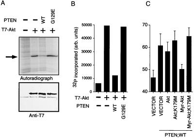
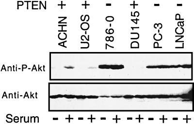
PTEN/MMAC1 is a tumor suppressor gene located on chromosome 10q23. Inherited PTEN/MMAC1 mutations are associated with a cancer predisposition syndrome known as Cowden's disease. Somatic mutation of PTEN has been found in a number of malignancies, including glioblastoma, melanoma, and carcinoma of the prostate and endometrium. The protein product (PTEN) encodes a dual-specificity protein phosphatase and in addition can dephosphorylate certain lipid substrates. Herein, we show that PTEN protein induces a G1 block when reconstituted in PTEN-null cells. A PTEN mutant associated with Cowden's disease (PTEN;G129E) has protein phosphatase activity yet is defective in dephosphorylating inositol 1,3,4,5-tetrakisphosphate in vitro and fails to arrest cells in G1. These data suggest a link between induction of a cell-cycle block by PTEN and its ability to dephosphorylate, in vivo, phosphatidylinositol 3,4,5-trisphosphate. In keeping with this notion, PTEN can inhibit the phosphatidylinositol 3,4, 5-trisphosphate-dependent Akt kinase, a downstream target of phosphatidylinositol 3-kinase, and constitutively active, but not wild-type, Akt overrides a PTEN G1 arrest. Finally, tumor cells lacking PTEN contain high levels of activated Akt, suggesting that PTEN is necessary for the appropriate regulation of the phosphatidylinositol 3-kinase/Akt pathway.


Figures

Figure 1
Figure 1
PTEN induces a G1 block. (A) PTEN, but not pVHL or cdc25C, induced a G1 block in 786-O cells. 786-O cells were transiently cotransfected with a plasmid encoding CD19 (pCD19) along with plasmids encoding the indicated proteins. Forty hours after transfection, cells were fixed and the cell-cycle distribution of the successfully transfected cells was determined by fluorescence-activated cell sorting analysis. The mean and SEM of two experiments are shown. (B) PTEN does not alter the cell-cycle profile of SAOS-2(RB−/−) cells. SAOS-2 cells were transiently transfected with pCD19 and the plasmids encoding the indicated proteins and analyzed as in Fig. 2A. The mean and SEM of two experiments are shown. (C) PTEN does not alter the cell-cycle profile of ACHN cells. ACHN cells were transiently transfected with pCD19 or plasmids encoding the indicated proteins and analyzed as in A. The mean and SEM of two experiments are shown. (D) Immunoblot detection of PTEN protein in 786-O, SAOS-2, and ACHN cells. C54 anti-PTEN antiserum was used to detect PTEN by immunoblot analysis of protein extracts from the indicated cell lines.
Figure 2
Figure 2
Substrate trapping variants of PTEN induce a G1 block but tumor-derived mutants do not. (A) Comparison of wild-type, substrate-trapping and C-terminal mutant forms of PTEN in the cell-cycle assay. 786-O cells were cotransfected with pCD19 and plasmids encoding the indicated proteins and analyzed as in Fig. 1A. (B) Expression of PTEN proteins in 786-O cells. 786-O cells were transfected with plasmids encoding the indicated proteins. Forty hours after transfection anti-HA immunoprecipitates of protein extracts prepared from metabolically labeled cells were separated by gel electrophoresis and subjected to fluorography. (C) Inositol phosphatase activity of GST-PTEN and mutant derivatives. The indicated GST-PTEN fusion proteins were used to dephosphorylate [3H]IP4. (D) Protein tyrosine phosphatase activity of GST-PTEN and mutant derivatives. The indicated GST-PTEN fusion proteins were used to dephosphorylate 33P-labeled poly(Glu4-Tyr1) copolymers. (A, C, and D) The mean and SEM of two experiments are shown.
Figure 3
Figure 3
CD mutant G129E preserves protein phosphatase activity but lacks inositol phosphatase activity and is incapable of inducing a G1 block. (A) Comparison of wild-type PTEN and the mutants G129R and G129E in the cell-cycle assay. 786-O cells were cotransfected with pCD19 and plasmids encoding the indicated proteins and analyzed as in Fig. 1A. (B) Comparison of the expression of wild-type PTEN and the mutants G129R and G129E in 786-O cells. 786-O cells were transfected with plasmids encoding the indicated proteins. Anti-HA immunoprecipitates of protein extracts prepared from metabolically labeled cells were separated by gel electrophoresis and subject to fluorography. (C) Inositol phosphatase activity of GST-PTEN and mutant derivatives. The indicated GST-PTEN fusion proteins were analyzed as in Fig. 2C. (D) Protein tyrosine phosphatase activity of GST-PTEN and mutant derivatives. The indicated GST-PTEN fusion proteins were analyzed as in Fig. 2D. (A, C, and D) The mean and SEM of two experiments are shown.
Figure 4
Figure 4
PTEN inhibits Akt kinase activity and an activated form of Akt can override a PTEN induced G1 block. (A) Inhibition of Akt kinase by wild-type PTEN. U2-OS cells were transfected with plasmids encoding the indicated proteins. After transfection, anti-T7 immunoprecipitates were prepared and used to phosphorylate a GST-peptide substrate in vitro. Autoradiography (Upper) and anti-T7 immunoblot (Lower) of the same membrane are shown. The black arrow indicates the position of the substrate. Results are representative of two experiments. (B) Quantitation of 32P incorporation in A with a PhosphoImager. (C) Myr-Akt overrides a PTEN-induced G1 block. 786-O cells were transiently cotransfected with pCD19 and plasmids encoding PTEN with empty vector (pLNCX) or with plasmids encoding the indicated Akt proteins. After transfection the cells were analyzed as in Fig. 1A. The mean and SEM of two experiments are shown.
Figure 5
Figure 5
Cells lacking PTEN protein contain high levels of Akt, phosphorylated on Ser-473. The indicated cells lines were serum-deprived for 24 h in duplicate. One p100 plate from each pair was refed with fresh medium containing 10% fetal calf serum for 90 min, after which protein extracts were prepared and subject to immunoblot analysis with an antibody directed at Akt phospho-Ser-473 (Upper) or an antibody that recognizes Akt independent of its phosphorylation status (Lower).

Similar articles

Cited by

References

    1. Gray I C, Phillips S M, Lee S J, Neoptolemos J P, Weissenbach J, Spurr N K. Cancer Res. 1995;55:4800–4803. - PubMed
    1. Rasheed B K, McLendon R E, Friedman H S, Friedman A H, Fuchs H E, Bigner D D, Bigner S H. Oncogene. 1995;10:2243–2246. - PubMed
    1. Steck P A, Pershouse M A, Jasser S A, Yung W K, Lin H, Ligon A H, Langford L A, Baumgard M L, Hattier T, Davis T, et al. Nat Genet. 1997;15:356–362. - PubMed
    1. Li J, Yen C, Liaw D, Podsypanina K, Bose S, Wang S I, Puc J, Miliaresis C, Rodgers L, McCombie R, et al. Science. 1997;275:1943–1947. - PubMed
    1. Li D M, Sun H. Cancer Res. 1997;57:2124–2129. - PubMed

Publication types

MeSH terms

Substances