. 1999 Mar 2;96(5):2141-6.
doi: 10.1073/pnas.96.5.2141.

Human deafness dystonia syndrome is a mitochondrial disease

Affiliations

Human deafness dystonia syndrome is a mitochondrial disease

C M Koehler et al. Proc Natl Acad Sci U S A. .

Abstract

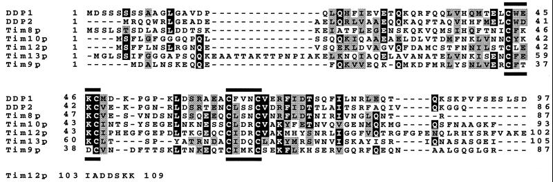
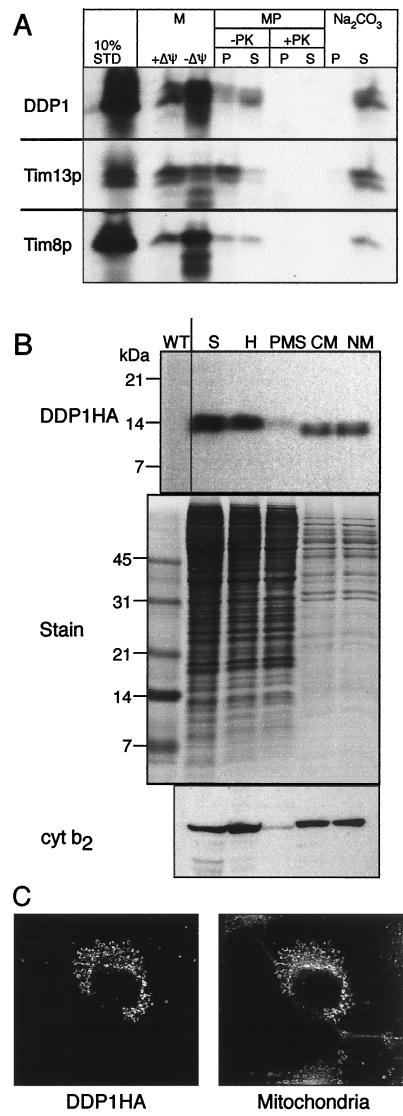
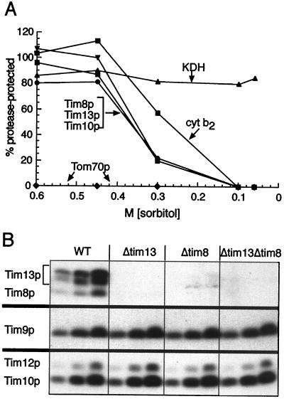
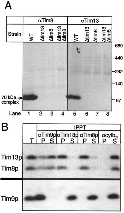
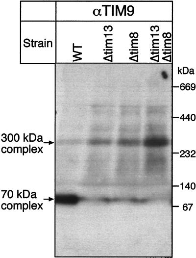
The human deafness dystonia syndrome results from the mutation of a protein (DDP) of unknown function. We show now that DDP is a mitochondrial protein and similar to five small proteins (Tim8p, Tim9p, Tim10p, Tim12p, and Tim13p) of the yeast mitochondrial intermembrane space. Tim9p, Tim10p, and Tim12p mediate the import of metabolite transporters from the cytoplasm into the mitochondrial inner membrane and interact structurally and functionally with Tim8p and Tim13p. DDP is most similar to Tim8p. Tim8p exists as a soluble 70-kDa complex with Tim13p and Tim9p, and deletion of Tim8p is synthetically lethal with a conditional mutation in Tim10p. The deafness dystonia syndrome thus is a novel type of mitochondrial disease that probably is caused by a defective mitochondrial protein-import system.


Figures

Figure 1
Figure 1
Members of the Tim family in the mitochondrial intermembrane space share a “twin CX3C” motif. Sequence comparison of DDP1, DDP2, Tim8p, Tim10p, Tim12p, Tim13p, and Tim9p. Black boxes, identical amino acids; shaded boxes, similar amino acids; black bars, “CX3C” motif.
Figure 2
Figure 2
DDP1 is located in the mitochondrial intermembrane space. (A) Import into isolated yeast mitochondria. Radiolabeled Tim8p, Tim13p, and DDP1 were synthesized in vitro and incubated for 10 min at 25°C with wild-type yeast mitochondria in the presence (+) or absence (−) of a membrane potential (Δψ). Mitochondria (M) were treated with trypsin to digest nonimported precursor and then with soybean trypsin inhibitor, and finally were analyzed by SDS/PAGE and fluorography. Equal aliquots of the mitochondria that had imported precursor in the presence of a membrane potential were subjected further to osmotic shock or alkali extraction (5, 6). Osmotic shock (that selectively ruptures the outer membrane and generates mitoplasts, MP) was performed in the absence or presence of 50 μg/ml proteinase K (PK). After addition of 1 mM phenylmethylsulfonyl fluoride (PMSF), samples were centrifuged at 14,000 × g for 10 min to separate pellet (P) and supernatant (S). For alkali extraction (Na2CO3), mitochondria were incubated with 100 mM Na2CO3 for 30 min, followed by centrifugation at 100,000 × g for 15 min to separate pellet (P) and supernatant (S). 10% STD, 10% of the radioactive precursor present in each assay. (B) Expression in yeast cells. A yeast transformant expressing DDP1 with a C-terminal HA tag from a multicopy plasmid was grown in semisynthetic lactate medium at 30°C, converted to spheroplasts (S), and fractionated into total homogenate (H), postmitochondrial supernatant (PMS), crude mitochondria (CM), and mitochondria purified on a Nycodenz gradient (NM). An equal amount of each fraction was analyzed by SDS/PAGE and immunoblotting with an mAb against HA followed by rabbit anti-mouse antibody or with a rabbit antiserum against cytochrome b2 (cyt b2). The immune complexes then were decorated with [125I]protein A. As a control, a Coomassie blue-stained gel of the fractions is included (Stain). WT, mitochondria from a strain not transformed with the tagged DDP1 gene. (C) Expression in monkey cells. DDP1 was transiently expressed in cultured COS7 cells under the control of the cytomegalovirus promoter. After staining, the cells were examined by indirect immunofluorescence (ref. ; primary antibody for DDP1HA, monoclonal 12CA5 antibody; secondary antibody, FITC-conjugated rabbit anti-mouse) as well as by staining with the mitochondria-specific fluorescent dye Mitotracker (Mitochondria).
Figure 3
Figure 3
Tim8p and Tim13p are located in the intermembrane space and stabilize each other. (A) Submitochondrial localization. Isolated yeast mitochondria were incubated in 20 mM Hepes-KOH, pH 7.4, 100 μg/ml proteinase K, and the indicated sorbitol concentrations on ice for 30 min. After addition of 1 mM PMSF and centrifugation, the pellet was analyzed by SDS/PAGE and immunoblotting for Tim8p, Tim10p, Tim13p, cytochrome b2 (cyt b2; intermembrane space marker), α-ketoglutarate dehydrogenase (KDH; matrix marker), and Tom70p (outer membrane marker). Antigen protected from protease was quantified by densitometry and expressed as a percentage of the antigen measured in the absence of protease. (B) Deletion of Tim8p causes loss of Tim13p, and vice versa. The parental wild-type strain (WT) and strains deleted for TIM13 (Δtim13), TIM8 (Δtim8), or both TIM13 and TIM8 (Δtim13Δtim8) were grown in lactate medium for 16 hr at 30°C. Three different amounts (50, 100, and 200 μg protein) of purified mitochondria were analyzed by SDS/PAGE and immunoblotting, with monospecific rabbit antisera for the proteins indicated on the left and (data not shown) for Tim22p, Tim54p, the ADP/ATP carrier, cytochrome b2, and porin. Blots were decorated with [125I]protein A and autoradiographed.
Figure 4
Figure 4
Tim8p, Tim13p, and a fraction of Tim9p form a 70-kDa complex in the intermembrane space. (A) Proteins solubilized from purified yeast mitochondria with 0.16% n-dodecylmaltoside were subjected to “blue native gel” electrophoresis (ref. ; 6–16% acrylamide) and analyzed for Tim8p and Tim13p by immunoblotting and decoration with [125I]protein A. WT, parental strain; Δtim13, TIM13 deleted; Δtim8, TIM8 deleted; Δtim13Δtim8, TIM13 and TIM8 deleted. All strains were haploid. (B) A soluble intermembrane space fraction was incubated at 4°C for 2 hr with protein A-Sepharose that had been coupled to IgGs against Tim9p (αTim9p), Tim13p (αTim13p), Tim8p (αTim8p), or cytochrome b2 (αcytb2). After centrifugation, aliquots (equivalent to 200 μg mitochondria) of unbound (S) and bound (P) proteins were analyzed by SDS/PAGE and immunoblotting for Tim13p and Tim8p (Upper) or for Tim9p (Lower). IPPT, immunoprecipitation; T, total mitochondrial intermembrane space fraction analyzed without immunoprecipitation.
Figure 5
Figure 5
Deletion of TIM8 or TIM13 causes redistribution of Tim9p from the 70-kDa complex to the 300-kDa complex. Proteins solubilized from purified yeast mitochondria with 0.16% n-dodecylmaltoside were subjected to “blue native gel” electrophoresis (ref. ; 6–16% acrylamide) and analyzed for Tim9p by immunoblotting and decoration with [125I]protein A. WT, parental strain; Δtim13, TIM13 deleted; Δtim8, TIM8 deleted; Δtim13Δtim8, TIM13 and TIM8 deleted.

Comment in

References

    1. Ryan K R, Jensen R E. Cell. 1995;83:517–519. - PubMed
    1. Schatz G, Dobberstein B. Science. 1996;271:1519–1526. - PubMed
    1. Neupert W. Annu Rev Biochem. 1997;66:863–917. - PubMed
    1. Pfanner N. Curr Biol. 1998;8:R262–R265. - PubMed
    1. Koehler C M, Jarosch E, Tokatlidis K, Schmid K, Schweyen R J, Schatz G. Science. 1998;279:369–373. - PubMed

Publication types

MeSH terms