. 1999 Mar 2;96(5):2233-8.
doi: 10.1073/pnas.96.5.2233.

Immune surveillance against a solid tumor fails because of immunological ignorance

Affiliations

Immune surveillance against a solid tumor fails because of immunological ignorance

A F Ochsenbein et al. Proc Natl Acad Sci U S A. .

Abstract

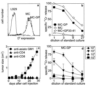
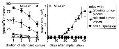
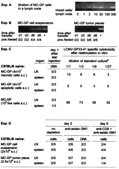
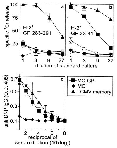
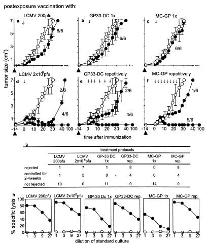
Many peripheral solid tumors such as sarcomas and carcinomas express tumor-specific antigens that can serve as targets for immune effector T cells. Nevertheless, overall immune surveillance against such tumors seems relatively inefficient. We studied immune surveillance against a s.c. sarcoma expressing a characterized viral tumor antigen. Surprisingly, the tumor cells were capable of inducing a protective cytotoxic T cell response if transferred as a single-cell suspension. However, if they were transplanted as small tumor pieces, tumors readily grew. Tumor growth correlated strictly with (i) failure of tumor cells to reach the draining lymph nodes and (ii) absence of primed cytotoxic T cells. Cytotoxic T cells were not tolerant or deleted because a tumor antigen-specific cytotoxic T cell response was readily induced in lymphoid tissue by immunization with virus or with tumor cells even in the presence of large tumors. Established tumors were rejected by vaccine-induced effector T cells if effector T cells were maintained by prolonged or repetitive vaccination, but not by single-dose vaccination. Thus, in addition to several other tumor-promoting parameters, some antigenic peripheral sarcomas-and probably carcinomas-may grow not because they anergize or tolerize tumor-specific T cells, but because such tumors are immunologically dealt with as if they were in a so-called immunologically privileged site and are ignored for too long.


Figures

Figure 1
Figure 1
Characterization of the MC-GP tumor cells, tumor growth, and immune response. (a) MHC class I (Db) expression of MC-GP cells vs. L929 (H-2k) cells; MC-GP cells were negative for intercellular adhesion molecule-1, B7.1, B7.2, and lymphocyte function-associated antigen-1 (not shown). (b) MC-GP and MC57 cells with or without peptide labeling (GP33–41) were used as target cells in vitro in a 51Cr-release assay. (c) MC-GP cells (2 × 10⁶) were injected s.c. in both flanks of C57BL/6 mice that were depleted of NK-cells [anti-asialo GM1 (Wako Biochemicals, Osaka)], 30 μl diluted in 200 μl balanced salt solution i.v. on day −1), of CD4+ T cells [200 μl i.p. of anti-CD4 antibodies (YTS191.1) on days −3 and −1], or of CD8+ T-cells [200 μl i.p of anti-CD8 antibodies (YTS169.4.2) on day −3 or −1]. Tumor growth was followed during 30 days. Tumor volume was calculated by the formula/V = πxabc/6, where a, b, and c are the orthogonal diameters. Titrated doses of (d) live MC-GP were injected s.c. into the flank of C57BL/6 animals. Eight days later, splenocytes were restimulated in vitro on irradiated GP33–41-pulsed splenocytes for 5 days and the CTL activity was determined then in a Cr51-release assay.
Figure 2
Figure 2
Comparison of tumor take vs. induction of CTL responses against MC-GP tumor cells or tumor pieces. MC-GP cells (2 × 10⁶) were injected as single cells s.c. in both flanks (●), or small tumor pieces containing comparable numbers of tumor cells were implanted at similar locations (▴, ▾). According to the results, secondary CTL activity (a) and tumor growth (b) were divided and presented as two groups: growing tumors (▴) and rejected tumors (▾). The induction of a CTL response was assessed by a 51Cr-release assay after 5 days of restimulation in vitro of local lymph node or spleen cells taken 8 days after injection of the tumor-cell suspension (●, ○). Open symbols represent killing against unpulsed EL-4 target cells, closed symbols against GP33–41-pulsed EL-4 target cells (a). CTL activity was similarly assessed 8 days (not shown) and 2 weeks (not shown) after implantation of tumor pieces and at the end of the experiment (after about 40 days). CTL activity is given for each group as mean ± SD. One of three comparable experiments is shown (error bars indicate SD). After each in vivo experiment, tumor cells were cultured and all tested positive for Db-GP33–41 expression (not shown).
Figure 3
Figure 3
Homing of MC-GP tumor cells into local lymph nodes and spleen. DNA was extracted from draining inguinal lymph nodes or spleen at the indicated time points after injection of a tumor cell suspension or after the implantation of small tumor pieces in both flanks of C57BL/6 mice. A LCMV GP-specific nested PCR was performed with the primers described in Materials and Methods. (A) The sensitivity of the assay was determined in vitro by mixing tumor cells with a constant number of 106 lymph node cells from untreated C57BL/6 mice. (B) The number of mice with positive lymph nodes over the total number of mice tested is given for each time point in each group. Lymph nodes from control C57BL/6 mice, which had not received tumor cells, and water were tested in parallel and were negative in all experiments shown. The integrity of the DNA extracted from lymph nodes was successfully tested by a perforin exon 3-specific PCR (not shown). (C) Dead tumor cells s.c. did not lead to a PCR signal or to a primed CTL response in contrast to live cells (106 s.c.). MC-GP cells (107) were either treated by freeze-thawing (necrotic, trypan blue positive, not shown) or kept on 42°C for 24 hr (apoptotic, trypan blue negative hypodiploid DNA peak in propidium iodide staining and flow cytometry, details not shown) and then injected repetitively (4 times) on alternate days in both flanks of C57BL/6 mice. One day after the last injection, DNA was prepared from spleen and from draining lymph nodes and tested by nested PCR for LCMV GP-specific DNA. Values indicate number of PCR-positive samples per total number of samples tested. At the same time point, splenocytes were restimulated in vitro for 5 days and then tested in a 51Cr-release assay. Values indicate percent specific 51Cr release as mean of three animals at the dilution of standard culture indicated. One of two comparable experiments is shown. (D) No live tumor cells could be isolated from the spleen or lymph nodes of untreated C57BL/6 mice on day 2, 4 (not shown), or 6 (not shown). Live tumor cells and GP-specific DNA could be detected in vitro after depletion of NK cells [30 μl of anti-asialo GM1 (Wako Biochemicals, Osaka) diluted in 200 μl balanced salt solution i.v. on day −1] on day 2 and after CD8 [200 μl i.p. anti-CD8 (YTS169.4.2) on days −3 and −1] plus NK depletion on day 6 after injection of MC-GP tumor cell suspensions, but not after transplantation of solid MC-GP tumor pieces. To detect live cells, lymph nodes (LN) or spleens were passed through a fine-mesh stainless steel grid, and the resulting single-cell suspension was cultured on selection medium/[0.8 mg/ml G-418 (GIBCO/BRL)]. Values indicate positive samples over total number of mice tested.
Figure 4
Figure 4
Examination of crosspriming vs. direct induction of a CTL response by MC-GP tumor cells. Tumor cells (5 × 10⁶ MC57 or MC-GP) were injected four times on alternate days i.p into (C57BL/6 H-2b × B10.D2 H2d)F1 animals. On day eight after the first injection, the splenocytes were restimulated on GP283–291 (H-2d)- or GP33–41 (H-2b)-labeled F1 splenocytes for 5 days. CTL activity was then tested on (a) GP283–291-labeled P815 (H-2d) or (b) GP33–41-labeled EL4 (H-2b) cells. Closed symbols represent peptide-labeled targets, open symbols represent unlabeled targets. (c) C57BL/6 (H-2b) mice treated with the same repetitive injection protocol were challenged with 20 μg of DNP coupled to P13 [helper epitope GP60–80 in H-2b (27)] 2 days after the last injection of cells. Seven days later, anti-DNP IgG titers were measured by ELISA. LCMV-primed (−60 days, 102 pfu LCMV i.v.) mice served as positive controls. The mean and the SD of three animals per group are shown. One of two comparable experiments is shown.
Figure 5
Figure 5
Immunotherapy of peripheral MC-GP tumors. Mice with growing MC-GP tumors of ≥5 mm on day 14 after transplantation were selected for immunotherapy experiments; tumor growth after the different priming strategies was followed up to 100 days. Closed circles indicate animals from the different experimental groups and open circles represent untreated control animals (a–f). The numbers shown indicate the proportion of tumor growth (or/rejection) per tumors tested. Each experiment was repeated twice with similar results. A low dose of LCMV (2 × 10² pfu) (a) or a high dose (2 × 10⁶ pfu) (d) was injected intravenously 14 days after transfer/transplantation of tumor cells; this time point is now taken as day 0 of therapy. The day of transplantation is indicated as ▴. Dendritic cells were isolated from GP33–41 transgenic mice (H-8) (22) (GP33-DC) and injected either once on day 0 (b) or repetitively during 3 weeks as indicated (e) (105 cells per injection). MC-GP cells were also injected once (c) or repetitively every other day during 3 weeks (2 × 10⁶ cells per injection) (f). The outcome of all performed experiments is summarized in g. All the treated animals mounted a CTL response when tested in a 51Cr release assay after in vitro restimulation, whereas the untreated controls did not (h). (●), CTL response of animals 40 days after the beginning of the immunotherapy; (○), animals without immunotherapy.

References

    1. Old L J, Boyse E A, Clarke D A. Ann N Y Acad Sci. 1962;101:80–106.
    1. Pardoll D M. Nat Med. 1998;4:525–531. - PubMed
    1. Boon T, Cerottini J C, van-den-Eynde B, van-der-Bruggen P, Van-Pel A. Annu Rev Immunol. 1994;12:337–365. - PubMed
    1. Möller G. Immunol Rev. 1995;145:1–250.
    1. Andrews E J. J Natl Cancer Inst. 1974;52:729–732. - PubMed

Publication types

MeSH terms

LinkOut - more resources