. 1999 Mar 2;96(5):2396-401.
doi: 10.1073/pnas.96.5.2396.

The Salmonella invasin SipB induces macrophage apoptosis by binding to caspase-1

Affiliations

The Salmonella invasin SipB induces macrophage apoptosis by binding to caspase-1

D Hersh et al. Proc Natl Acad Sci U S A. .

Abstract

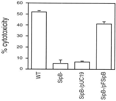
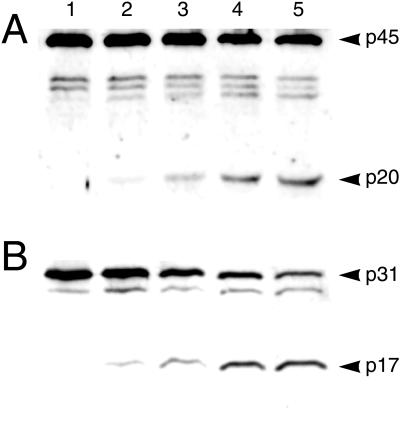
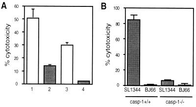
Recently, Salmonella spp. were shown to induce apoptosis in infected macrophages. The mechanism responsible for this process is unknown. In this report, we establish that the Inv-Spa type III secretion apparatus target invasin SipB is necessary and sufficient for the induction of apoptosis. Purified SipB microinjected into macrophages led to cell death. Binding studies show that SipB associates with the proapoptotic protease caspase-1. This interaction results in the activation of caspase-1, as seen in its proteolytic maturation and the processing of its substrate interleukin-1beta. Caspase-1 activity is essential for the cytotoxicity. Functional inhibition of caspase-1 activity by acetyl-Tyr-Val-Ala-Asp-chloromethyl ketone blocks macrophage cytotoxicity, and macrophages lacking caspase-1 are not susceptible to Salmonella-induced apoptosis. Taken together, the data demonstrate that SipB functions as an analog of the Shigella invasin IpaB.


Figures

Figure 1
Figure 1
SipB dependence of the cytotoxicity of Salmonella on the J774A.1 macrophage cell line. Cells were infected at an moi of 100:1 for 10 hr and then processed for a LDH-release assay. The wild-type S. typhi (WT) was cytotoxic to J774A.1 cells. An insertion mutant of sipB (SipB-) did not retain the cytotoxic phenotype, which can be complemented by sipB in trans (SipB−/pFSipB). The negative control (SipB−/pUC19) did not restore the killing activity. The means and standard deviations of three experiments are shown.
Figure 2
Figure 2
Macrophages are killed after microinjection of SipB. Every macrophage in the field was microinjected with purified GST (A and B, same field), GST–IpaB (C and D, same field), or GST–SipB (E and F, same field) and then stained with PI in PBS without fixation. Apoptotic cells were then scored for PI uptake and cellular morphology. Many more cells are positive for PI uptake and apoptotic cellular alterations in GST–SipB (E and F) and GST–IpaB (C and D) injected cells than found in GST-injected cells (A and B). Arrowheads highlight some of the cells undergoing apoptosis. See text for quantification of the levels of apoptosis.
Figure 3
Figure 3
Subcellular localization of SipB in Salmonella-infected macrophages. RAW264.7 cells infected with S. typhi Ty233 transformed by pFSipB (A, C, and E), pSipB (D and F), or vector alone (B) for 40 min. Indirect immunofluorescence (A and B): Both macrophage nuclei and the bacteria were stained with the DNA binding dye PI. FSipB was detected by using a fluorescein-labeled secondary antibody (green) and shown to be dispersed in the cytoplasm of infected macrophages. Immunoelectron microscopy: Gold particles show that the majority of FSipB is localized in the cytoplasm of the macrophage (C and E). There appears to be an association between FSipB and endomembranes, most notably endoplasmic reticulum. (E) Computer enhancement of signal found in C. SipB lacking the FLAG epitope was not detected (D and F). (F) Computer enhancement of signal found in D. (Bars = 10 μm.)
Figure 4
Figure 4
Binding assays to determine if SipB binds to caspase-1. (A) Western blot analysis of GST (lane 1) and GST–SipB (lane 2) affinity-purified proteins from J774A.1 cell lysates resolved on 15% SDS/PAGE gel with anti-mouse caspase-1 rabbit serum. GST–SipB specifically bound to two proteins of 45 and 33 kDa recognized by the anti-caspase-1 antibody. These proteins are the precursor of caspase-1 and one of its intermediate forms. (B) Western blot analysis of proteins coimmunoprecipitated by anti-FLAG antibody from J774A.1 cells infected for 40 min with either SF620/pSipB (lane 2) or SF620/pFSipB (lane 3). Included as control is lysate from uninfected equivalent number of cells (lane 1). Samples were resolved on 15% SDS/PAGE and immunoblotted with an anti-caspase-1 antibody. The precursor form of caspase-1 (p45) was coimmunoprecipitated with FSipB, demonstrating that SipB binds to caspase-1 during infection of macrophages. No proteins immunoprecipitated from the lysate of cells exposed to SipB lacking the FLAG epitope were recognized by the anti-caspase-1 antibody.
Figure 5
Figure 5
Caspase-1 is activated during infection of macrophages infected with Salmonella. (A and B) LPS-activated murine peritoneal macrophages were infected with S. typhimurium C5. After 10 min (lane 2), 25 min (lane 3), 40 min (lane 4), and 70 min (lane 5) of infection, cell lysates were resolved on an 15% SDS/PAGE gel and immunoblotted with either an anti-caspase-1 antibody (A) or an anti-IL-1β antibody (B). As a control, macrophages were infected with the sipB mutant ST100 for 70 min (lane 1 in each gel) and processed in parallel. Both caspase-1 and its specific substrate IL-1β undergo catalytic maturation within 10 min of infection with mature forms accumulating throughout the course of infection observed.
Figure 6
Figure 6
Caspase-1 is essential for the induction of apoptosis. (A) Protection by YVAD of RAW264.7 macrophages from Salmonella-induced cell death. At 1 hr before infection with S. typhimurium SL1344 at an moi of either 100:1 (1 and 2) or 50:1 (3 and 4), cells were preincubated with 50 μM Ac-YVAD-CMK (2 and 4) or media alone (1 and 3). The presence of the caspase-1 inhibitor led to a decrease in cytotoxicity. (B) Caspase-1 is necessary for Salmonella-induced cytotoxicity. Peritoneal macrophages from either wild-type or caspase-1 −/− mice were infected for 8 hr with S. typhimurium SL1344 or BJ66. SL1344 killed the macrophages from the wild-type mice, whereas neither SL1344 nor BJ66 were able to kill macrophages lacking caspase-1. The means and SDs of three experiments are shown.

References

    1. Keusch G T. In: Harrison’s Principles of Internal Medicine. 13th Ed. Isselbacher K J, Braunwald E, Wilson J D, Martin J B, Fauci A S, Kasper D L, editors. New York: McGraw–Hill; 1994. pp. 671–676.
    1. Finlay B B. Curr Top Microbiol Immunol. 1994;192:163–185. - PubMed
    1. Jones B D, Ghori N, Falkow S. J Exp Med. 1994;180:15–23. - PMC - PubMed
    1. Clark M A, Jepson M A, Simmons N L, Hirst B H. Res Microbiol. 1994;145:543–553. - PubMed
    1. Fields P I, Swanson R V, Haidaris C G, Heffron F. Proc Natl Acad Sci USA. 1986;83:5189–5193. - PMC - PubMed

Publication types

MeSH terms