

The biological clock of very premature primate infants is responsive to light
- PMID: 10051658
- PMCID: PMC26800
- DOI: 10.1073/pnas.96.5.2426
The biological clock of very premature primate infants is responsive to light
Abstract
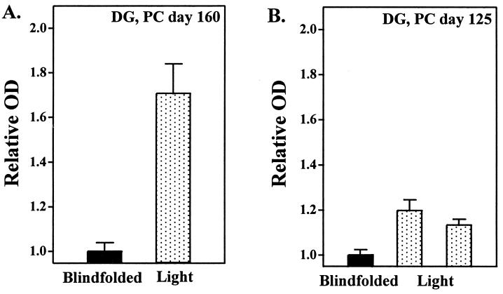
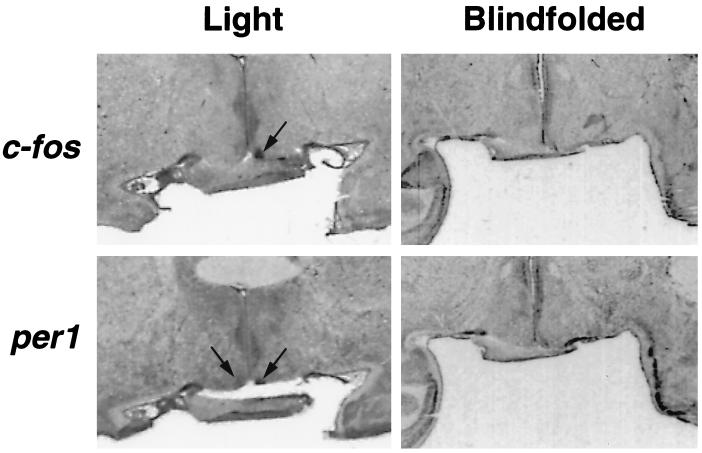
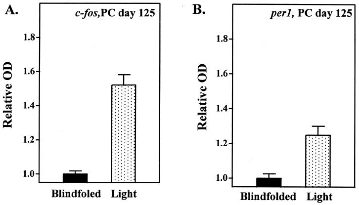
Each year more than 250,000 infants in the United States are exposed to artificial lighting in hospital nurseries with little consideration given to environmental lighting cycles. Essential in determining whether environmental lighting cycles need to be considered in hospital nurseries is identifying when the infant's endogenous circadian clock becomes responsive to light. Using a non-human primate model of the developing human, we examined when the circadian clock, located in the hypothalamic suprachiasmatic nuclei (SCN), becomes responsive to light. Preterm infant baboons of different ages were exposed to light (5,000 lux) at night, and then changes in SCN metabolic activity and gene expression were assessed. After exposure to bright light at night, robust increases in SCN metabolic activity and gene expression were seen at ages that were equivalent to human infants at 24 weeks after conception. These data provide direct evidence that the biological clock of very premature primate infants is responsive to light.
Figures

References
-
- Moore R Y. J Biol Rhythms. 1993;8:S3–S9. - PubMed
-
- Moore-Ede M C, Czeisler C A, Richardson G S. N Engl J Med. 1983;309:469–476. - PubMed
-
- Moore-Ede M C, Czeisler C A, Richardson G S. N Engl J Med. 1983;309:530–536. - PubMed
-
- Klein D C, Moore R Y, Reppert S M. Suprachiasmatic Nucleus: The Mind’s Clock. New York: Oxford Univ. Press; 1991.
Publication types
MeSH terms
Substances
Grants and funding
LinkOut - more resources
Full Text Sources
Other Literature Sources
Medical
